1/19
BIOLOGIC BIOMATERIALS: TISSUE DERIVED BIOMATERIAL
(COLLAGEN)
[
Adopsi dari:
Joseph D. Bronzino, “Biomedical Engineering Fundamentals”, CRC Press, third edition, 2006, Section V, CHAPTER 43, by Shu Tung Li.]43.1 STRUCTURE AND PROPERTIES OF COLLAGEN AND COLLAGEN-RICH TISSUES
43.1.1 Structure of Collagen
Collagen is a multifunctional familyof proteins of unique structural characteristics. It is the most abundant and ubiquitous protein in the body, its functions ranging from serving crucial Biomechanical functions in bone, skin, tendon, and ligament to controlling cellular gene expressions in development [Nimni and Harkness, 1988]. Collagen molecules like all proteins are formed in vivo by enzymatic regulated step-wise polymerization reaction between amino and carboxyl groups of amino acids, where R is a side group of an amino acid residue.
The simplest amino acid is glycine (Gly) (R=H ), where a hypothetical flat sheet organization of polyglycine molecules can form and be stabilized by intermolecular hydrogen bonds (Figure 43.la). However, when R is a large group as in most other amino acids, the stereochemical constraints frequently force the polypeptide chain to adapt a less constraining conformation by rotating the bulky R groups away from the crowded interactions, forming a helix, where the large R groups are directed toward the surface of the helix (Figure 43.lb). The hydrogen bonds are allowed to form within a helix between the hydrogen attached to nitrogen in one amino acid residue and the oxygen attached to a second amino acid residue. Thus, the final conformation of a protein, which is directly related to its function, is governed primarily by the amino acid sequence of the particular protein.
Collagen is a protein comprised of three polypeptides (a chains), each having a general amino acid sequence of (—.Gly—X—Y—)n where X is any other amino acid and is frequently proline
2/19
FIGURE 43.1 (a) Hypothetical flat sheet structure of a protein. (b) Helical arrangement of a protein chain.
TABLE 43.1 Amino Acid Content of Collagen
To date, 19 proteins can be classified as collagen [Fukai et al., 1994]. Among the various collagens, type I collagen is the most abundant and is the major constituent of bone, skin, ligament, and tendon. Due to the abundance and ready accessibility of these tissues, they have been frequently used as a source for the preparation of collagen. This chapter will not review the details of the structure of the different collagens. The readers are referred to recent reviews for a more in-depth discussion of this subject [Nimni, 1988; van der Rest et al., 1990; Fukai et al., 1994; Brodsky and Ramshaw, 1997]. It is, however, of particular relevance to review some salient structural features of the type I collagen in order to facilitate the subsequent discussions of properties and its relation to biomedical applications.
A type I collagen molecule (also referred to as tropocollagen) isolated from various tissues has a molecular weight of about 283,000 daltons. It is comprised of three left-handed helical polypeptide chains (Figure 43.2a) which are intertwined forming a right-handed helix around a central molecular axis (Figure 43.2b). Two of the polypeptide chains are identical (α1) having
1056 amino acid residues, and the third polypeptide chain (α2) has 1029 amino acid residues
3/19
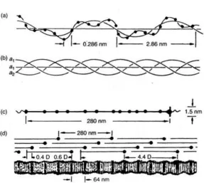
FIGURE 43.2 Diagram depicting the formation of collagen, which can be visualized as taking place in several steps: (a) single chain left-handed helix; (b) three single chains intertwined into a triple stranded helix; (c) a collagen (tropocollagen) molecule; (d) collagen molecules aligned in D staggered
fashion in a fibril producing overlap and hole regions.
FIGURE 43.3 A space-filling model of the collagen triple helix, showing all the atoms in a ten-residue segment of repeating triplet sequence (Gly—Pro—Hyp)N. The arrow shows an interchain hydrogen bond.
The arrow heads identify the hydroxy groups of hydroxyproline in one chain. The circle shows a hydrogen-bonded water molecule. The short white lines identify the ridge of amino acid chains. The short
4/19
The triple-helical structure of a collagen molecule is stabilized by several factors (Figure 43.3): (1) a tight fit of the amino acids within the triple-helix — this geometrical stabilization factor can be appreciated from a space-tilling model constructed from a triple helix with (Gly—Pro—Hyp) sequence (Figure 43.3); (2) the interchain hydrogen bond formation between the backbone carbonyl and amino hydrogen interactions; and (3) the contribution of water molecules to the interchain hydrogen bond formation.
The telopeptides are regions where intermolecular crosslinks are formed in vivo. A common inter molecular crosslinks is formed between an allysine (the ε-amino group of lysine or hydroxy-lysine has been converted to an aldehyde) of one telopeptide of one molecule and an ε-amino group of a lysine or hydroxylysine in the triple helix or a second molecule (43.2). Thus the method commonly used to solubilize the collagen molecules from crosslinked fibrils with proteolytic enzymes such as pepsin removes the telopeptides (cleaves the intermolecular crosslinks) from the collagen molecule. The pepsin solubilized collagen is occasionally referred to as atelocollagen [Stenzl, 1974].
Since the presence of hydroxyproline is unique in collagen elastin contains a small amount the determination of collagen content in a collagen-rich tissue is readily done by assaying the hvdroxyproline content.
5/19
FIGURE 43.4 (a) Scanning electron micrograph of the surface of an adult rabbit bone matrix, showing how the collagen fibrils branch and interconnect in an intricate. woven pattern (x4800) [Tiffit, 1980]. (b)
Transmission electron micrographs of (x 24,000) parallel collagen fibrils in tendon I Fung. 1992). (c) Transmission electron micrographs of (x24,000) mesh work of fibrils in skin [Fung, 1993].
6/19
One interesting and important structural aspect of collagen is its approximate equal number of acidic (aspartic and glutamic acids) and basic (lysines and arginines) side groups. Since these groups are charged under physiological conditions, the collagen is essentially electrically neutral [Li and Katz. 1976]. The packing of collagen molecules with a D staggering results in clusters of regions where the charged groups are located [Hofmann and Kuhn, 1981]. These groups therefore are in close proximity to form intra- and intermolecular hydrogen-bonded salt-linkages of the form (Pr — COO- H3N — Pr) [Li et al., 1975]. In addition, the side groups of many amino
acids are nonpolar [alanine (Ala), valine (Val), leucine (Leu), isoleucine (Ile), proline (Pro), and phenolalanine (Phe)] in character and hence hydrophobic, therefore, chains with these amino acids avoid contact with water molecules and seek interactions with the nonpolar chains of amino acids. In fact, the result of molecular packing of collagen in a fibril is such that the nonpolar groups are also clustered, forming hydrophobic regions within collagen fibrils [Hofmann and Kuhn, 1981]. Indeed, the packing of the collagen molecules in various tissues is believed to be a result of intermolecular interactions involving both the electrostatic and hydrophobic interactions [Hofmann and Kuhn, 1981; Katz and Li, 1981; Li et al., 1975].
The three-dimensional organization of type I collagen molecules within a fibril has been the subject of extensive research over the past 40 years [Fraser et al, 1983; Katz and Li, 1972, 1973a, b, 1981; Miller, 1976; Ramachandran, 1967; Yamuchi et al., 1986]. Many structural models have been proposed based on
an analysis of equatorial and off-equatorial x-ray diffraction patterns of rat-tail-tendon collagen [Miller, 1976; North et al., 1954]. intrafibrillar volume determination of various collagenous tissues [Katz and Li, 1972, 1973a,b], inermnlecular side chain interaction [Hofmann and Kuhn, 1981; Katz and Li, 1981; Li et al., 1981], and intermolecular crosslinking patterns studies [Yamuchi et al.. 1986]. The general understanding of the three-dimensional molecular packing in type I collagen fibrils is that the collagen molecules are arranged in hexagonal or near hexagonal arrays [Katz and Li, 1972, 1981; Miller. 1976]. Depending on the tissue, the intermolecular distance varies from about 0.15 nm in rat tail tendon to as large as 0.18 nm in bone and dentin [Katz and Li. l973b]. The axial staggering of the molecules by 1 ∼ 4D with respect to one another is tissue-specific and has not yet been fully elucidated.
There are very few interspecies differences in the structure of type I collagen molecule. The extensive homology of the structure of type I collagen may explain why this collagen obtained from animal species is acceptable as a material for human implantation.
43.1.2 Properties of Collagen-Rich Tissue
The function of collagenous tissue is related to its structure and properties. This section reviews some important properties of collagen-rich tissues.
43.1.2.1 Physical and Biomechanical Properties. The physical properties of tissues vary according to the amount and structural variations of the collagen fibers. In general, a collagen-rich tissue contains about 75 to 90% of collagen on a dry weight basis. Table 43.2 is a typical composition of a collagen-rich soft tissue such as skin. Collagen fibers (bundles of collagen fibrils) are arranged in different configurations in different tissues for their respective functions at specific anatomic shes. For example, collagen fibers are arranged in parallel in tendon (Figure 43.4b) and ligament for their high-tensile strength requirements, whereas collagen fibers in skin are arranged in random arrays (Figure 43.4c) to provide the resiliency of the tissue under stress. Other structure-supporting functions of collagen such as transparency for the lens of the eye and shaping of the ear or tip of the nose can also be provided by the collagen fiber. Thus, an important physical property of collagen is the three-dimensional organization of the collagen fibers.
7/19
crystalline collagen fibrils at about 56°C. The melting temperature of crystalline collagen fibrils is referred to as the denaruraijo,, temperature of collagenous tissues.
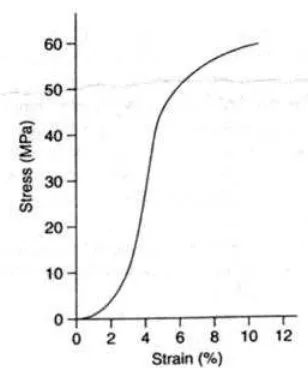
The stress—strain curves of a collagenous tissue such as tendon exhibit nonlinear behavior (Figure 43.6). This nonlinear behavior of stress—strain of tendon collagen is similar to that observed in synthetic fibers. The initial toe region represents alignment of fibers in the direction of stress. The steep rise in slope represents the majority of fibers stretched along their long axes. The decrease in slope following the steep rise may represent the breaking of individual fibers prior to the final catastrophic failure. Table 43.3 summarizes some mechanical properties of collagen and elastic fibers. The difference in biomechanical properties between collagen and elastin is a good example of the requirements for these proteins lo serve their specific functions in the body.
TABLE 43.2 Composition of Collagen-Rich Soft Tissues
FIGURE 43.6 A typical stress-strain curve for tendon I Rigby et aI.. 19591.
TABLE 43.3 Elastic Properties of Collagen and Eiasic Fibers
8/19
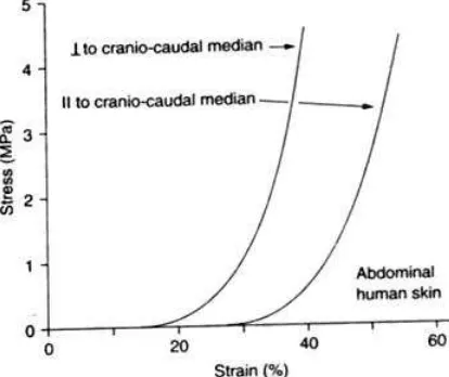
small load the fibers are straightened and aligned rather than stretched. Upon further stretching the fibrous lamellae align with respect to each other and resist further extension. When the skin is highly stretched the modulus of elasticity approaches that of tendon as expected of the aligned collagen fibers.
Cartilage is another collagen-rich tissue which has two main physiological functions One is the maintenance of shape (ear, tip of nose, and rings around the trachea), and the other is to provide bearing surfaces at joints. It contains very large and diffuse proteoglycan (protein-polysaccharide) molecules which form a gel in which the collagen-rich molecules entangled. They can affect the mechanical properties of the collagen by hindering the movements through the interstices of the collagenous matrix network.
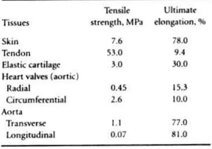
The joint cartilage has a very low coefficient of friction (<0.01). This is largely attributed to the squeeze-film effect between cartilage and synovial fluid. The synovial fluid can be squeezed out through highly fenestrated cartilage upon compressive loading, and the reverse action will take place in tension. The lubricating function is carried out in conjunction with glycosaminoglycans (GAG), especially chondroitin sulfates. The modulus of elasticity (10.3 to 20.7 MPa) and tensile strength (3.4 MPa) are quite low. However, wherever high stress is required the cartilage is replaced by purely collagenous tissue. Mechanical properties of some collagen-rich tissues are given in Table 43.4 as a reference.
43.1.2.2 Physiochemical Properties.
Electrostatic properties: A collagen molecule has a total of approximately 240 ε-amino and guanidine groups of lysines, hydroxylysines, and arginines and 230 carboxyl groups of aspartic and glutamic acids.
These groups are charged under physiological conditions. In a native fibril, most of these groups interact either intra- or intermolecularly forming salt-linkages providing significant stabilization energy to the collagen fibril [Li et al. 1975]. Only a small number of charged groups are free. However, the electrostatic state within a collagen fibril can be altered by changing the pH of the environment. Since the pK for an amino group is about 10 and about 4 for a carboxyl group, the electrostatic interactions are significantly perturbed at a pH below 4 and above ¡0. The net result of the pH change is a weakening of the intra- and intermolecular electrostatic interactions, resulting in a swelling of the fibrils. The fibril swelling can be prevented by chemically introducing covalent intermolecular crosslinks. Any bifunctional reagent which reacts with amino, carboxyl, and hydroxyl groups can serve as a crosslinking agent. The introduction of covalent intermolecular crosslinks fixes the physical state of the fibrilar structure and balances the swelling pressures obtained from any pH changes.
9/19
TABLE 43.4 Mechanical Properties of Some Nonmineralized Human Tissues
Another way of altering the electrostatic state of a collagen fibril is by chemically modifying the electrostatic side groups. For example, the positively charged ε-amino groups of lysine and hydroxylysine can be chemically modified with acetic anhydride, which converts the ε-amino groups to a neutral acetyl group [Green et al., 1954]. The result of this modification increases the number of the net negative charges of the fibril. Conversely, the negatively charged carboxyl groups of aspartic and glutamic acid can be chemically modified to a neutral group by methylation [Fraenkel-.Conrat, 1944]. Thus, by adjusting the pH of the solution and applying chemical modification methods, a range of electrostatic properties of collagen can be obtained.
Ion and macromolecular binding properties: In the native state and under physiological conditions, a collagen molecule has only about 60 free carboxyl groups [Li et al., 1 975]. These groups have the capability of binding cations such as calcium with a free energy of formation for the protein-COO-Ca++ of about 1.2 kcal/mol. This energy is not large enough to compete for the hydrogen bonded salt-linkage interactions, which have a free energy of formation of about -1.6 kcal/mol. The extent of ion binding, however, can be enhanced in the presence of lyotropic salts such as KCNS, which breaks the salt-linkages, or by shifting the pH away from the isoelectric point of collagen. Macromolecules can bind to collagen via covalent bonding, cooperative ionic binding, entrapment, entanglement, and a combination of the above. In addition, binding of charged ions and macromolecules can be significantly increased by modifying the charge profile of collagen as described previously. For example,-a complete N-acetylation of collagen will eliminate all the positively charged ε-amino groups and, thus, will increase the free negatively charged groups. The resulting acetylated collagen enhances the binding of positively charged ions and macromolecules. On the other hand, the methylation of collagen will eliminate the negatively charged carboxyl groups and, thus, will increase the free positively charge moieties. The methylated collagen, therefore, enhances the binding of negatively charged ions and macromolecules [Li and Katz, l976].
10/19
electron microscopy. The collagen molecules can be induced to aggregate into other polymorphic forms such as the segment-long-spacing (SES) form where all heads are aligned in parallel and the fibrous-long-spacing (FLS) form where the molecules are randomly aligned in either a head-to-tail, tail-head-to-tail, or head-to-head orientation.
43.1.2.3 Biologic Properties
Hemostatic properties: Native collagen aggregates are intrinsically hemostatic. The mechanism of collagen-induced hemostasis has been the subject of numerous investigations [Wilneret al., 1968; Jafïe and Deykin,¡974; Wang et al., 1978]. The general conclusion from these studies is that platelets first adhere to a collagen surface. This induces the release of platelet contents, followed by platelet aggregation, leading to the eventual hemostatic plug. The hemostatic activity of collagen is dependent on the size of the collagen aggregate and the native organization of the molecules [Wang et al, 1978]. Denatured collagen (gelatin) is not effective in inducing hemostasis [Jonas et al., 1988].
Cell interaction properties: Collagen forms the essential framework of the tissues and organs. Many cells, such as epithelial and endothelial cells, are found resting on the collagenous surfaces or within a collagenous matrix such as that of many connective tissue cells. Collagen-cell interactions are essential features during the development stage and during wound healing and tissue remodeling in adults [Kleinman et al, 1981; Linbold and Kormos, 1991]. Studying collagen-cell interactions is useful in developing simulated tissue and organ structures and in investigating cell behavior in the in vivo simulated systems. Numerous studies have aimed at developing viable tissues and organs in vitro for transplantation applications [Bell et al., 1981; Montesano et al., 1983; Silbermann, 1990; Bellamkonda and Aebischer, 1994; Hubbel, 1995; Moghe et al., 1996; Sittinger et al., l996].
Immunologic properties: Soluble collagen has long been known to be a poor immunogen [Timpl, 1982]. A significant level of antibodies cannot be raised without the use of Freund’s complete adjuvant (a mixture of mineral oil and heat-killed mycobacteria) which augments antibody response. It is known that insoluble collagen is even less immunogenic [Stenzel et al., 1974]. Thus, xenogeneic collagenous tissue devices such as porcine and bovine pericardial heart valves are acceptable for long-term implantation in humans. The reasons for the low antibody response against collagen are not known. It may be related to the homology of the collagen structure from different species (low level of foreignness) or to certain structural features associated with collagen [Timpi, 1982].
43.2 BIOTECHNOLOGY OF COLLAGEN
43.2.1 Isolation and Purification of Collagen
There are two distinct ways of isolating and purifying collagen material. One is the molecular technology and the other is the fibrillar technology. These two technologies are briefly reviewed here.
11/19
43.2.1.2 Isolation and Purification of Fibrillar Collagen. The isolation and purification of collagen fibers relies on the removal of noncollagenous materials from the collagenous tissue. Salt extraction removes the newly synthesized collagen molecules that have not been covalently incorporated into the collagen fibrils. Salt also removes the noncollagenous materials that are soluble in aqueous conditions and are bound to collagen fibrils by nonspecific interactions. Lipids are removed by low-molecular-weight organic solvents such as low-molecular-weight ethers and alcohols. Acid extraction facilities the removal of acidic proteins and glycosaminoglycans due to weakening of the interactions between the acidic proteins and collagen fibrils. Alkaline extraction weakens the interaction between the basic proteins and collagen fibrils and thus facilitates the removal of basic proteins. In addition, various enzymes other than collagenase can be used to facilitate the removal of the small amounts of glycoproteins. proteoglycans and elastins from the tissue. Purified collagen fibers can be obtained through these sequential extractions and enzymatic digestions from the collagen-rich tissues.
43.2.2 Matrix Fabrication Technology
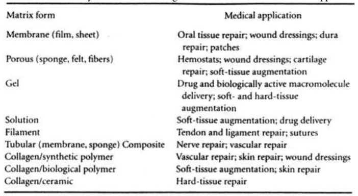
The purified collagen materials obtained from either the molecular technology or from the fibrillar technology are subjected to additional processing to fabricate the materials into useful devices for specific medical applications. The different matrices and their medical applications are summarized in Table 43.5. The technology in fabricating these matrices are briefly outlined below.
43.2.2.1 Membranous Matrix. Collagen membranes can be produced by drying a collagen solution or a fibrillar collagen dispersion cast on a nonadhesive surfáce. The thickness of the membrane is governed by the concentration and the initial thickness of the cast solution or dispersion. In general. membrane thickness of up to 0.5 mm can be easily obtained by air drying a cast collagen material. Additional chemical crosslinking is required to stabilize the membrane from dissolution or dissociation. The membrane produced by casting and air drying does not permit manipulation of the pore structure. Generally, the structure of a cast membrane is dense and amorphous with minimal permeability to macromolecules [Li et al., 1991]. Porous membranes may be obtained by freeze-drying a cast solution or dispersion of a predetermined density or by partially compressing a preformed porous matrix to a predetermined density and pore structure.
TABLE 43.5 Summary of Different Collagen Matrices and Their Medical Applications
12/19
of the collagen in the solution or dispersion. Other factors that contribute to the pore structure include the rate of freezing, the size of fibers in the dispersion, and the presence and absence of other macromolecules. Apparent densities from 0.05 to 0.3 g matrix per cubic centimeter matrix volume can be obtained. These porous matrices generally have pores from about 50 µm to as large as 1500µm.
43.2.2.3 Gel Matrix. A gel matrix may be defined as a homogeneous phase between a liquid and a solid. As such, a gel may vary from a simple viscous fluid to a highly concentrated puny-like material. Collagen gels may be formed by shifting the pH of a dispersion away from its isoelectric point. Alternatively, the collagen material may be subjected to a chemical modification procedure to change its charge profile to a net positively charged or a net negatively charged protein before hydrating the material to firm a gel matrix. For example, native fibers dispersed in water at pH 7 will be in the form of two phases. The dispersed fibers become gel when the pH changes from 7 to 3. Succinylating the primary amino groups of collagen, which converts the positively charged amino groups to negatively charged carboxyl groups, changes the isoelectric point of collagen from about 7 to about 4.5. Such a collagen material swells to a gel at a pH of 7.
43.2.2.4 Solution Matrix. A collagen solution is obtained by dissolving the collagen molecules in an aqueous solution. Collagen molecules are obtained by digesting the insoluble tissue with pepsin to deave the crosslinking sites of collagen (telopeptides) as previously described. The solubility of collagen depends on the pH, the temperature, the ionic strength of the solution, and the molecular weight. Generally, collagen is more soluble in the cold. Collagen molecules aggregate into fibrils when the temperature of the solution increases to the body temperature. pH plays an important role in solubilizing collagen. Collagen is more soluble at a pH away from the isoelectric point of the protein. Collagen is less soluble at higher ionic strength of a solution. The solubility of collagen decreases J1th increasing the size of molecular aggregates. Thus, collagen becomes increasingly less soluble with increasing the extent of crosslinking [Bailey et al., 1970].
43.2.2.5 Filamentous Matrix. Collagen filaments can be produced by extrusion techniques [Kemp et al, 1995; Li and Stone, 1993; Schimpf and Rodriquez, 1976]. A collagen solution or dispersion having a concentration in the range of 0.5 to 1.5% (w/v) is first prepared. Collagen is extruded into a coacervation bath containing a high concentration of a salt or into an aqueous solution at a pH of the isoelectric point of the collagen. Tensile strength of 30 MPa has been obtained for the reconstituted filaments.
43.2.2.6 Tubular Matrix. Tubular matrices may be formed by extrusion through a coaxial cylinder (Stenzl et al., I 974J, or by coating collagen onto a mandrel [Li, 1990]. Different properties of the tubular membranes can be obtained by controlling the drying properties.
43.2.2.7 Composite Matrix. Collagen can form a variety of homogeneous composites with other water-soluble materials. Ions, peptides, proteins, and polysaccharides can all be uniformly incorporated into a collagen matrix. The methods of homogeneous composite formation include ionic and covalent bonding, entrapment, entanglement, and coprecipitation. A heterogeneous composite can be formed between collagen, ceramics, and synthetic polymers that have distinct properties for medical applications [Li, 1988].
43.3 DESIGN OF A RESORBABLE COLLAGEN-BASED MEDICAL IMPLANT
13/19
fabricated either from synthetic or natural materials. Implants for blood vessel, heart valve, and most soft tissue repair fall into this class. Permanent implants, particularly those made of synthetic and biological materials, frequently suffer from the long-term effects of material degradation.
Material degradation can result from biological processes such as enzymatic degradation or environmentally induced degradation from mechanical, metal-catalyzed oxidation, and from the permeation of body fluids into the polymeric devices [Bruck, 1991]. The material degradation is particularly manifested in applications where there is repetitive stress-strain on the implant, such as artificial blood vessels and heart valves.
As a result of the lack of suitable materials for long-term implantation, the concept of using a resorbable template to guide host tissue regeneration (guided tissue regeneration) has received vigorous attention in recent years. This area of research can be categorized into synthetic and biological templates. Polyglycolic acid (PGA), polylactic acid (PLA), polyglycolic-polylactic acid copolymers, and polydioxanone are among the polymers most selected for resorbable medical implant development. Among the biological materials used for resorbable medical implant development, collagen has been one of the most popular materials in this category. Collagen-based templates have been developed for skin [Yannas and Burke, 1981], peripheral nerve [Li et al., 1990; Yannas et al., 1985],oral tissue [Altman and Li, 1990; Blumenthal, 1988], and meniscat regeneration [Li et al., 1994; Stone et al., 1997]. A variety of other collagen based templates are being developed for tissue repair and regeneration applications [Goldstein et al., 1989; Ma et al., 1990; Li,et al., 1997].
The following discussion is useful in designing a template for tissue repair1and regeneration applications. By way of an example, the design parameters listed below are specifically applied to the development of a resorbable collagen based template for guiding meniscal tissue repair and regeneration in the knee joint.
Menisci are semilunar fibrocartilages that are anatomically located between the femoral condyles and tibial plateau, providing stability, weight bearing, shock absorption and assisting in lubrication of the knee joint. A major portion of the meniscal tissue is avascular except the peripheral rim, which comprises about 10 to 30% of the total width of the structure and which is nourished by the peripheral vasculature [Arnoczky and Warren, 1982]. Collagen is the major matrix material of the meniscus, and the fibers are oriented primarily in the circumferential direction in the line of stress for mechanical function. Repair of damaged meniscal tissue in the peripheral vascular rim can be accomplished with sutures. However, in cases where the injured site is in the avascular region, partial or total removal of the rneniscal tissue is often indicated. This is primarily due to the inadequacy of the fibrochondrocytes alone to self-repair the damaged meniscal tissue. Studies in animals and humans have shown that removal of the meniscus is a prelude to degenerative knees manifested by the development of osteoarthritis [Hede and Sarberg, 1992; Shapiro and Glimcher, 1980]. At present there is no suitable permanent substitute for meniscal tissue.
43.3.1 Biocompatibility
14/19
types such as polymorphonuclcar leukocytes, fibroblasts, and macrophages, during the wound healing period, are capable of secreting enzyme collagenases which cleave a collagen molecule at 1/4 position from the C-terminal end of the molecule [Woolley, 1984]. The enzyme first reduces a collagen molecule to two smaller triple helices which are not stable at body temperature and are subsequently denatured to random coiled polypeptides. These polypeptides are further degraded by proteases into amino acids and short peptides that are metabolized through normal metabolic pathways [Nimni and Harkness, 1988].
Despite the safety record of collagen materials for implantation, during the process of preparing the collagen template, small amounts of unwanted noncollagenous materials could be incorporated into the device such as salts and crosslinking agents. Therefore, a series of biocompatibility testing must be conducted to ensure the residuals of these materials do not cause any safety issues. The FDA has published a new guideline for biocompatibility testing of implantable devices [Biological Evaluation of Medical Devices, 1995].
43.3.2 Physical Dimension
The physical dimension of a template defines the boundary of regeneration. Thus, the size of the collagen template should match the tissue defect to be repaired. A properly sized meniscal substitute has been found to function better than a substitute which mismatches the physical dimension of the host meniscus [Rodkey et al., 1998; Sommerlath et al., 1991]. For a porous, elastic matrix such as the one designed from collagen for meniscal tissue repair, the shape of the meniscus is further defined in vivo by the space available between the femoral condyles and tibial plateau within the synovial joint.
43.3.3 Apparent Density
The apparent density as defined as the weight of the dry matrix in a unit volume of matrix. Thus, the apparent density is a direct measure of the empty space which is not occupied by the matrix material per se in the dry state. For example, for a collagen matrix of an apparent density 0.2
The dimension of a mammalian fibrogenic cell body is on the order of 10 to 50 µm, depending on the substrate to which the cell adheres [Folkman et ai, 19781. In order for cells to infiltrate into the interstitial space of a matrix, the majority of the pores must be significantly larger than the dimension of a cell such that both the cell and its cellular processes can easily enter the interstitial space. In a number of studies using collagen-based matrices for tissue regeneration, it has been found that pore size plays an important role in the effectiveness of the collagen matrix to induce host tissue regeneration [Chvapil, 1982; Dagalailis et al., 1980; Yannas, 1996]. It was suggested that pore size in the range of 100 to 400 µm was optimal for tissue regeneration. Similar observations were also found to be true for porous metal implants in total hip replacement [Cook et al., 1991]. The question of interconnecting pores may not be a critical issue in a collagen template as collagenases are synthesized by most inflammatory cells during wound healing and remodeling processes. The interporous membranes which exist in the noninterconnecting pores should be digested as part of resorption and wound healing processes.
43.3.5 Mechanical Property
15/19
template has to be compensated by the strength increase from the regenerated tissue such that at any given time point, the total mechanical properties of the template are maintained. In order to accomplish this goal, one must first be certain that the initial mechanical properties are adequate for supporting the weight-bearing application. For example, compressing the implant with multiple body weights should not cause fraying of the collagen matrix material. It is also of particular importance to design an implant having an adequate and consistent suture pullout strength in order to reduce the incidence of detachment of the implant from the host tissue. The suture pullout strength is also important during surgical procedures as the lack of suture pull strength may result in retrieval and reimplantation of the template. In meniscal tissue repair the suture pullout strength of 1 kg has been found to be adequate for arthroscopically assisted surgery in simulated placement procedures in human cadaver knees, and this suture pullout strength should be maintained as the minimal strength required for this particular application.
43.3.6 Hydrophilicity
Hydration of an implant facilitates nutrient diffusion. The extent of hydration would also provide information on the space available for tissue ingrowth. The porous collagen matrix is highly hydrophilic and therefore facilitates cellular ingrowth. The biomechanical properties of the hydrophilic collagen matrix such as fluid outflow under stress, fluid inflow in the absence of stress, and the resiliency for shock absorption are the properties also found in the weight-bearing cartilagenous tissues.
43.3.7 Permeability
The permeability of ions and macromolecules is of primary importance in tissues that do not rely on vascular transport of nutrients to the end organs. The diffusion of nutrients into tle interstitial space ensures the survival of the cells and their continued ability of growth and synthesis of tissue specific extracellular matrix. Generally, the permeability of a macromolecule the size of the bovine serum albumin (MW 67,000) can be used as a guideline for probing accessibility of the interstitial space of a collagen template [Li et al., 1994].
43.3.7.1 In Vivo Stability. As stated above, the rate of template resorption and the rate of new tissue regeneration have to be balanced so that the adequate mechanical properties are maintained at all times. The rate of in vivo resorption of a collagen-based implant can be controlled by controlling the density of the implant and the extent of intermolecular crosslinking. The lower the density, the greater the interstitial space and generally the larger the pores for cell infiltration, leading to a higher rate of matrix degradation. The control of the extent of intermolecular crosslinking can be accomplished by using bifunctional crosslinking agents under conditions that do not denature the collagen. Glutaraldehyde, formaldehyde, adipyl chloride, hexamethylene diisocyanate, and carbodiimides are among the many agents used in crosslinking the collagen-based implants. Crosslinking can also be achieved through vapor phase of a crosslinking agent. The vapor phase crosslinking is effective using crosslinking agents of high vapor pressures such as formaldehyde and glutaraldehyde. The vapor crosslinking is particularly useful for thick implants of vapor permeable dense fibers where crosslinking in solution produces nonuniform crosslinking. In addition, intermolecular crosslinking can be achieved by heat treatment under high vacuum. This treatment causes the formation of an amide bond between an amino group of one molecule and the carboxyl group of an adjacent molecule and has often been referred to in the literature as dehydrothermal crosslinking.
16/19
A second method of assessing the in vivo stability is to determine the crosslinking density by applying the theory of rubber elasticity to denatured collagen [Wiederhorn and Beardon, 1952]. Thus, the in vivo stability can be directly correlated with the number of intermolecular crosslinks introduced by a given crosslinking agent.
Another method that has been frequently used in assessing the iii vivo stability of a collagen-based implant is to conduct an in vitro collagenase digestion of a collagen implant. Bacterial collagenase is generally used in this application. The action of bacterial collagenase on collagen is different from that of mammalian collagenase [Woolley, 1984]. In addition, the enzymatic activity used in in vitro studies is arbitrarily defined. Thus, the data generated from the bacterial collagenase should be viewed with caution. The bacterial collagenase digestion studies, however, are useful in comparing a prototype with a collagen material of known rate of in vivo resorption. Each of the above parameters should be considered in designing a resorbable implant. The interdependency of the parameters must also be balanced for maximal efikacy of the implant.
43.4 TISSUE ENGINEERING FOR TISSUE AND ORGAN REGENERATION
Biomedical applications of collagen have entered a new era in the past decade. The potential use of collagen materials in medicine has increasingly been appreciated as the science and technology advances. One major emerging field of biomedical research which has received rigorous attention in recent years is tissue engineering. Tissue engineering is an interdisciplinary science of biochemistry, cell and molecular biology, genetics, materials science, biomedical engineering, and medicine to produce innovative three-dimensional composites having structure/function properties that can be used either to replace or correct poorly functioning components in humans and animals or to introduce better functional components into these living systems. Thus, the field of tissue engineering requires a close collaboration among various disciplines for success.
Tissue engineering consists primarily of three components: (1) extracellular matrix. (2) cells, and (3) regulatory signals (e.g.. tissue specific growth factors). One of the key elements in tissue engineering is the extracellular matrix which either provides a scaffolding for cells or acts as a delivery vehicle for regulatory signals such as growth factors.
Type I collagen is the major component of the extracellular matrix and is intimately associated with development, wound healing, and regeneration. The development of the type I collagen based matrices described in this review article will greatly facilitate the future development of tissue engineering products for tissue and organ repair and regeneration applications.
17/19
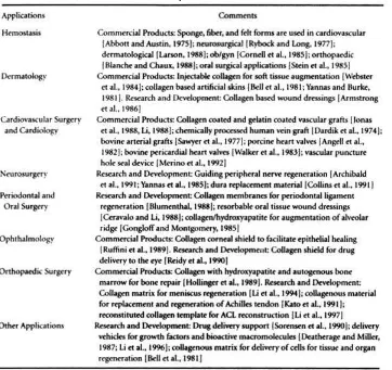
TABLE 43.6 Survey of Collagen.Based Medical Products, and Research and Development Activities
DEFINING TERMS
Alanine (Ala): One of the amino acids in collagen molecules.
Allysine: The ε-amino group of lysine has been enzymatically modified to an aldehyde group.
Apparent density: Calculated as the weight of the dry collagen matrix per unit volume of matrix.
Arginine (Arg): One of the amino acids in collagen molecules.
Aspartic acid (Asp): One of the amino acids in collagen molecules.
Atelocollagen: A collagen molecule without the telopeptides.
Chondroitin sulfate: Sulfated polysaccharide commonly found in cartilages,bone, corea, tendon, and skin.
Collagen: A family fibrous insoluble proteins having a triple helical conformation extending over a major part of the molecule. Glycine is present at every third amino acid in the triple helix and proline and hydroxyproline are required in the triple helix.
Collagenase: A proteolytic enzyme that specifically catalyzes the degradation of collagen molecules.
Dehydrohydroxylysinonorleucine (deH-HLNL): A covalently crosslinked product between an allysine and a hydroxylysine residues in collagen fibrils.
D spacing: The repeat distance observed in collagen fibrils by electron microscopic and x-ray diffraction methods.
Elastin: One of the proteins in connective tissue. It is highly stable at high temperatures and in chemicals. It also has rubberlike properties.
18/19 Fibril: A self-assembled group of collagen molecules.
Fibroblast: Any cell from which connective tissue is developed.
Fibrochondrocyte: Type of cells that are associated with special types of cartilage tissues such as men iscus of the knee and intervertebral disc of the spine.
Fibrous long spacing (FLS): One of the polymorphic forms of collagen where the collagen molecules are randomly aligned in either head-to-tail, tail-to-tail, or head-to-head orientation.
Gelatin: A random coiled form (denatured form) of collagen molecules.
Glu tamic acid (Glu): One of the amino acids in collagen molecules.
Glycine (Gly): One of the amino acids in collagen molecules having the simplest structure.
Glycoprotein: A compound consisting of a carbohydrate protein. The carbohydrate is generally hexosamine, an amino sugar.
Glycosaminoglycan (GAG): A polymerized sugar (see polysaccharide) commonly found in various connective tissues.
Helical pitch: Repeating distance within a single polypeptide chain ¡ri a collagen molecule.
Hemostat: Device or medicine which arrests the flow of blood.
Hydrophilicity: The tendency to attract and hold water.
Hydrophobicity: The tendency to repel or avoid contact with water. Substances generally are nonpolar in character, such as lipids and nonpolar amino acids.
Hydroxylysine (Hyl): One of the amino acids in collagen molecules.
Hydroxyproline (Hyp): One of the amino acids uniquely present in collagen molecules.
Inflammatory cell: Cells associated with the succession of changes which occur in living tissue when it is injured. These include macrophages, polymorphonuclear leukocytes, and lymphocytes.
Intermolecular crosslink: Covalent bonds formed in vivo between a side group of one molecule and a side group of another molecules, covalent bonds formed between a side group of one molecule and one end of a bifunctional agent and between a side group of a second molecule and the other end of a bifunctional agent.
lntraflbrillar volume: The volume of a flbril excluding the volume occupied by the collagen molecule.
In vitro: In glass, as in a test tube. An in vitro test is one done in the laboratory, usually involving isolated tissues, organs, or cells.
In vivo: In the living body or organism. A test performed in a living organism.
Isoelectric point: Generally used to refer to a particular pH of a protein solution. At this pH, there is no net electric charge on the molecule.
Isoleucine (Ile): One of the amino acids in collagen molecules.
Leucine (Leu): One of the amino acids in collagen molecules.
Lipidi: Any one of a group of fats or fat-like substances, characterized by their insolubility in water and solubility in fat solvents such as alcohol, ether, and chloroform.
Lysine (Lys): One of the amino acids in collagen molecules.
Meniscus: A C-shaped fibrocartilage anatomically located between the femoral condyles and tibial plateau providing stability and shock absorption and assisting in lubrication of the knee joint.
Macrophage: Cells of the reticuloendothelial system having the ability to phagocytose particulate substances and to store vital dyes and other colloidal substances. They are found in loose connective tissues and various organs of the body.
19/19
Osteoarthritis: A chronic disease involving the joint, especially those bearing the weight, characterized by destruction of articular cartilage, overgrown of bone with impaired function.
Permeability: The space within a collagen matrix, excluding the space occupied by collagen molecules, which is accessible to a given size of molecule.
Pepsin: A proteolytic enzyme commonly found in the gastric juice. It is formed by the chief cells of gastric glands and produces maximum activity at a pH of 1.5 to 2.0.
Phenolalanine (Phe): One of the amino acids in collagen molecules.
Platelet: A round or oval disk, 2 to 4 ¡m in diameter, found in the blood of vertebrates. Platelets contain no hemoglobin.
Polydioxanone: A synthetic polymer formed from dioxanone monomers which degrades by hydrolysis.
Polyglycolic acid (PGA): A synthetic polymer formed from glycolic acid monomers which degrades by hydrolysis.
Polylactic acid (PL4): A synthetic polymer formed from lactic acid monomers which degrades by hydrolysis.
Polymorphism: Different types of aggregated states of the collagen molecules.
Polymorphonudear leukocyte: A white blood cell which possesses a nucleus composed of two or more lobes or parts; a granulocyte (neutrophil, eosinophil, basophil).
Polypeptide: Polymerized amino acid molecules formed by enzymatically regulated stepwise polymer ization in vivo between the carboxyl group of one amino acid and the amino group of a second amino acid.
Polysaccharide: Polymerized sugar molecules found in tissues as lubricant (synovial fluid) or cement (between osteons, tooth root attachment) or complexed with proteins such as glycoproteins or proteoglycans.
Proline (Pro): One of the amino acids commonly occurring in collagen molecules.
Proteolytic enzyme: Enzymes which catalyze the breakdown of native proteins.
Resorbable collagen: Collagen which can be biodegraded in vivo.
Saltlinkage: An electrostatic bond formed between a negative charge group and a positive charge group in collagen molecules and fibrils.
Segment-long-spacing (SLS): One of the polymorphic forms of collagen where all heads of collagen molecules are aligned in parallel.
Soluble collagen: Collagen molecules that can be extracted with salts and dilute acids. Soluble collagen molecules contain the telopeptides.
Telopeptide: The two short nontriple helical peptide segments located at the ends of collagen molecules.