Investigating Brain Networks in Anxious-Misery and Fear Symptom Dimensions Julia Pines
Dr. Kaczkurkin PSY4998-01
Spring 2022
Abstract
Anxiety symptoms can be dissociated into anxious-misery and fear components;
however, little is known about how these two symptom dimensions differ in terms of brain network properties. Thus, the purpose of this study was to compare the local efficiency of the default mode network (DMN) and frontoparietal network (FPN) in anxious-misery and fear symptom dimensions, with examination of six additional networks and small-worldness as exploratory analyses. To do this, we used data from children ages 9-10 years old from the Adolescent Brain Cognitive Development (ABCD) Study database. Our primary measure of interest was local efficiency, which measures the efficiency of information exchange between nodes of a network when one node is removed. We hypothesized that the DMN would exhibit increased local efficiency and the FPN would show decreased local efficiency in anxious-misery symptoms, and both networks would have lower local efficiency in fear symptoms. We found no significant associations between local efficiency and anxious-misery and fear dimensions or between small-world omega and the dimensions. However, we found a significant positive association between anxious-misery symptoms and local efficiency in the FPN and a significant negative association between fear symptoms and local efficiency in the FPN at uncorrected levels. These results suggest the need for further study of local efficiency in the FPN in an older population and the use of other network metrics in anxious-misery and fear symptom
dimensions.
Investigating Brain Networks in Anxious-Misery and Fear Symptom Dimensions Anxiety and depressive disorders are some of the most common psychiatric illnesses, and they make up the larger category of internalizing disorders for their shared symptoms of
disordered mood or emotion (Kovacs & Devlin, 1998). According to the Diagnostic and Statistical Manual of Mental Disorders, Fifth Edition (DSM-5), common features of depressive disorders include the presence of sad, empty, or irritable mood, accompanied by somatic and cognitive changes that significantly affect the individual's ability to function (American Psychiatric Association, 2013). Anxiety disorders have typical features of excessive fear and anxiety beyond normal developmental periods and in greater magnitude than the situation warrants (American Psychiatric Association, 2013). Anxiety and depressive disorders share many similar symptoms or may co-occur in one patient, a phenomenon known as comorbidity (Regier et al., 2013; Krueger et al., 1999). Depressive disorders and anxiety disorders, for example, are in DSM sections next to each other, and their comorbidity has been supported by studies showing that these two types of disorders overlap in symptoms (Krueger et al., 1999).
Previous research using exploratory factor analysis, which reveals patterns or categories in data, has shown that symptoms of these internalizing disorders can be separated into anxious- misery and fear disorders (Kaczkurkin et al., 2019; Krueger et al., 1999). Anxious-misery
disorders include major depressive disorder (MDD), dysthymia, and generalized anxiety disorder (GAD) (Kaczkurkin et al., 2019). Fear disorders include anxiety disorders such as social anxiety disorder (SAD), specific phobias, agoraphobia, panic disorder, and separation anxiety disorder (Kaczkurkin et al., 2019). Using these categories, we assigned symptoms characteristic of these disorders to anxious-misery and fear dimensions. Based on prior neuroimaging studies
examining individual disorders, we hypothesized a model of the neurobiological mechanisms
underlying anxious-misery and fear symptoms. A widely accepted theory in neuroscience is that brain functions are not carried out by one brain region alone, but through the interaction of multiple parts of the brain. The current study will examine two brain networks implicated in anxious-misery and fear-based dimensions: the frontoparietal network (FPN) and the default mode network (DMN). Analysis of six additional brain networks will be exploratory.
Importantly, this model will highlight both the brain network patterns hypothesized to be
common across anxious-misery and fear symptoms, and the brain network characteristics that are hypothesized to be unique to each class of symptoms.
Neural networks associated with anxious-misery disorders
The brain regions included in the FPN and DMN vary; here we use the definitions provided by Mulders and colleagues (Mulders et al., 2016). The major brain regions of the FPN are the dorsolateral prefrontal cortex (dlPFC) and the posterior parietal cortex (PPC). The FPN is implicated in cognitive tasks like working memory and attention (Mulders et al., 2016). The DMN includes the medial prefrontal cortex (mPFC), the posterior cingulate cortex (PCC) and precuneus cortex (PCu). The DMN is likely involved in emotional control and self-referential thought (Mulders et al., 2016).
Multiple studies have found decreased functional connectivity within the FPN in depressed patients (Alexopoulos et al., 2012; Kaiser et al., 2015; Liston et al., 2014; Lui et al., 2011) and patients with GAD (Andreescu et al., 2015). Functional connectivity is the strength that the activity of a region of a brain network is correlated with the activity of another region of that network (Mohanty et al., 2020). This reduced activity was restored after transcranial
magnetic stimulation (TMS) treatment (Liston et al., 2014). In addition, reduced resting-state connectivity in the FPN predicted poorer treatment response including lower remission rates and
the persistence of depressive symptoms (Alexopoulos et al., 2012). However, there are some studies with contradictory findings; Etkin et al. (2009) found increased connectivity in the FPN of GAD patients. Overall, much of the literature on the connectivity within the FPN in depressed patients suggests that there is decreased activity in the FPN in anxious-misery disorders
compared to controls.
The second network relevant in anxious-misery disorders is the default mode network (DMN). Several studies found higher functional connectivity within the DMN in patients with MDD (Andreescu et al., 2013, Berman et al., 2010; Greicius et al., 2007, Li et al., 2013, Zhu et al., 2012) and those with GAD (Qiao et al., 2017). In one study, increased activity of a region of the DMN was correlated with the duration of the depressive episode (Greicius et al., 2007).
Antidepressant treatment decreased the activity of the DMN to normal levels (Daws et al., 2021).
In contrast, other studies found reduced connectivity in the DMN in MDD (Cullen et al., 2009;
Kaiser et al., 2015; Yan et al., 2019) and GAD (Andreescu et al., 2014). Thus, the literature on the DMN in anxious-misery disorders remains mixed.
Taken together, the major findings within the research on brain networks in anxious- misery disorders show that there is reduced connectivity in the FPN and heightened connectivity within the DMN. However, there is still a gap in the research since these studies focused on single disorders only (such as MDD), rather than a dimension of anxious-misery symptoms. The current study will investigate associations between these two networks in a large sample of participants with anxious-misery symptoms.
Neural networks associated with fear-based disorders
In a literature review focused on brain networks in fear disorders, Sylvester et al., (2012) proposed that anxiety disorders are associated with decreased functional connectivity in the FPN
and DMN. Most of the previous literature studied these networks separately, but Dixon et al., (2021) looked at the FPN and DMN together in patients with social anxiety disorder (SAD).
Behaviorally, this study found a significant difference in self-referential processing between SAD patients and controls, which is the main function of the DMN but also involves the FPN.
Dixon et al., (2021) found no significant differences in brain activation in the DMN or FPN of SAD patients compared to controls. However, the researchers suggested that their sample size was too small, and the findings do not conclusively reflect that there was no difference in DMN or FPN functioning between anxious or non-anxious patients.
From previous literature, there is likely decreased functional connectivity in the FPN in patients with fear disorders. Decreased functioning in the FPN has been found in patients with SAD (Manning et al., 2015). Much of the previous literature on the FPN in anxiety has looked at trait anxiety rather than specific anxiety disorders. Many studies have found that individuals with high trait anxiety measured by the State-Trait Anxiety Inventory (STAI) showed decreased activation in regions of the FPN compared to controls (Bishop et al., 2009; Comte et al., 2015;
Forster et al., 2013; Modi et al., 2015). Since the decrease in FPN activation was associated with trait and not state anxiety, it is most likely a vulnerability to anxiety rather than an outcome of it (Bishop et al., 2009). Individuals with high trait anxiety needed higher levels of FPN activity to achieve the same degree of cognitive control as adults with low anxiety (Basten et al., 2011; Li et al., 2020). However, studies suggest that the STAI actually confounds both fear and anxious- misery because it includes many depressive items (Bados et al., 2010; Bieling et al., 1998;
Nitschke et al., 2001). Another study that looked at children with SAD, separation anxiety disorder, and GAD found significant changes in the FPN only during a stimulus-driven attention task, and no difference between those with anxiety disorders and healthy controls during the
resting-state fMRI scan (Perino et al., 2021). In all, past research suggests that the FPN may show decreased functional connectivity in fear disorders.
The DMN was also hypothesized by Sylvester et al., (2012) to be involved in fear disorders. In general, there seems to be dysfunction in emotion regulation in many anxiety disorders including panic disorder and SAD (Cisler et al., 2010). Since the DMN is activated during self-focused attention and worry, there may be abnormal activity in the DMN in fear disorders. Multiple studies have found decreased functional connectivity in the DMN in individuals with anxiety symptoms (De Micco et al., 2020; Modi et al., 2015; Simmons et al., 2008) and in patients with SAD (Hahn et al., 2011). However, some studies found no significant difference in the DMN between SAD patients and controls (Dixon et al., 2021, Fang et al., 2021).
In all, previous studies of brain networks in fear disorders suggest that there is decreased connectivity in the FPN and DMN (Sylvester et al., 2011). Similar to the literature on depressive disorders, most of these studies focused on single anxiety disorders instead of a dimension of fear symptoms. Additionally, much of the research on trait anxiety confounded fear and anxious- misery symptoms. Further research is needed to compare the FPN and DMN between anxious- misery and fear symptoms.
Network Metrics
Previous literature has employed mostly functional connectivity fMRI data to compare the connectivity of the FPN and DMN in these two groups of symptoms. However, more specific graph theory metrics can also be used to measure structural changes in network communication in certain disorders. According to graph theory, brain networks can be parcellated into nodes, which are distinct brain regions that are involved in a network (Stanley et al., 2013). The current
study will use the graph theory metric of local efficiency to examine group differences. Local efficiency measures the fault tolerance of a network, meaning that it shows how well the other nodes of a network exchange information when one node is removed (Tao et al. 2015). High local efficiency occurs when the network can successfully transfer information even in the absence of a functioning node and suggests that neighboring nodes of the network are highly interconnected (Cohen & D’Esposito, 2016). Studies have found increased local efficiency within the DMN in depressive disorders (Luo et al., 2015, Meng et al., 2014). This compensation of the DMN suggests that there may be stronger communication between nodes of the DMN in depressed patients which could lead to the symptom of rumination (Luo et al., 2015). However, other studies found reduced local network function in regions of the DMN in GAD (Makovac et al., 2018). There is less information available about local efficiency in the FPN in depressed patients. One study found less connections in the FPN in MDD patients (Luo et al., 2015). Other studies looked at global rather than local efficiency to investigate the overall information transfer capacity of the FPN and found reduced global efficiency of the FPN in MDD (Tan et al., 2020).
From past research, the DMN may have increased local efficiency while the FPN may have decreased local efficiency in anxious-misery disorders. Regarding fear, there is insufficient literature on local efficiency in these kinds of disorders. One study found decreased local efficiency in the DMN in SAD (Zhu et al., 2017). Lower local efficiency in the DMN was negatively correlated with anxiety levels in healthy participants (Tao et al., 2015). To our knowledge, there was no previous research on local efficiency in the FPN in people with fear symptoms. Although the previous research is sparse, there may be lower local efficiency in both the DMN and FPN in fear disorders based on previous local efficiency and functional
connectivity findings. In all, previous literature concerning local efficiency in anxious-misery
and fear-based disorders in the DMN and FPN is deficient, especially with fear disorders.
Therefore, the local efficiency information used to analyze the DMN and FPN in this study will serve to fill these gaps.
We also conducted exploratory analyses on other measures of graph theory including small-world omega. Small-worldness is a measure of the interconnectedness among nodes of a network, with high small-worldness meaning the nodes are densely packed and highly
connected. Specifically, small-world omega compares the tightness of the clusters of nodes in a network to a model graph of ‘perfect’ small-worldness. Previous studies have looked at small- worldness in the DMN and FPN in depressive disorders through varying metrics. A few studies found lower small-worldness in the DMN in patients with MDD (Li et al., 2017) and remitted depression (Zhu et al., 2018) compared to healthy controls. However, another study found smaller distances between nodes of the DMN in MDD, which implies higher small-worldness (Hou et al., 2016). There was little information on the small-worldness metric in the FPN in patients with anxious-misery symptoms. In addition, there is sparse information on small- worldness in the DMN and FPN in fear symptoms. One study found increased density of nodes in the DMN in patients with higher anxiety (Tao et al., 2015), implying higher small-worldness.
Another study found lower small-worldness in SAD patients but looked at the whole brain rather than particular networks (Zhu et al., 2017). Overall, past research on the small-worldness of the DMN and FPN in anxious-misery and fear symptoms is sparse and warrants further
investigation.
Current Study
The current study aims to examine two network metrics (local efficiency and small-world omega) of the DMN and FPN in dimensions of anxious-misery and fear. Research on the
dysfunction of brain networks in internalizing symptoms so far has focused on depressive disorders (Mulders et al., 2016) or anxiety disorders (Sylvester et al., 2012) separately. This is a limitation because psychiatric disorders are highly comorbid (Kaczkurkin et al., 2019) and prior factor analytic research suggests that internalizing symptoms are more accurately grouped into anxious-misery and fear categories (Krueger et al., 1999). There are also some discrepancies in past research regarding whether these networks have more or less effective communication in anxious-misery and fear dimensions. In addition, much of the previous literature on brain network communication in patients with internalizing symptoms has focused on adults. We propose to advance this research by directly comparing anxious-misery and fear symptom dimensions using network metrics derived from resting-state data in a sample of children between 9 and 10 years old. We hypothesize that the default mode network (DMN) will exhibit increased local efficiency and the frontoparietal network (FPN) will show decreased local efficiency in participants with anxious-misery symptoms, and both networks will have lower efficiency in those with fear symptoms. Small world omega analyses were exploratory.
Method Participants
We used data collected from the Adolescent Brain Cognitive Development (ABCD) Study database. Vanderbilt University’s Institutional Review Board approved the use of this publicly available, de-identified dataset. The ABCD Study includes data from 11,876 children between 9 and 10 years of age (Karcher & Barch 2020). These children are followed over the course of ten years beginning in 2018 with data released annually (Garavan et al. 2018). Annual lab-based assessments were conducted, including fMRI scanning (Durham et al. 2021). The ABCD Study attempted to reflect the representation of the U.S. population by recruiting through
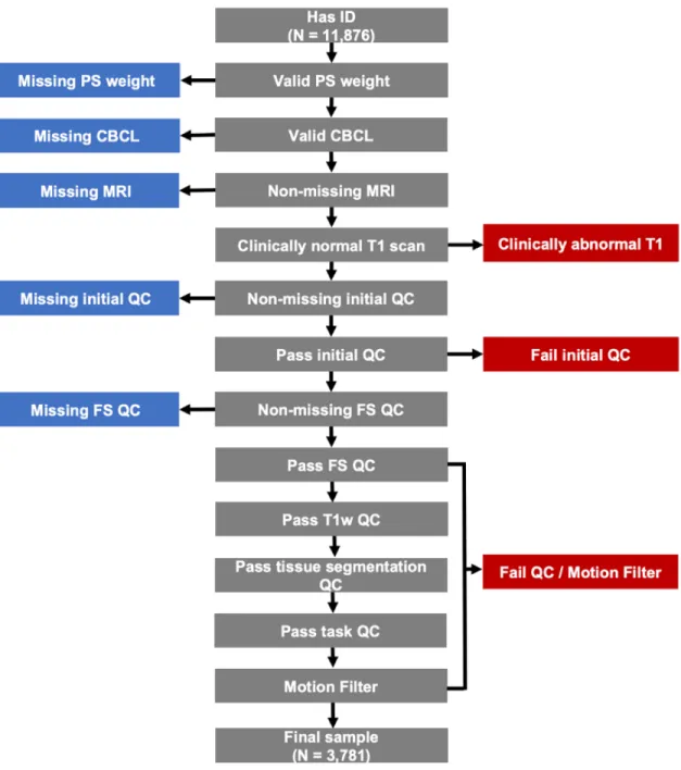
multistage probability sampling (Garavan et al. 2018). This sampling included selecting a nationally distributed set of 21 study sites, using probability sampling of schools within the defined areas for each site, and recruiting eligible children in each sample school. The present analyses used baseline data from Wave 1 (release 4.0) of the ABCD Study. We excluded participants for missing data on the variables of interest. We also excluded participants if there were abnormal structural images or if there was failure to meet quality assurance protocols.
Finally, because network metrics are highly sensitive to motion artifacts, we used a stringent criterion for motion (Figure 1). The final sample size for our analyses was 3,781.
Materials
The psychopathology symptoms of the participants were assessed in the ABCD Study using the Child Behavior Checklist (CBCL) (Karcher & Barch, 2020). This assessment evaluated parent-report of youth behavioral and emotional problems and contains 119 items (Achenbach, 2009). Specific probes and scoring criteria were provided to assess each symptom, and
symptoms have been categorized into syndromes (Achenbach, 1991). For example, some items to evaluate the internalizing syndrome of anxious/depressed were “cries a lot,” “feels or
complains that no one loves him/her,” and “unhappy, sad, or depressed” (Achenbach, 2009).
Parents were asked to rate the extent that the behavior is characteristic of their child over the past six months, with the following scale: 0 = not true, 1 = somewhat or sometimes true, and 2 = very true or often true.
Figure 1. Participant flowchart of resting-state data. Flowchart indicating exclusions for primary analyses with dimensions of anxious-misery and fear and network efficiency metrics of resting-state data. PS = poststratification weights (makes the sample more representative of the general US population); CBCL = Child Behavior Checklist; QC = quality control; FS =
Freesurfer (the fMRI processing software used).
Design
This was a cross-sectional study using quasi-independent variables collected through an observational design. The independent variables were the dimensional measures of anxious- misery and fear, and our primary dependent variable of interest was local efficiency in each network, which measures how well the other nodes of a network exchange information when one node is removed. We used the Shen atlas (Shen et al., 2013) to parcellate the brain into eight networks: the frontoparietal network (FPN), the default mode network (DMN), the medial frontal network (MFN), the subcortical and cerebellar regions (SC), the motor network (MON), the visual I network (VisI), the visual II network (VisII), and the visual association network (VA).
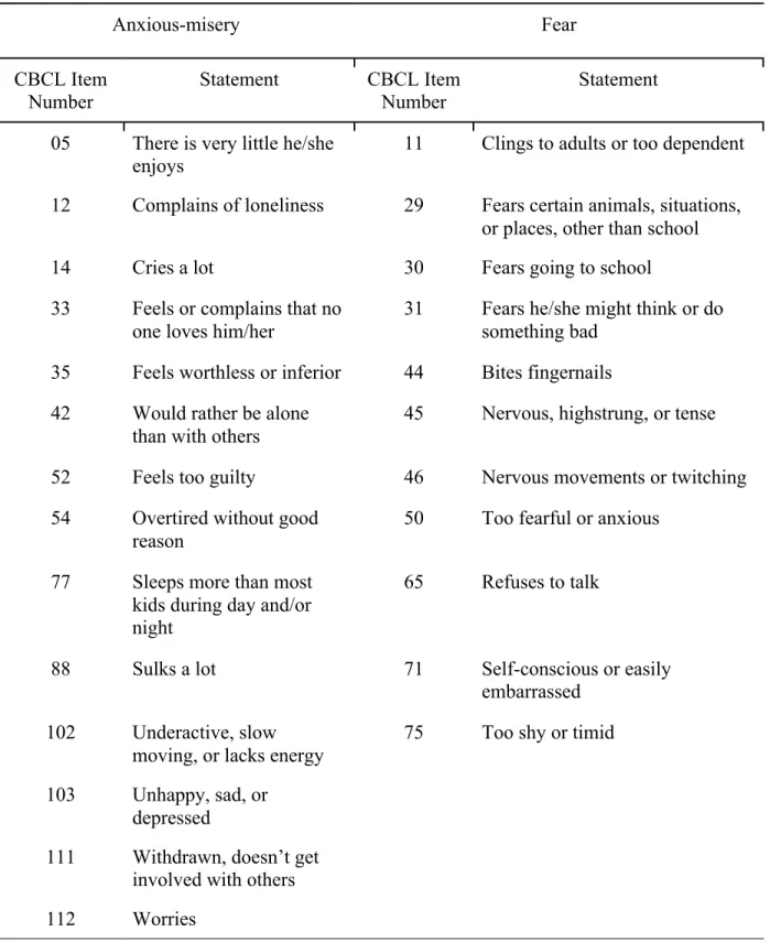
Our primary interest was in the FPN and DMN networks. We did not have predictions about the other networks provided by the Shen atlas (MFN, SC, MON, VisI, VisII, and VA), so analyses with these networks were exploratory. We also conducted exploratory analyses of another measures of graph theory, small-world omega, which measures the interconnectedness among nodes of a network. The participants were grouped into anxious-misery or fear dimensions based on responses to statements in the CBCL (Table 1).
Image acquisition, processing, and quality assurance
The current study examined resting-state data collected on multiple models of 3 tesla (3T) scanners: General Electric Discovery MR750, Siemens Prisma, Siemens Prisma Fit, Phillips Achieva dStream, and Philips Ingenia. Because of the use of more than one type of scanner in this data, we added the scanner model as an additional covariate to control for differences between scanners. When the participants first entered the scanner, a child-friendly movie was played (Casey et al. 2018). The functional scans include twenty minutes of resting-state data acquired with eyes open and passive viewing of a crosshair. The ABCD Study detected and
corrected for motion in the scanner. Minimally processed functional MRI scans were used for the current study. Processing included correction for head motion, B0 distortion correction, gradient nonlinearity distortion correction, resampling, and registration to T1 structural images. For additional details on the image acquisition, processing, and quality assurance procedures, see Stier et al. (2021).
Data analysis
We investigated the local efficiency of the eight functional networks outlined in the Shen atlas in dimensions of anxious-misery and fear symptoms through resting-state fMRI data. To derive functional networks from correlation matrices of signals between brain regions, we applied a threshold of 30% which kept only the strongest 30% of connections between node pairs. The measure of local efficiency quantifies how well a node’s neighbors can exchange information if that node is removed; higher efficiency suggests that the remaining nodes
communicated efficiently despite the loss of one node and relates to better cognitive functioning (Cohen & D’Esposito, 2016). We included age, sex, race/ethnicity, and MRI scanner model as covariates. We controlled for age because the brain changes as we grow older, especially in children. Sex was controlled for because males and females differ in brain size. We controlled for race/ethnicity based on prior work showing that this is an important variable to control for in this sample (Assari & Boyce, 2021). MRI scanner was included as a covariate because there are known differences between the scanners used for this study (Moore et al., 2020). For each brain network, we examined associations between dimensional measures of anxious-misery and fear and local efficiency through structural equation modeling (SEM) in Mplus as follows:
Network = β × age + β × sex + β × race/ethnicity + β × MRI scanner model + β × anxious misery + β × fear
As exploratory analysis, we also looked at the association between the anxious-misery and fear dimensions and small-world omega for each of the eight brain networks. All analyses used post-stratification weights to make the sample more representative of the U.S. population in terms of demographics like race/ethnicity. The analyses were stratified by site to control for differences between the 21 data sites. Analyses were also clustered by family ID to account for relatedness between pairs of twins and siblings, with families being modeled with a random intercept. Multiple comparisons were accounted for using false discovery rate (FDR) correction.
Results Sample characteristics
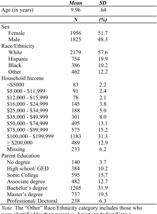
Table 2 shows a summary of demographics based on the final sample of 3,781 participants. The average age of participants was 9.96 years (SD = 0.64). The sample was
predominantly White (58%) with slightly more females (52%) than males. A large portion of the sample had a household income above $100,000 (44%) and a majority of the parents of the participants had a bachelor’s degree or higher (58%).
No significant association between anxious-misery symptoms and local efficiency across the eight networks
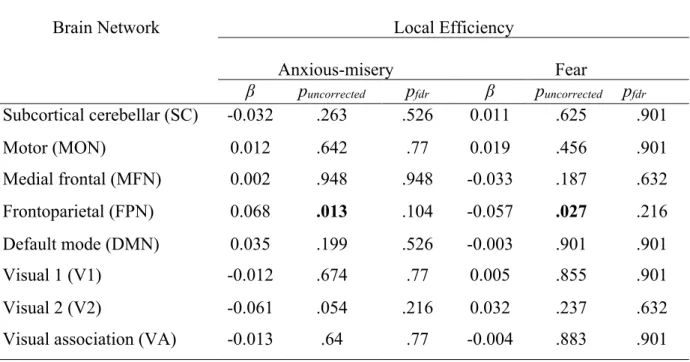
First, we examined the relationship between our dimensional measure of anxious-misery and local efficiency in the eight networks (FPN, DMN, MFN, SC, MON, VisI, VisII, and VA), while controlling for age, sex, race/ethnicity, and MRI scanner model. The results showed that the anxious-misery dimension was not significantly associated with local efficiency in the DMN (pfdr = .53) or the FPN (pfdr = .10) at rest. See Table 3 for estimates for all networks. There were also no significant associations between anxious-misery symptoms and the local efficiency of the six exploratory brain networks of the MFN, SC, MON, VisI, VisII, and VA (Table 3).
No significant association between fear symptoms and local efficiency across the eight networks
Next, we repeated these analyses for the fear dimension using the same covariates. The fear dimension was not significantly associated with local efficiency in the DMN (pfdr = .90) or the FPN (pfdr = .22) (Table 3). In addition, there were no significant associations between the fear dimension and the local efficiency of the exploratory brain networks (Table 3).
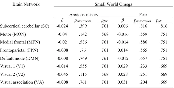
No significant association between anxious-misery or fear symptoms and small-world omega across the eight networks
Small-world omega measures the interconnectedness among nodes of a network and was examined in exploratory analyses. The anxious-misery dimension was not significantly
associated with small-world omega in the DMN (pfdr = .76) or the FPN (pfdr = .71) (Table 4).
There was also no significant association between the fear dimension and small-world omega in the DMN (pfdr = .75) or the FPN (pfdr = .75) (Table 4). Finally, there was no significant
associations between either dimension and small-world omega in any of the exploratory brain networks (Table 4).
Uncorrected results reveal an opposite association for anxious-misery and fear in the local efficiency of the FPN
Lastly, we examined the uncorrected p-values to determine whether there was any signal apparent in the data that might warrant further investigation in a sample with greater power. The results showed that there was a significant positive association between anxious-misery
symptoms and local efficiency in the FPN (p = .013) and a significant negative association between fear symptoms and local efficiency in the FPN (p = .027) at uncorrected levels.
Discussion
The current study utilized data from the ABCD Study to examine the association between anxious-misery and fear symptom dimensions and the local efficiency of the frontoparietal (FPN) and default mode (DMN) networks. The results showed no significant associations between the local efficiencies of the FPN and DMN networks in the anxious-misery and fear symptom dimensions after FDR correction. Exploratory analyses also revealed no significant results for small-world omega. The results of this study could imply that internalizing
symptomatology may not be associated with local efficiency or small-world omega differences in the brain, and it may be advantageous to investigate other network metrics in future studies.
However, some interesting results were found when examining the uncorrected p-values. We found an opposite relationship between anxious-misery and fear in the local efficiency of the FPN. Specifically, the results showed that there was a significant positive association between anxious-misery symptoms and local efficiency in the FPN and a significant negative association between fear symptoms and local efficiency in the FPN at uncorrected levels. The fact that the raw p-values did not survive FDR correction shows that these results are relatively weak, but they do suggest there may be some signal in the data. It is possible that we were underpowered to find such a small effect. Or it may be the case that the associations between these dimensions and local efficiency in the FPN were not yet apparent in this age range but will become stronger with time. Future work could examine the longitudinal data points from this study to see if this
association increases across development.
The finding of no significant association between anxious-misery symptoms and local efficiency of the FPN and DMN networks contradicts previous studies that found increased local efficiency in the DMN (Luo et al., 2015, Meng et al., 2014) and decreased local efficiency in the
FPN (Luo et al., 2015) in people with anxious-misery symptoms. There was a scarcity of
previous research on local efficiency in these networks in fear symptoms, but some studies found lower local efficiency in the DMN (Tao et al., 2015; Zhu et al., 2017) in fear symptoms.
Interestingly, our finding of a significant positive correlation between anxious-misery symptoms and local efficiency in the FPN at uncorrected p-values contradicts previous findings and our hypothesis. One explanation for this contradiction and the null results in the DMN after FDR correction is that distinct sections of this network may be differently affected by anxious-misery symptoms. For example, one previous study found that higher depression scores were positively correlated with activity in the orbitofrontal gyrus and middle frontal gyrus of the DMN, but negatively correlated with activity of other regions such as the PCC and precuneus (Coutinho et al., 2014). Opposing changes in functional connectivity in two major sections of the DMN have been found in other studies of anxious-misery symptoms (Li et al. 2013; Mulders et al. 2016;
Zhu et al. 2012). Higher anxiety scores were positively correlated with activity in the DMN regions of the mPFC and ACC and negatively with parietal and temporal areas (Coutinho et al., 2014), but research on differences in DMN sections in fear symptoms is generally sparse. It is possible that a difference in activity of regions of the DMN affected the local efficiency; perhaps different sections of the DMN experienced higher and lower local efficiency respectively and these measures canceled out. Differences in activity of distinct sections of the FPN have not yet been investigated in anxious-misery or fear symptoms. Overall, future research on local
efficiency in subsections of the DMN and FPN in people with anxious-misery and fear symptoms may illuminate whether there are local efficiency changes in these networks.
In addition, there were many previous studies that found an increase in functional
connectivity in the DMN (Andreescu et al., 2013, Berman et al., 2010; Greicius et al., 2007, Li et
al., 2013, Qiao et al., 2017; Zhu et al., 2012) and decreased connectivity in the FPN
(Alexopoulos et al., 2012; Andreescu et al., 2015; Kaiser et al., 2015; Liston et al., 2014; Lui et al., 2011) in people with anxious-misery symptoms, as well as decreased functional connectivity in the DMN (De Micco et al., 2020; Hahn et al., 2011; Modi et al., 2015; Simmons et al., 2008) and FPN (Comte et al., 2015; Modi et al., 2015) in people with fear symptoms. Since significant differences in the local efficiency of these networks in anxious-misery and fear symptom
categories were not found in this study, the measure of local efficiency may not reflect changes in these networks due to anxious-misery or fear symptoms. Local efficiency measures
communication between nodes of a network while functional connectivity measures the
relationship between the activity of two brain regions in a network (Eickhoff & Muller, 2015). It is possible that only the functional connectivity of these networks was changed in patients with anxious-misery symptoms and these symptoms did not affect the local efficiency. This may be due to the negative correlation between the distance between the nodes and local efficiency which is not present with the measure of functional connectivity (Stanley et al., 2015).
Additionally, functional connectivity is a more general measure of communication within a network, and it may be that local efficiency was too specific to reflect the effect of anxious- misery or fear symptoms on the DMN and FPN. However, our finding of a significant negative correlation between fear symptoms and local efficiency in the FPN at uncorrected p-values supports our hypothesis and previous functional connectivity research in the FPN, so there may be some merit to the local efficiency measure in this context. Future research on an older population will be crucial to understanding if there is a significant change in local efficiency in the FPN in patients with fear symptoms.
Additionally, there was no significant association between the small-world omega measure in the DMN and FPN and both anxious-misery and fear symptoms. Small-world omega measures the interconnectedness of nodes in a network, considering the density of the nodes and the ability of one node to reach other nodes in a network. Some previous studies found lower small-worldness in the DMN in people with anxious-misery symptoms (Li et al., 2017; Zhu et al., 2018) while another found higher small-worldness in the DMN compared to healthy controls (Hou et al., 2016). There were also conflicting findings concerning the DMN in fear symptoms, with one study finding higher small-worldness (Tao et al., 2015) and another lower (Zhu et al., 2017). The discrepancies in the direction of change of small-worldness in the DMN in anxious- misery and fear symptoms from past research could support the lack of change in small-
worldness found in this paper. There was also little past research on small-worldness in the FPN in patients with anxious-misery symptoms or fear symptoms.
Ultimately, future research is needed on anxious-misery and fear symptom dimensions in the DMN and FPN in an older population. It is possible that associations between these
symptoms and local efficiency in these networks were not strong enough in our sample of young children to lead to significant results after correction. Thus far, graph theory metrics such as local efficiency and small-worldness have not been extensively researched in anxious-misery or fear symptomology. Future research into these measures could support the creation of new methods of treatment that target communication deficits other than functional connectivity in brain networks of patients with severe anxious-misery or fear symptoms.
There were several limitations of the current study which could provide direction for future investigations of the DMN and FPN in anxious-misery and fear dimensions. First, the Child-Behavior Checklist (CBCL) scores used in this study were determined by parent-reported
symptoms that their child was experiencing, so the reported symptoms may not be exactly true to the child’s experience. This limitation may diminish for future studies that use an older
population who are able to self-report their symptoms. In addition, this study was confined to the eight networks outlined by the Shen Atlas used in the ABCD dataset. These networks may not have corresponded to the regions studied in previous studies of local efficiency in the DMN and FPN in anxious-misery or fear symptoms, which could have impacted the results and their agreement with past research.
In all, the role of graph theory metrics such as local efficiency and small-worldness in brain networks such as the DMN and FPN in participants with internalizing symptoms has not yet been extensively researched. Further study of the local efficiency in these networks in an older population of participants could open new pathways for treatment of network
communication deficits to relieve people of these symptoms.
References
Achenbach T. (1991). Integrative guide to the 1991 CBCL, YSR, and TRF profiles.
Department of Psychiatry, Burlington University, Vermont.
Achenbach, T. (2009). Achenbach System of Empirically Based Assessment (ASEBA):
Development, Findings, Theory, and Applications. Encyclopedia of Autism Spectrum Disorders.
Alexopoulos, G.S., Hoptman, M.J., Kanellopoulos, D., Murphy, C.F., Lim, K.O., Gunning, F.M.
(2012). Functional connectivity in the cognitive control network and the default mode network in late-life depression. Journal of Affective Disorders, 139(1): 56-65.
doi:10.1016/j.jad.2011.12.002
American Psychiatric Association (2013). Diagnostic and statistical manual of mental disorders (5th ed).
Andreescu, C., Sheu, L.K., Tudorascu, D., Gross, J.J., et al. (2015). Emotion
reactivity and regulation in late-life generalized anxiety disorder: Functional connectivity at baseline and post-treatment. American Journal of Geriatric Psychiatry, 23(2):200-214.
doi:10.1016/j.jagp.2014.05.003
Andreescu, C., Sheu, L.L., Tudorascu, D., Walker, S., & Aizenstein, H. (2014). The ages of anxiety - differences across the lifespan in the default mode network functional connectivity in generalized anxiety disorder. International Journal of Geriatric Psychiatry, 29(7):704-712. doi:10.1002/gps.4051
Andreescu, C., Tudorascu, D., Butters, M.A., Tamburo, E., et al. (2013). Resting state functional connectivity and treatment response in late-life depression. Psychiatric Research,
214(3):313-321. doi:10.1016/j.pscychresns.2013.08.007
Assari, S. & Boyce, S. (2020). Race, Socioeconomic Status, and Cerebellum Cortex Fractional Anisotropy in Pre-Adolescents. Adolescents, 1(2):70-94.
doi:10.3390/adolescents1020007
Bados, A., Gómez-Benito, J., & Balaguer, G. (2010). The state-trait anxiety inventory, trait version: Does it really measure anxiety? J Pers Assess, 92:560-567.
10.1080/00223891.2010.513295
Basten, U., Stelzel, C., & Fiebach, C.J. (2011). Trait Anxiety Modulates the Neural Efficiency of Inhibitory Control. Journal of Cognitive Neuroscience, 23(10):3132-3145.
doi:10.1162/jocn_a_00003
Berman, M.G., Pelteir, S., Nee, D.E., Kross, E., Deldin, P.J., & Jonides, J. (2010). Depression, rumination, and the default network. Social Cognitive and Affective Neuroscience, 6(5):548-555. doi:10.1093/scan/nsq080
Bieling, P.J., Antony, M.M., & Swinson, R.P. (1998). The state-trait anxiety inventory, trait version: Structure and content re-examined. Behav Res Ther, 36:777-788.
10.1016/S0005-7967(98)00023-0
Bishop, S.J. (2009). Trait anxiety and impoverished prefrontal control of attention. Nature Neuroscience, 12: 92-98.
Casey, B.J., Cannonier, T., Conley, M.I., et al. (2018). The Adolescent Brain Cognitive Development (ABCD) study: Imaging acquisition across 21 sites. Developmental Cognitive Neuroscience, 32:43-54.
Cisler, J.M., Olatunji, B.O., Feldner, M.T., Forsyth, J.P. (2009). Emotion Regulation and the Anxiety Disorders: An Integrative Review. Journal of Psychopathology and Behavioral Assessment, 32: 68-82.
Cohen, J.R., & D’Esposito, M. (2016). The Segregation and Integration of Distinct Brain Networks and Their Relationship to Cognition. The Journal of Neuroscience, 36(48):12083-12094. doi:10.1523/JNEUROSCI.2965-15.2016
Comte, M., Cancel, A., Coull, J.T., Schön, D., Reynaud, E., et al. (2015). Effect of trait anxiety on prefrontal control mechanisms during emotional conflict. Human Brain Mapping, 36(6):2207-2214. doi:10.1002/hbm.22765
Coutinho, J., Goncalves, O., Fernandes, S.V., Soares, J.M., Maia, L., & Sampaio, A. (2014).
EPA-0263 - Default mode network activation in depressive and anxiety symptoms, 29(1):1. doi:10.1016/S0924-9338(14)77711-9
Cullen, K.R., Gee, D.G., Klimes-Dougan, B., Gabbay, V., Hulvershorn, L. et al. (2009). A preliminary study of functional connectivity in comorbid adolescent depression.
Neuroscience Letters, 460(3):227-231. doi:10.1016/j.neulet.2009.05.022
Daws. R., Timmerman, C., Giribaldi, B., Sexton, J., Wall, M., et al. (2021). Decreased brain modularity after psilocybin therapy for depression. PREPRINT (Version 1).
doi:10.21203/rs.3.rs-513323/v1
De Micco, R., Satolli, S., Siciliano, M., Di Nardo, F., Caiazzo, G., et al. (2020). Connectivity Correlates of Anxiety Symptoms in Drug-Naive Parkinson’s Disease Patients. Movement Disorders, 36(1):96-105. doi:10.1002/mds.28372
Dixon, M.L., Moodie, C.A., Goldin, P.R., Farb, N., Heimberg, R.G., Zhang, J., & Gross, J.J.
(2021). Frontoparietal and Default Mode Network Contributions to Self-Referential Processing in Social Anxiety Disorder.
Durham, E.L., Jeong, H.J., Moore, T.M., et al. (2021). Association of gray matter volumes with general and specific dimensions of psychopathology in children.
Neuropsychopharmacology, 46(7):1333-1339. doi:10.1038/s41386-020-00952-w
Eickhoff, S.B. & Muller, V.I. (2015). Functional Connectivity. Brain Mapping: An Encyclopedic Reference, 2:187-201. doi:10.1016/B978-0-12-397025-1.00221-8
Etkin, A., Prater, K.E., Schatzberg, A.F. et al. (2009). Disrupted Amygalar Subregion Functional Connectivity and Evidence of a Compensatory Network in Generalized Anxiety
Disorder. Archives of General Psychiatry, 66(12): 1361-1372.
doi:10.1001/archgenpsychiatry.2009.104
Fang, A., Baran, B., Beatty, C.C., Mosley, J.D., Feusner, K., Phan, L.,Wilhelm, S., & Manoach, D.S. (2021). Maladaptive Self-Focused Attention and Default Mode Network
Connectivity: A Transdiagnostic Investigation Across Social Anxiety and Body Dysmorphic Disorders. medRxiv. doi:10.1101/2021.05.25.21257688
Forster, S., Nunez Elizalde, A.O., Castle, E., Bishop, S.J. (2013). Unraveling the Anxious Mind:
Anxiety, Worry, and Frontal Engagement in Sustained Attention Versus Off-Task Processing. Cerebral Cortex, 25(3):609-618. doi:10.1093/cercor/bht248
Garavan, H., Bartsch, H., Conway, K., et al. (2018). Recruiting the ABCD sample: Design considerations and procedures. Developmental Cognitive Neuroscience, 32:16-22.
doi:10.1016/j.dcn.2018.04.004
Greicius, M.D., Flores, B.H., Menon, V., Glover, G.H., et al. (2007). Resting-State Functional Connectivity in Major Depression: Abnormally Increased Contributions from Subgenual Cingulate Cortex and Thalamus. Biological Psychiatry, 62(5): 429-437.
doi:10.1016/j.biopsych.2006.09.020
Hahn, A., Stein, P., Windischberger, C., Weissenbacher, A. et al. (2011). Reduced resting-state functional connectivity between amygdala and orbitofrontal cortex in social anxiety
disorder. NeuroImage, 56(3): 881-889. doi:10.1016/j.neuroimage.2011.02.064 Hou, Z., Wang, Z., Jiang, W., Yin, Y., Yue, Y., Zhang, Y., Song, X., & Yuan, Y. (2016).
Divergent topological architecture of the default mode network as a pretreatment predictor of early antidepressant response in major depressive disorder. Scientific Reports, 6:39243. doi:10.1038/srep39243
Kaczkurkin A.N., Park, S.S., Sotiras, A., Moore, T.M., Calkins, M.E., Cieslak, M. et al. (2019):
Evidence for dissociable linkage of dimensions of psychopathology to brain structure in youths. American Journal of Psychiatry, 176: 1000–1009.
doi:10.1176/appi.ajp.2019.18070835
Kaiser, R.H., Andrews-Hana, J.R., Wager, T.D., & Pizzagalli, D.A. (2015). Large-Scale Network Dysfunction in Major Depressive Disorder. A Meta-analysis of Resting-State Functional Connectivity. JAMA Psychiatry, 72(6):603-611.
doi:10.1001/jamapsychiatry.2015.0071
Karcher, N.R. & Barch, D.M. (2021). The ABCD study: understanding the development of risk for mental and physical health outcomes. Neuropsychopharmacology, 46:131-142.
doi:10.1038/s41386-020-0736-6
Kaufman, J., Birmaher, B., Axelson, D., Perepletchikova, F., Brent, D., & Ryan, N. (2013).
KSADS-PL
Kovacs, M., & Devlin, B. (1998). Internalizing Disorders in Childhood. The Journal of Child Psychology and Psychiatry and Allied Disciplines, 39(1), 47-63.
doi:10.1017/S0021963097001765
Krueger, R.F. (1999). The Structure of Common Mental Disorders. Archives of General Psychiatry, 56(10): 921-926. doi:10.1001/archpsyc.56.10.921
Li, B., Liu, L., Friston, K.J., Shen, H. et al. (2013). A Treatment-Resistant Default Mode Subnetwork in Major Depression. Biological Psychiatry, 74(1): 48-54.
doi:10.1016/j.biopsych.2012.11.007
Li, H., Lin, X., Liu, L., Su, S., Zhu, X., et al. (2020). Disruption of the structural and functional connectivity of the frontoparietal network underlies symptomatic anxiety in late-life depression. Neuroimage Clinical, 28. doi:10.1016/j.nicl.2020.102398
Li, H., Zhou, H., Yang, Y., Wang, H., Zhong, N. (2017). More randomized and resilient in the topological properties of functional brain networks in patients with major depressive disorder. Journal of Clinical Neuroscience, 44:274-278. doi:10.1016/j.jocn.2017.06.037 Luo, Q., Deng, Z., Win, J., Wei, D., Cun, L., Qiu, J., Hitchman, G., & Xie, P. (2015). Frequency
Dependant Topological Alterations of Intrinsic Functional Connectome in Major Depressive Disorder. Scientific Reports, 5:9710. doi:10.1038/srep09710
Liston, C., Che, A.C., Zebley, B.D., Drysdale, A.T. et al. (2014). Default Mode Network Mechanisms of Transcranial Magnetic Stimulation in Depression. Biological Psychiatry, 76(7): 517-526. doi:10.1016/j.biopsych.2014.01.023
Lui, S., Wu, Q., Qiu, L., Yang, X., Kuang, W. et al. (2011). Resting-State Functional
Connectivity in Treatment-Resistant Depression. The American Journal of Psychiatry.
doi:10.1176/appi.ajp.2010.10101419
Makovac, E., Mancini, M., Fagioli, S., Watson, D.R., Meeten, F., et al. (2018). Network abnormalities in generalized anxiety pervade beyond the amygdala-prefrontal cortex circuit: Insights from graph theory. Psychiatry Research: Neuroimaging, 281(3):107-116.
doi:10.1016/j.pscychresns.2018.09.006
Manning, J., Reynolds, G., Saygin, Z.M., Hofmann, S.G., Pollack, M., Gabrieli, J.D.E., et al.
(2015). Altered Resting-State Functional Connectivity of the Frontal-Striatal Reward System in Social Anxiety Disorder. PLoS ONE, 10(4): e0125286.
Modi, S., Kumar, M., Kumar, P., & Khushu, S. (2015). Aberrant functional connectivity of resting state networks associated with trait anxiety. Psychiatry Research: Neuroimaging, 234(1):25-34. doi:10.1016/j.pscychresns.2015.07.006
Mohanty, R., Sethares, W.A., Nair, V.A., & Prabhakaran, V. (2020). Rethinking Measures of Functional Connectivity via Feature Extraction. Scientific Reports, 10:1298.
doi:10.1038/s41598-020-57915-w
Moore, T.M., Kaczkurkin, A.N., Durham, E.L., Jeong, H.J., McDowell, M.G., Dupont, R.M., et al. (2020). Criterion validity and relationships between alternative hierarchical
dimensional models of general and specific psychopathology. Journal of Abnormal Psychology, 129(7):677-688. doi:10.1037/abn0000601
Mulders, P.C., Van Eijndhoven, P.F., & Beckmann, C.F. (2016). Identifying Large-Scale Neural Networks Using fMRI. Elsevier Inc. doi:10.1016/B978-0-12-802456-0.00007-8.
Perino, M.T., Myers, M.J., Wheelock, M.D., Yu, Q., Harper, J.C., et al. (2021). Whole-Brain Resting-State Functional Connectivity Patterns Associated With Pediatric Anxiety and Involuntary Attention Capture. Biological Psychiatry, 1(3):229-238.
doi:10.1016/j.bpsgos.2021.05.007
Qiao, J., Li, A., Cao, C., Wang, Z., Sun, J., & Xu, G. (2017). Aberrant Functional Network Connectivity as a Biomarker of Generalized Anxiety Disorder. Frontiers Human Neuroscience, 11:626. doi:10.3389/fnhum.2017.00626
Regier, D.A., Kuhl, E.A., & Kupfer, D.J. (2013). The DSM-5: Classification and criteria changes. World Psychiatry, 12(2): 92-98. doi:10.1002/wps.20050
Shen, X., Tokoglu, F., Papdemetris, X., Constable, R.T. (2013). Groupwise whole-brain parcellation from resting-state fMRI data for network node identification. NeuroImage, 82: 403-415. doi:10.1016/j.neuroimage.2013.05.081
Simmons, A., Matthews, S.C., Feinstein, J.S., Hitchcock, C. et al. (2008). Anxiety vulnerability is associated with altered anterior cingulate response to an affective appraisal task.
Neuroreport, 19(10): 1033-1037. doi:10.1097/WNR.0b013e328305b722
Stanley, M.L., Moussa, M.N., Paolini, B.M., Lyday, R.G., Burdette, J.H., & Laurienti, P.J.
(2013). Defining nodes in complex brain networks. Frontiers in Computational Neuroscience, 7:169. doi:10.3389/fncom.2013.00169
Stanley, M.L., Simpson, S.L., Dagenbach, D., Lyday, R.G., Burdette, J.H., & Laurienti, P.J.
(2015). Changes in Brain Network Efficiency and Working Memory Performance in Aging. PLoS One, 10(4):e0123950. doi:10.1371/journal.pone.0123950
Stier, A.J., Cardenas-Iniguez, C., Kardan, O., Moore, T.M., Meyer, F.A.C., Rosenberg, M.D., et al. (2021). A Scale-Free Gradient of Cognitive Resource Disruptions in Childhood Psychopathology. bioRxiv. doi:10.1101/2021.08.24.457554
Sylvester C.M., Corbetta M., Raichle M.E., et al. (2012). Functional network dysfunction in anxiety and anxiety disorders. Trends in Neurosciences, 35(9): 527-535.
doi:10.1016/j.tins.2012.04.012
Tan, W., Liu, Z., Xi, C., Deng, M., Long, Y., Palaniyappan, L., & Yang, J. (2020). Decreased integration of the frontoparietal network during a working memory task in major
depressive disorder. Australian and New Zealand Journal of Psychiatry, 55(6):577-587.
doi:10.1177/0004867420978284
Tao, Y., Liu, B., Zhang, X., Li, J., Qin, W., Yu, C., & Jiang, T. (2015). The Structural
Connectivity Pattern of the Default Mode Network and its Associations with Memory and Anxiety. Frontiers in Neuroanatomy, 9:152. doi:10.3389/fnana.2015.00152 Yan, C. -G., Chen, X., Li, L., Castellanos, F.X., et al. (2019). Reduced default mode network
functional connectivity in patients with recurrent major depressive disorder. PNAS, 116(18):9078-9083. doi:10.1073/pnas.1900390116
Zhu, H., Qiu, C., Meng, Y., Yuan, M., Zhang, Y., Ren, Z., Li, Y., Huang, X., Gong, Q., Lui, S.,
& Zhang, W. (2017). Altered Topological Properties of Brain Networks in Social Anxiety Disorder: A Resting-state Functional MRI Study. Scientific Reports, 7:43089.
doi:10.1038/srep43089
Zhu, X., Wang, X., Xiao, J., Liao, J. et al. (2012). Evidence of a Dissociation Pattern in Resting-State Default Mode Network Connectivity in First-Episode, Treatment-Naive Major Depression Patients. Biological Psychiatry, 71(7): 611-617.
doi:10.1016/j.biopsych.2011.10.035
Zhu, Y., Wang, D., Liu, Z., & Li, Y. (2018). Aberrant topographical organization in default- mode network in first-episode remitted geriatric depression: a graph-theoretical analysis.
International Psychogeriatrics, 30(5):619-628. doi:10.1017/S1041610218000054
Table 1: Symptoms from the Child Behavior Checklist (CBCL) assigned to anxious-misery and fear symptom dimensions
Anxious-misery Fear CBCL Item
Number
Statement CBCL Item
Number
Statement 05 There is very little he/she
enjoys
11 Clings to adults or too dependent 12 Complains of loneliness 29 Fears certain animals, situations,
or places, other than school
14 Cries a lot 30 Fears going to school
33 Feels or complains that no one loves him/her
31 Fears he/she might think or do something bad
35 Feels worthless or inferior 44 Bites fingernails 42 Would rather be alone
than with others
45 Nervous, highstrung, or tense 52 Feels too guilty 46 Nervous movements or twitching 54 Overtired without good
reason
50 Too fearful or anxious 77 Sleeps more than most
kids during day and/or night
65 Refuses to talk
88 Sulks a lot 71 Self-conscious or easily
embarrassed 102 Underactive, slow
moving, or lacks energy 75 Too shy or timid 103 Unhappy, sad, or
depressed
111 Withdrawn, doesn’t get involved with others 112 Worries
Table 2. Demographics of the sample (N = 3781)
Mean SD
Age (in years) 9.96 .64
N (%)
Sex
Female 1956 51.7
Male 1825 48.3
Race/Ethnicity
White 2179 57.6
Hispanic 754 19.9
Black 386 10.2
Other 462 12.2
Household Income
<$5000 83 2.2
$5,000 - $11,999 91 2.4
$12,000 - $15,999 78 2.1 $16,000 - $24,999 145 3.8 $25,000 - $34,999 188 5.0 $35,000 - $49,999 301 8.0 $50,000 - $74,999 495 13.1 $75,000 - $99,999 575 15.2 $100,000 - $199,999 1183 31.3
> $200,000 489 12.9
Missing 233 6.2
Parent Education
No degree 140 3.7
High school/ GED 384 10.2
Some College 595 15.7
Associate degree 482 12.7 Bachelor’s degree 1205 31.9 Master’s degree 737 19.5 Professional/ Doctoral 238 6.3
Note. The “Other” Race/Ethnicity category includes those who were identified by their parent as American Indian/Native
American, Alaska Native, Native Hawaiian, Guamanian, Samoan, Other Pacific Islander, Asian Indian, Chinese, Filipino, Japanese, Korean, Vietnamese, Other Asian, or Other Race.
SD, Standard Deviation; GED, General Education Development
Table 3: Estimates for the results examining the relationship between anxious-misery and fear symptom dimensions and local efficiency across eight brain networks.
Brain Network Local Efficiency
Anxious-misery Fear
β puncorrected pfdr β puncorrected pfdr
Subcortical cerebellar (SC) -0.032 .263 .526 0.011 .625 .901
Motor (MON) 0.012 .642 .77 0.019 .456 .901
Medial frontal (MFN) 0.002 .948 .948 -0.033 .187 .632 Frontoparietal (FPN) 0.068 .013 .104 -0.057 .027 .216
Default mode (DMN) 0.035 .199 .526 -0.003 .901 .901
Visual 1 (V1) -0.012 .674 .77 0.005 .855 .901
Visual 2 (V2) -0.061 .054 .216 0.032 .237 .632
Visual association (VA) -0.013 .64 .77 -0.004 .883 .901
Table 4: Results examining the relationship between anxious-misery and fear symptom dimensions and small-world omega across eight brain networks.
Brain Network Small World Omega
Anxious-misery Fear
β puncorrected pfdr β puncorrected pfdr
Subcortical cerebellar (SC) -0.024 .399 .761 0.006 .816 .816
Motor (MON) -0.04 .142 .568 -0.016 .559 .751
Medial frontal (MFN) -0.02 .586 .761 -0.014 .586 .751
Frontoparietal (FPN) -0.008 .76 .761 0.014 .565 .751
Default mode (DMN) -0.008 .749 .761 -0.012 .657 .751
Visual 1 (V1) -0.014 .555 .761 0.029 .233 .669
Visual 2 (V2) -0.045 .115 .568 0.028 .251 .669
Visual association (VA) -0.008 .761 .761 0.031 .204 .669