In these series are published original articles and monoraphs dealing with the collections and work of the Museum and which set forth newly acquired facts in the fields of anthropology^ biology, geology, history,. Since 1902 papers relating to the botanical collections of the Museum have been published in the Bulletin series under the heading Contributions of the United States National Herbarium.
THE SKELETON OF A MIOCENE CHOPTANK CETOTHERE
The humerus of the North American arcliaeocetes Basilosauruscetoides and Zygorhizakochiii is elongated, the length of the radius Ijeing equal, respectively, to one-half-seven-tenths of the length of the humerus. Conversely, the humerus of the large Calvert ceto-resis is shorter than theradius, which is equal to 66 percent of the length of the radius of Pelocetufi calvertensis, as opposed to a ratio of 74 percent (USNM. The length ratio of the humerus to the radius of one 45)-foot adult sei male is 49 percent.
CETOTHERES FROM THE MIOCENE CHOPTANK FORMATION
SKULL
UNITED STATES NATIONAL MUSEUM BULLETIN 294
TYIVIPANIC BULLA
PERIOTIC
CETOTHERES FROM THE MIOCENE CHOPTAJTK FORMATION
AUDITORY OSSICLES
6 UNITED STATES NATIONAL MUSEUM BULLETIN 294
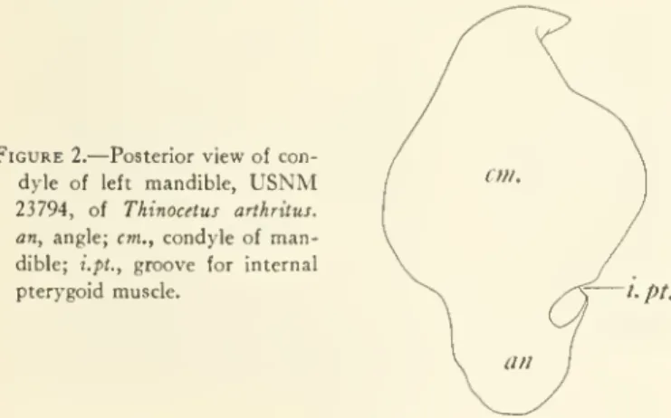
MANDIBLE
CETOTERES FROM THE MIOCENE CHOPTAISTK FORMATION above and in front of the anterointernal rim of.
CETOTHERES FROM THE MIOCENE CHOPTAISTK FORMATION above and in front of the anterointernal rim of the
VSNM USNM
8 UNITED STATES NATIONAL MUSEUM BULLETIN 294 have been about one-fiftli (19.5 percent) of tlie entire
SCAPULA
The transversely flattened radial facet is deposited by a ridge-like ridge from the more convex transverse surface of the ulnar facet, which extends upward on the posterior surface of the shaft. The . the vertical portion of this articular surface ends approximately 20 mm. below the apex of the olecranon process. The . The distal epiphysis of the right ulna is quite narrow, weakening towards the posterior end and measuring 84 mm. . in length; the largest thickness is 25 mm.
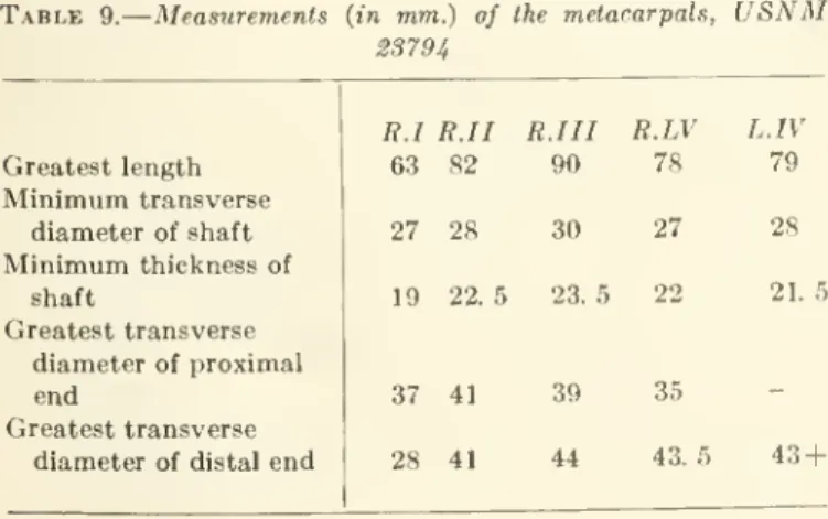
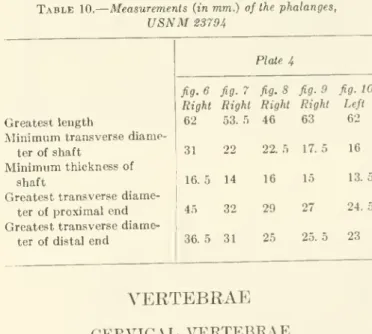
MANUS
Below its anterior ventral border, the articular surface of the greater sigmoid fossa meets the semicircular face at a right angle for articulation with the opposite face at the proximal end of the radius; The vertical diameter of this page is 30 mm. The back edge is . somewhat thinner than the anterior edge of the shaft, and this condition rests on the rough distal edge, which has the posterior angle extended backward. The first metacarpal was found distalto theradial. The second metacarpal was malpositioned and rested on the adjacent ulna.
12 UNITED STATES NATIONAL MUSEUM BULLETIN 29 4
CETOTHERES FROM THE MIOCENE CHOPTANK FORMATION 13
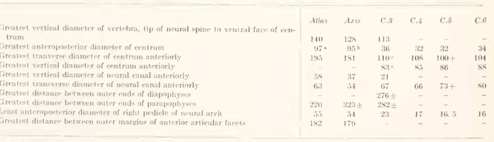
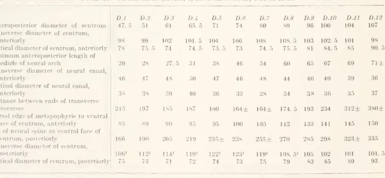
DORSAL VERTEBRAE
strontium; the pedicles of the neural arch are broad (right, niininuiin transverse diameter, 41 mm.) and very short; they support the roof of the neural canal as well as contribute to part of the base of the diapophysis. The broad pedicles (36 mm.) of the neural arch support the low roof of the neural canal as well as contributing part of the base of the diapophysis. No material change in the transverse or vertical diameter of the neural canal is apparent.
CETOTHERES FROM THE MIOCENE CHOPTANK FORMATION 15
Each parapophysis has a thick concave curved posterior margin, a thin rounded anterior margin on the distal half of its length, and a novoidal (right, length 50 mm) facet for the head of the eleventh rib, posterior to the extremity. The elliptical profiles of the anterior (pi.6,fig. 10) and posterior parts of the center are almost identical, as are their vertical and transverse diameters. Twelfth dorsal. Fairly broad (minimum diameter, 0.65 mm) and dorsoventrally compressed parapophyses project outward from near the center of the outer surface of the centrum (USNM 23794); the anterior and posterior margins of this process are thin and the end is truncated obliquely from the anterior margin to the posterodistal angle, which is thickened but not otherwise adapted for attachment of the head of the twelfth rib.
LUMBAR VERTEBRAE
The elongate nietapophyses, which rise 50 mm. above the floor of the neural canal, project considerably as-. The median longitudinal ridge is more strongly developed on the ventral surface of the centrum than on the floor of the neural canal. A very slight reduction in the length of the compressed subspatulat parapophysis dorsoventrally on the anteroposterior diameter of the slender pedicle of the neural arch is discernible.
The median longitudinal edge does not extend the length of the ventral surface of the center; there are no minor excesses. 23694). Most of the right parapophysis (Fig. 10,Fig. 8) and the distal end of the neural spine are lost. Eighth lumbar. The convex curvature of the posterior edge of the thin outwards and downwards.
A median longitudinal ridge with sharp edges appears on the ventral side of the centrum; the corresponding ridge on the floor of the neural canal is low. ward than in the upper part of the middle; these processes are also slightly shorter and wider than those on the middle eighth. The dorsoventrally appressed parapophyses (p. 11, fig. 10) are approximately lateral (75 mm.), but are slightly shorter than those of the anterior middle and are less obliquely deflected. A sharp-edged median longitudinal ridge is present on the ventral surface of the centrum, and a similar less elevated ridge runs the length of the floor of the neural canal.
This lumbar spine was detached from the vertebral column before the accumulation of the protective covering sediments and was found to be unrelated to the skeleton.
CETOTHERES FROM THE MIOCENE CHOPTANK FORMATION 19
CAUDAL VERTEBKAE
An approximately equal development of the front and back sides of the haemal tubercles, . and an increase in the width of the mid-ventral-longitudinal haemal groove occurs first in this caudal. Sixth caudal.— In this caudal (USNM 23794) the profile of the posterior end of the centrum is almost ovoid in contrast to the hexagonal anterior end (pi. . 12, fig.6) and are also somewhat narrower. Segmental blood vessels (pi. Abovethisoda vessels in their upward direction pass through a vertical aqueduct in the lateral part of the center to reach the left side of the foramen in the pedicle of the neural arch.
The very low neural spine (pi.. 7) extends almost the entire length of the roof of the neural spine.
CETOTHERES FROM THE MIOCENE CHOPTANK FORMATION 21
CHEVRONS
22 UNITED STATES NATIONAL MUSEUM BULLETIN 294
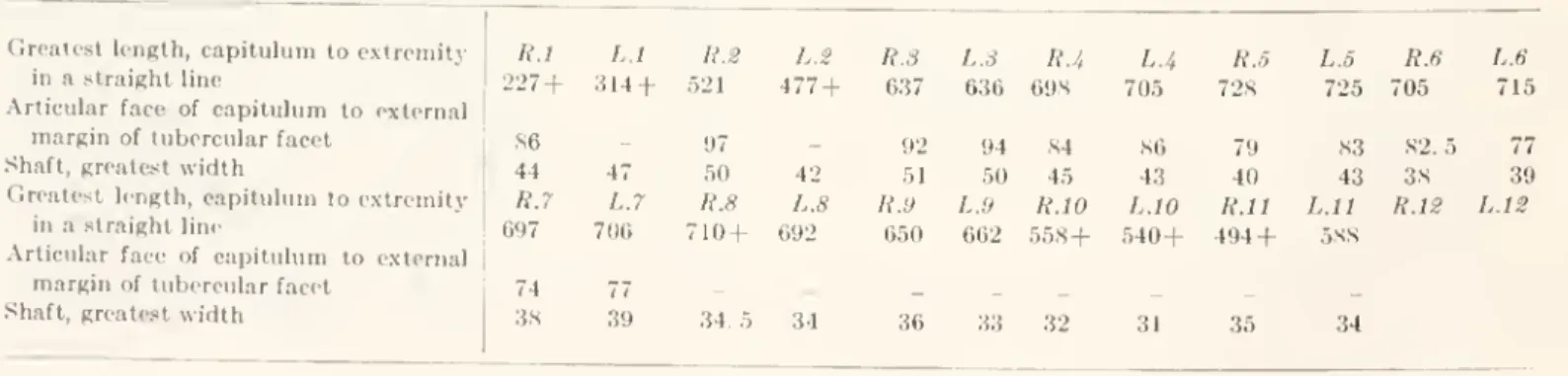
RIBS
CETOTHERES FROM THE MIOCENE CHOPTANK FORMATION 23
STERNUM
THE VERTEBRAE OF A SECOND MIOCENE CHOPTANK CETOTHERE
Reduction of cervical length attributed to the mechanical compression of the neck between the head and thorax by water pressure from the front during swimming, as suggested by Winge (1918, p. Including Calvert and Choptankctotheres fusion of the cervical vertebrae involving the coalescence) opposite the centra and pedicles of the neural arches , occurs first between the axis and the third cervical (USNM. The major dorsal neck muscles are considered an integral part of the musculature of the trunk and function as such during swimming.
On these dorsals, a well-defined articular facet, located at the postcrodorsal angle of the outer surface of the centrum below the base of the neural canal, articulates with the capitulum of the fol-. On the posterior dorsal, the transverse processes are the same size and length as the corresponding processes in the lumbar region and are considered serially homologous. Atrophy of the hind limb and the accompanying degeneration of the pelvis had been carried out in some at least of the cetotheres before the close of the Miocene.
Retrogressive remodeling of unresolved Middle Miocene (Astoria fm.) bones. 24) had continued as far as the acetabulum was. Four five modified terminal caudals are embedded in the caudal processes of recent mysticetes, and the corresponding caudals are similarly degenerate.
CETOTHERES FROM THE MIOCENE CHOPTANK FORMATION 25
TYMPANIC BULLA
USNM SS6S6
26 XJNITED STATES NATIONAL MUSEUM BULLETIN 294 in front of the sigmoid process; the anterior pedicle has
VERTEBRAE
CETOTHERES FROM THE MIOCEISTE CHOPTANK FORMATION 27
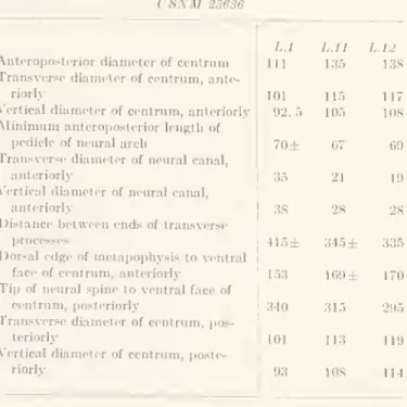
CEKVICAL VEETEBRAE
The ends of the diapophyses and parapophyses are not joined distally to completely close the fora-. Fifth cervical.— In contrast to the fourth cervical, the vertical diameter of the anterior surface of the centrum is increased, the neural canal is wider, and the projection for the cervical extension (pi. The ventral surface of the centrum is pressed on each side of the lower longitudinal median.
Each pedicle supports an elongated concave prezygapophysial facet that projects anteriorly beyond the level of the anterior surface of the center. The post-zygapophysial facets are oval, but slope less prominently downward from the external to the internal margins; they project posteriorly beyond the level of the posterior face of the center. The median longitudinal ridge on the ventral side of the center is much narrower and more prominent than the corresponding broad ridge on the preceding cervical vertebra.
The neural arch pedicles are low and support forwardly projecting elongated concave prezygapophyseal facets that are not symmetrical. The diapophyses are dorsoventrally expanded at the base and project outward from the dorso-external part of the centrum anteriorly. On the anterior right back, the pedicles of the neural arch are massive and wide.
The diapophysis increases progressively in width by the eight anterior dorsals, and on the whole these processes arise partly from the pedicle of the neural arch and partly from the dorso-external part of the centrum anteriorly.
30 UNITED STATES NATIONAL MUSEUM BULLETIN 294 the twelfth dorsals. The width of the gap between the
CETOTHERES FROM THE MIOCENE CHOPTANK FORMATION 31
The basal part of each diapophysis arises from the thick pedicle (minimum transverse width, 32 mm.) and from the dorso-external part of the anterior centrum. The low blunt metapophyses project strongly beyond the level of the anterior face of the centrum;. each is traversed anteroposteriorly by a rounded ridge which bounds the prezygapopiicial facet externally. The narrow elongate prezygapopiicial facets are deeply concave from side to side. The ial facets are very narrow and protrude strongly posteriorly beyond the level of the posterior face of the centrum.
An increase in the length (81 mm.) of the centrum as well as in its width anteriorly (108 mm.) is not unusual in this part of the dorsal series. Thick pedicles (riglite, minimum transverse diameter, 34 mm.) of the neural arch provide origin. lt;) the transverse processes (diapophyses), which pro-. The p^ach prezy-gapopiiysial facet is very narrow anteriorly, but increases inward and concavity toward the base of the neural spine.
The postero-external facet of the capitulum of the eighth rib is located mainly on the posterior surface of the prominent process. Each broad transverse process (diapophysis) projects outward from the transversely dilated (40 mm) robust pedicle of the neural arch from the dorso-external portion of the center forward and is curved upward, very slightly beyond the level of the neural arch bow. front face of center. The rough facet of the tubercle of the eighth rib at the tip of each diapophysis is elongated (length 54 mm; vertical diameter anteriorly 21 mm), subcrescentic in circumference and deeply excavated medially.
Each metapophysis is laterally compressed, pointed anterodorsally, extends anteriorly beyond the level of the anterior face of the centrum, and contributes to the outer wall of the rather narrow prezygapophyseal facet.
CETOTHERES FROM THE MIOCENE CHOPTANK FORMATION 33
The initial development of the median longitudinal keel is observable on the ventral surface of the first lumbar centrum, and in the twelfth and eleventh this keel is low, rounded, and inconspicuous. The nerve lances decrease in height from first to last, and were apparently thrown widely in the middle of the first eleven after the last. However, the anterior border of the neural spine is eroded; the edge of it .. maybe could have widened a bit.
CETOTHERES FROM THE MIOCENE CHOPTANK FORMATION 35
CAUDAL VERTEBRAE
Table 22. Dimensions {inmm.) of the caudal vertebrae, USNMS36S6. Table 22. Dimensions {inmm.) of the caudal vertebrae, USNMS36S6.
CETOTHERES FROM THE MIOCENE CHOPTANK FORMATION 37 The rather thick pedicles of the neural arch (length,
38 UNITED STATES NATIONAL MUSEUM BULLETIN 294
39 Fifth chevron. — The anterior projection of the
S. NATIONAL MUSEUM BULLETIN 294, PLATE 1
S. NATIONAL MUSEUM BULLETIN 294, PLATE 3
S. NATIONAL MUSEUM BULLETIN 294, PLATE 5
S. NATIONAL MUSEUM BULLETIN 294, PLATE 7
S. NATIONAL MUSEUM BULLETIN 294, PLATE 9
S. NATIONAL MUSEUM BULLETIN 294, PLATE 13
S. NATIONAL MUSEUM BULLETIN 294, PLATE 15
S. NATIONAL AIUSEUM BULLETIN 294, PLATE 17
S. NATIONAL MUSEUM BULLETIN 294, PLATE 21
S. NATIONAL MUSEUM BULLETIN 294, PLATE 23
S. NATIONAL MUSEUM BULLETIN 294, PLATE 25