CHAPTER 1
Introduction to Sensitized Lanthanide Luminescence
and the Detection of Bacterial Spores
1.1 Lanthanides – Relevance and History
The lanthanides, or lanthanoids in IUPAC terminology, comprise the fifteen elements of the top row in the ‘f-block’ of the periodic table and have the electronic configuration [Xe] 4fn 5s2 5p6 where n varies from 0 to 14. Also known as ‘rare earth elements’ due to the etymology of the term ‘lanthanide’ (derived from the Greek lanthanein, meaning ‘to lie hidden’) and the uncommon oxides from which they were
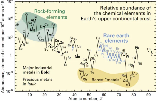
first isolated, lanthanides are in actuality neither ‘rare’ nor ‘earths’, an old term used to describe certain metal oxides such as lime and magnesia.1 Even the rarest lanthanides – thullium and lutetium – are nearly 200 times more abundant than gold (Figure 1.1).2 Yet, the name ‘rare earths’ has persisted, perhaps due to the enigmatic nature of these unusual metals, and their ability to ‘hide’ behind each other in minerals. Indeed, the similar chemical properties of lanthanides make their separation quite difficult, even today.
Lanthanides have found uses in a wide variety of industries and materials, such as catalysts, glasses, ceramics, permanent magnets, optics and electronics.3, 4 Solid phosphors containing europium, cerium and terbium are major contributors to commercial markets in fluorescent lighting and color displays. Various lanthanide ions can be used in lasers, with neodymium as the most famous in yttrium aluminum garnet (Nd-YAG). The green, blue and red luminescent bands in Euro banknotes are attributed to europium complexes.5 Certain lanthanides (Eu, La, Lu, Nd, Pr, Sm, Th, Tm and Yb) are used as tracers in wine chemistry to discriminate wines according to geographical region.6 The ratio of europium, which is almost entirely (~ 97%) formed in stars, to other rare earth elements in meteorites has helped us decipher much of the history of processes in our solar system, such as the early development of the feldspar-rich lunar crust.7
In aqueous solution, lanthanides are most stable in the +3 oxidation state, leading to high coherent behavior and hence making them difficult to separate and purify. The preference for the trivalent oxidation state is due in part to the energy of the 4f electrons being below those of the 5d and 6s electrons (except in the cases of La and Ce). When forming ions, electrons from the 6s and 5d orbitals are lost first, so that all Ln3+ ions have [Xe] 4fn electronic configurations. This, coupled to the high enthalpies of hydration for trivalent lanthanides, results in the stability of the +3 oxidation state. In reducing conditions, europium, samarium and ytterbium can be stable in the divalent form; cerium has also been known to adopt a +4 oxidation state.
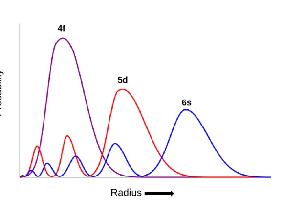
Lanthanide ions possess relatively high charge densities and have a strong electrostatic nature in their bonding, as the ions are polarizing and can be classified as hard Lewis acids. The 4f orbitals in Ln3+ ions are well shielded by the 5s and 6p orbitals, and therefore do not participate directly in bonding (Figure 1.2). Therefore, π-bonding is not possible, and no Ln=O or Ln≡N multiple bonds are known for lanthanide complexes.
Coordination to trivalent lanthanides tends to be more ionic in character, which leads to a strong preference for negatively charged or neutral donor groups possessing large ground state dipole moments. Therefore, combinations of amines and carboxylic acid groups are often used in lanthanide complexation.8, 9 This ionic character of binding also means lanthanide complexes tend to undergo facile exchange of ligands.5 Coordination geometries in lanthanides are determined by ligand steric factors as opposed to orbital overlap or crystal field effects.10, 11 In aqueous solution, donor groups containing neutral oxygen or nitrogen atoms generally bind when present in multidentate ligands (podands, crown ethers, cryptates, etc.).12-15 Relatively few complexes of
monodentate nitrogen donors exist, reinforcing the oxophilic tendency of lanthanide binding. This preference for oxygen donors also makes lanthanides quite lithophilic, and explains their occurrence in silicates as opposed to metallic or sulphidic minerals.16
The coordination number of [Ln(H2O)n]3+ is normally 9 for the early lanthanides (La-Eu) and 8 for those later in the series (Dy-Lu), with the intermediate metals (Sm-Dy) exhibiting a mixture of species. However, the coordination number can be dictated by the steric bulk of the coordinating ligands, and species with coordination numbers as low as 2 and as high as 12 are known.5, 17
As the 4f electrons of the lanthanides are well shielded from the environment, the spectroscopic and magnetic properties of these ions (e.g., electronic spectra and crystal- field splittings) are largely independent of environment (solvent, coordinated ligands, etc.). The number of configurations for n electrons rapidly increases with the number of unpaired electrons:
n)!
- (14 n!
14! [1.1]
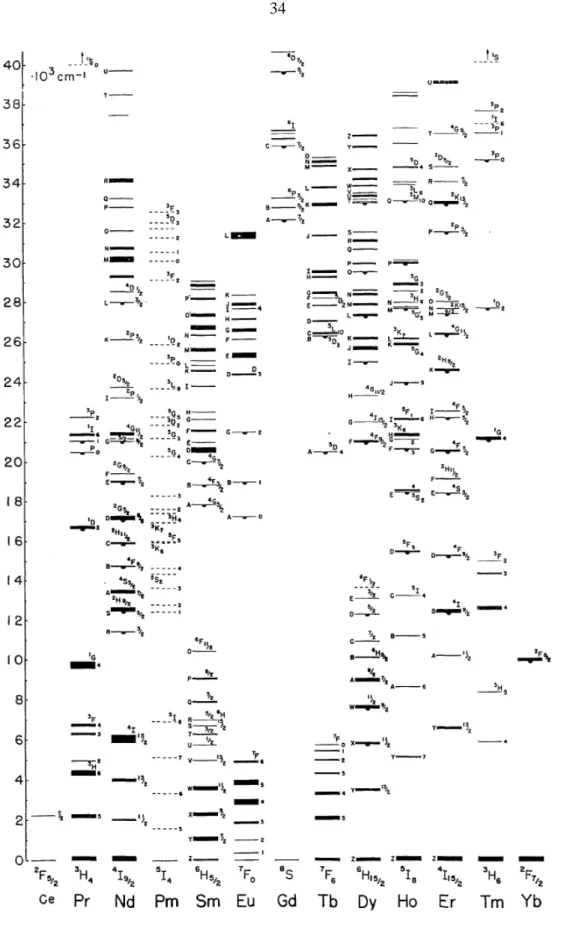
where 0 ≤ n ≤ 14, with the lowest energy term for each ion consistent with the predictions of Hund’s first and second rules.18, 19 Since all configurations have different energies, the lanthanides tend to exhibit rich and complex energy level structure (Figure 1.3).20 Due to spin-orbit coupling, the excited states of the lanthanides are well separated from the ground state manifold. Thus, the excited states are thermally inaccessible and ideal for electronic transitions. With the exceptions of the 4f0, 4f1, 4f13 and 4f14 species (La3+, Ce3+, Yb3+ and Lu3+, respectively), all lanthanide ions absorb electromagnetic radiation, primarily in the visible region, which is manifested in f-electrons from the partially filled
4f subshell being excited from the ground state to an excited state. These f–f transitions can be excited by both magnetic dipole and electric dipole radiation. Magnetic dipole transitions are Laporte (parity) allowed, while electric dipole transitions are Laporte- forbidden.21, 22 Electric dipole transitions are much weaker in lanthanides (ε ~ 0.1 mol-1 dm3 cm-1) than in the transition metals, meaning magnetic dipole transitions can often be seen.5, 23 Electronic transitions must involve promotion of an electron without a change in its spin (∆S = 0) and with a variation of either the total angular momentum and the total angular quantum number of one unit at most (∆L = ±1,0; ∆J = ±1,0). Though absorption of radiation can in theory promote the lanthanide ion to any energetically accessible state, emission normally occurs only from the lowest lying spectroscopic level of the first excited term due to rapid internal conversion.19 In cases of low symmetry or vibronic coupling, the f–f transitions can gain intensity through f- and d-state mixing with higher electronic states of opposite parity. Broad 4fn → 4fn-1 5d1 transitions can also be seen in the infrared region for some lanthanides.
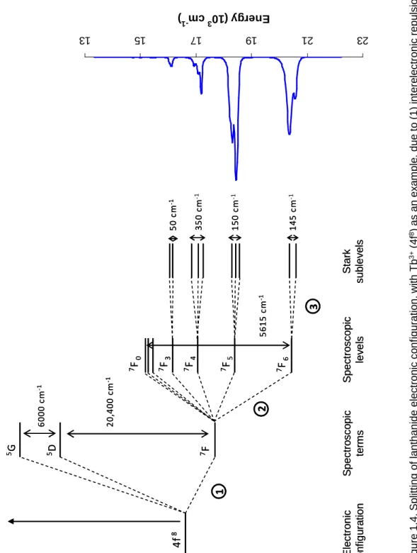
The electronic configuration of lanthanides is split due to a variety of interactions.
The initial configuration is split into spectroscopic terms by electronic repulsion, with separations on the order of 104 cm-1.24 These terms can be further split into spectroscopic levels, or J states, due to spin-orbit coupling effects. The energy differences between split J states lies in the range of 103 cm-1.25 These levels, in turn, can be split again into what are termed Stark sublevels due to ligand field effects from the coordination sphere around the lanthanide; Stark sublevel splitting is on the order of 102 cm-1 (Figure 1.4).26 This results in the overall emission peak position remaining largely unchanged as the f-electrons remain shielded, but the emission profile of a lanthanide (defined as the
relative intensity and degree of splitting of emission peaks) can vary greatly depending on modulation of these influences.27, 28 The number of Stark sublevels depends on the site symmetry of the lanthanide ion, and these can be thermally populated at room temperature, yielding more complex emission spectra.
Filling of the inner 4f electron shell across the lanthanide series results in a diminuation of the ionic radius by as much as 15% from lanthanum to lutetium, referred to as the lanthanide contraction.29 Though atomic radius contraction is not unique across a series (i.e., the actinides and the first two rows of the d-block), the fact that all lanthanides primarily adopt the trivalent oxidation state means that this particular row of elements exhibits a traceable change in properties in a way that is not observed elsewhere in the periodic table. Lanthanides behave similarly in reactions as long as the number of 4f electrons is conserved.30 Thus, we can use lanthanide substitution as a tool to tune the ionic radius in a lanthanide complex without changing its chemistry, to better understand how the size of the metal cation affects various properties.
1.2 Sensitized Lanthanide Luminescence
Though alone lanthanide ions have very low molar absorptivities and can only be effectively excited by lasers, lanthanide luminescence can be significantly enhanced by chelating ligands in a process called ‘sensitization’. The first demonstration of sensitized lanthanide luminescence was due to the efforts of Bhaumik and El-Sayed, who showed that if the lowest triplet level of europium tris hexafluoroacetylacetonate, or Eu(HFA)3, was excited by triplet-to-triplet intermolecular energy transfer from another donor (benzophenone), the energy could be transferred to the lanthanide cation.31, 32 This
indirect sensitization bypasses the selection rules that normally limit f–f excitation in lanthanides, and can result in luminescence enhancement by three orders of magnitude or more.24, 33, 34
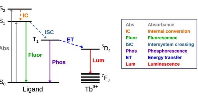
In sensitized lanthanide luminescence, the chromophore is normally an aromatic or unsaturated organic molecule that is either anionic or has a strong dipole moment to coordinate to the Ln3+ ion. In order to act as an efficient energy harvester or ‘antenna’ as they are often termed,35 the chromophore must absorb radiation effectively and pass as much of this energy as possible, nonradiatively, to the lanthanide for emission. This process, known as the absorption-energy transfer-emission (AETE) mechanism, has several steps. First, the light-harvesting ligand is excited from the ground state S0 to the singlet excited state S1 (Figure 1.5). Some chromophores have several singlet excited states; nonradiative relaxation from these higher singlet excited states (S2, S3, etc.) to the lowest singlet excited state (S1) via internal conversion (IC) can occur readily. Second, a triplet excited state (T1) is formed through intersystem crossing (ISC), a process that is more efficient near heavy atoms such as lanthanides, which promote spin-orbit coupling.36 Third, intramolecular energy transfer (ET) from the ligand triplet excited state to the lanthanide excited state occurs, resulting in a populated emittive level in the lanthanide. The efficiency of this step, the intramolecular energy transfer from chromophore to lanthanide, is the most important factor influencing the luminescence properties of rare earth complexes.37 The final step is the luminescence observed as the excited state in the lanthanide decays radiatively to the ground state manifold.38, 39
However, there are other pathways, both radiative and nonradiative, that can reduce the efficiency of sensitized lanthanide luminescence. Chromophores can lose
energy from the singlet excited state by two mechanisms: (1) fluorescence, where they radiatively decay from the singlet excited state to the ground state; or (2) the excited state can be nonradiatively quenched by photoinduced electron transfer or other means.12 The triplet excited state of the chromophore can also radiatively decay as phosphorescence, or be nonradiatively quenched by oxygen, though oxygen has been found to have little or no quenching effect on visible-emitting luminescent lanthanide complexes.40, 41 These mechanisms can be mitigated or minimized by judicial choice of chromophore.
The greatest vulnerability of sensitized lanthanide luminescence lies in nonradiative deactivation or relaxation due to solvent interactions, which can reduce emission intensity significantly through energy dissipation by vibronic modes.24, 42 Typically, this occurs by harmonic oscillators in the lanthanide coordination sphere, though outer-sphere quenching has also been observed.40 Vibronic quenching depends on both the number of oscillators close to the first coordination sphere and the R parameter:
κ µ
∆E ω R ∆E
ℏ ℏ =
= [1.2]
where ∆E is the energy gap between the emitting state and the higher energy J state of the ground multiplet, ħω is the oscillator vibrational quantum, µ is the oscillator reduced mass and κ is the oscillator force constant.43, 44 The R parameter represents the number of vibrational quanta between ∆E; the lower the value of R, the higher the rate of vibronic coupling and the more pronounced the emission quenching will be.
The most common and efficient quencher of lanthanide luminescence is the O–H oscillator.45 For the four lanthanides that luminesce in the visible region (Sm, Eu, Tb and
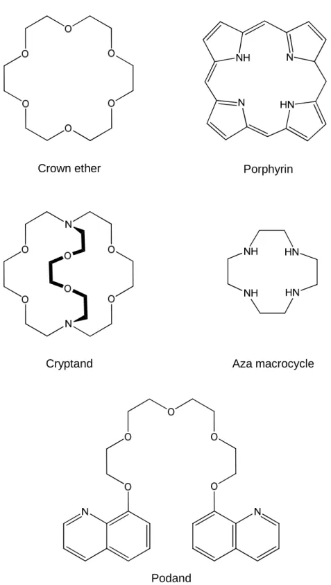
Dy), the R parameter lies in the range of 3 to 6, meaning quenching by OH vibrations is a significant mode of radiationless transitions.44 Gadolinium, in contrast, which is relatively unquenched by OH vibrational overtones, has an R value of ~10. In order to reduce or eliminate this pathway for nonradiative decay, the lanthanide ion must be effectively shielded from the solvent. This can be accomplished using various chelating ligands containing hard donors that bind to the lanthanide ion with high affinity and contain a cavity to encapsulate the ion and prevent solvent coordination. The first
‘insulating sheath’ for lanthanide ions was developed by Halverson in 1964 using fluorinated 1-diketonates.46 Since then a variety of successful ligands have been identified for this purpose, including cyclodextrins, cryptands, podands, calixarenes, porphyrins, crown ethers, and aza-crown macrocyclic and bicylic ligands (Figure 1.6).12,
13, 47-51
Triplet-mediated energy transfer in sensitized lanthanide luminescence has two proposed mechanisms. The Dexter energy transfer mechanism involves the transfer of an electron from donor (D) to acceptor (A). In the Dexter model, a resonant transfer of energy can be obtained between an allowed transition in the donor and a forbidden transition in the acceptor.52 The acceptor can therefore be made to luminesce after exposure to a given region of radiation where it would not normally, due to energy transfer from the donor. The Dexter mechanism is highly dependent on orbital overlap, and therefore efficient energy transfer is only seen at very small distances (~ 10 Å).53 The other proposed mechanism is the Förster, or Coulombic, energy transfer mechanism, in which energy transfer is dipole-induced and the electrons do not physically transfer.54 Energy transfer in the Förster model is highly dependent on the spectral overlap of the
emission spectrum of the donor and the absorbance spectrum of the acceptor, and can occur over longer distances. (Figure 1.7). However, it must be noted that both the Dexter and Förster mechanisms describe only single-step, photoinduced, nonradiative energy transfers, whereas sensitized lanthanide luminescence often involves multiple steps such as intersystem crossing and internal conversion prior to energy transfer.
The AETE mechanism depends on appropriate alignment of the ligand donating triplet energy level and the lanthanide accepting excited level for efficient energy transfer. The intramolecular energy transfer efficiency depends mainly on two energy transfer processes: Dexter resonant exchange interaction theory and thermal deactivation.55 The first describes the rate of energy transfer from the lowest triplet state to the resonant energy level as follows:56
( )
(
Ζ) ∫
=
−
=
(E)dE (E)E
F R 2π P
L R 2 exp KP k
a d 2
da
da da
ET [1.3]
where kET is rate constant of intermolecular energy transfer (ET), Pda is the transition probability of the resonant exchange interaction, Fd(E) is the experimental luminescence spectrum of E-donor (ligand), Ea(E) is the experimental absorption spectrum of the E-acceptor (Ln3+), Rda is the intermolecular distance between donor and acceptor atoms, L is the Van der Waals radius and 2πZ2/R is a constant relating to the specific mutual distance between the E-acceptor (Ln3+) and coordinated atoms (N and O). This is in competition with the reverse energy transition due to thermal deactivation:52
(
∆E RT)
Aexp
k(T)= − [1.4]
In other words, the donor and acceptor levels must be close enough to allow for efficient energy transfer, but if they are too close energy is lost due to thermal deactivation. The
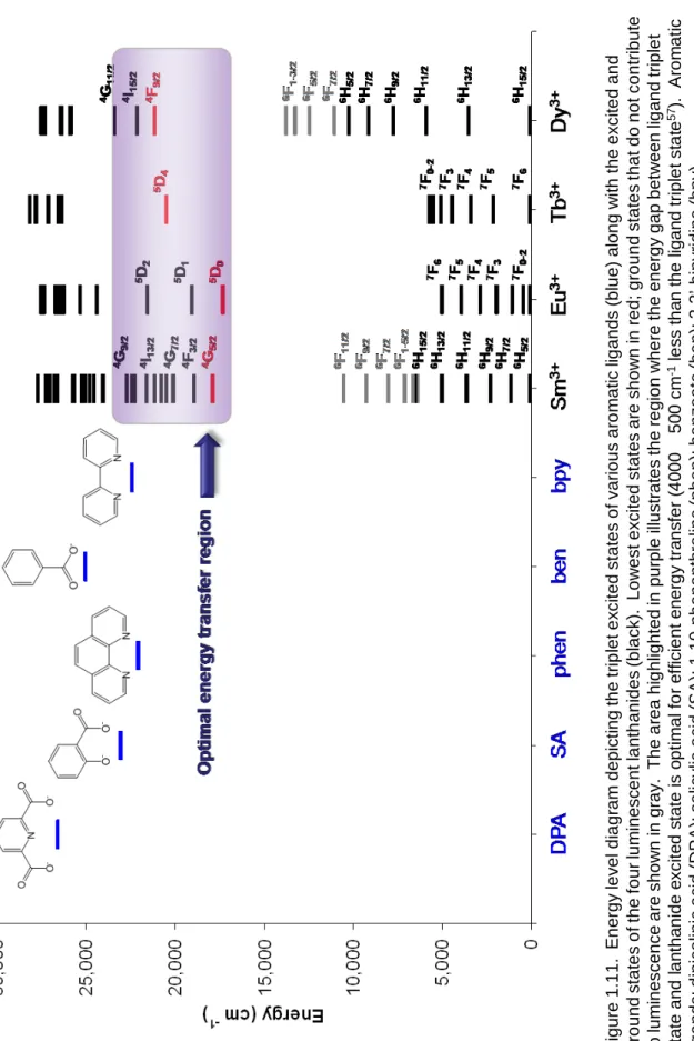
optimal energy gap between chromophore triplet state and lanthanide excited state is approximately 4000 ± 500 cm-1, meaning that the pairing of lanthanide and chromophore is of the utmost importance for achieving efficient energy transfer.37, 57
Sensitized lanthanide luminescence is a unique process, and as such these lanthanide complexes demonstrate some interesting features not found in other species that are known to luminesce. Emission spectra tend to exhibit narrow line-like bands because both excited and ground states have the same fn configuration.12, 24 These electronic f–f transitions are also largely independent of the chemical environment of the lanthanide ion, though the peak splitting and relative intensities can vary significantly in some cases. Due to the various energy transfer steps that occur in lanthanide luminescence (internal conversion, intersystem crossing, etc.), there is usually a very large difference between the absorption and emission maxima in these complexes, known as the Stokes shift.40, 58 The larger the Stokes shift, the less overlap between absorbance and emission bands and less energy is lost to reabsorption. Further, as the major transitions in these complexes are electric dipole ‘forced’ and normally forbidden, the excited state lifetimes tend to be very long, on the order of micro- to milliseconds.27 The triplet state of the ligand can have its own lifetime on the order of nano- to microseconds, so the energy transfer step from ligand to lanthanide can also lengthen the observed lifetime.59 Though the property of long luminescence lifetime is not necessarily unique, the fact that this occurs under ambient conditions for lanthanides is unusual. Most organic species that exhibit phosphorescence only do so at low temperatures and/or in the absence of oxygen.19 The observed excited state lifetime (τ) of a luminescent lanthanide complex can be expressed as follows:
nr
r k
k τ 1
= + [1.5]
where kr is the rate constant for radiative transition of the lanthanide ion and knr is the sum of rate constants for all nonradiative relaxation processes, which can include energy back-transfer and vibronic coupling. The excited state lifetime can also be regarded as a function of luminescence quantum yield (ΦLum):
nr r
r
Lum k k
Φ k
= + [1.6]
The overall quantum yield is dependent on the energy transfer efficiency (probability) from the ligand to the lanthanide ion (see equations 1.3 and 1.4).
Due to these unique properties, sensitized lanthanide luminescence has found a variety of applications.60 Europium and terbium chelates, due to their line-type emissions and long decay times, are used in time-resolved fluorescence resonance energy transfer (TR-FRET) immunoassays.61, 62 The lanthanide chelate is used as a donor, with some visible-absorbing dye such as Alexa® 647 or a rhodamine derivative used as the acceptor. Quenching of emission due to donor-acceptor proximity then operates as a tool to determine, for instance, protein-protein interaction or enzymatic reaction time. When concentrated as phosphors or in beads, lanthanides have been used in imaging microscopy in the millisecond time domain.63 Use of two or more lanthanides simultaneously is advantageous for PCR detection of diabetes risk,64 gene detection of bacterial toxins65 and an assay of papilloma virus.66 Sensitized lanthanide luminescence also has drug discovery applications, such as high-throughput screening or pharmacokinetic analysis of drug transportation and accumulation.40 However, the use of lanthanide complexes as tailored receptor sites has not been fully explored.
1.3 Lanthanides, Dipicolinic Acid and Bacterial Spores
Dipicolinic acid (DPA, pyridine-2,6-dicarboxylic acid) was originally used in a fluorescence spectroscopy method to detect trace amounts of terbium for probing alkaline earth metal ion interactions in biological systems.67 It was realized over twenty years later that this detection technique based on intense luminescence could be used in reverse – to detect dipicolinate as a marker of bacterial spores.
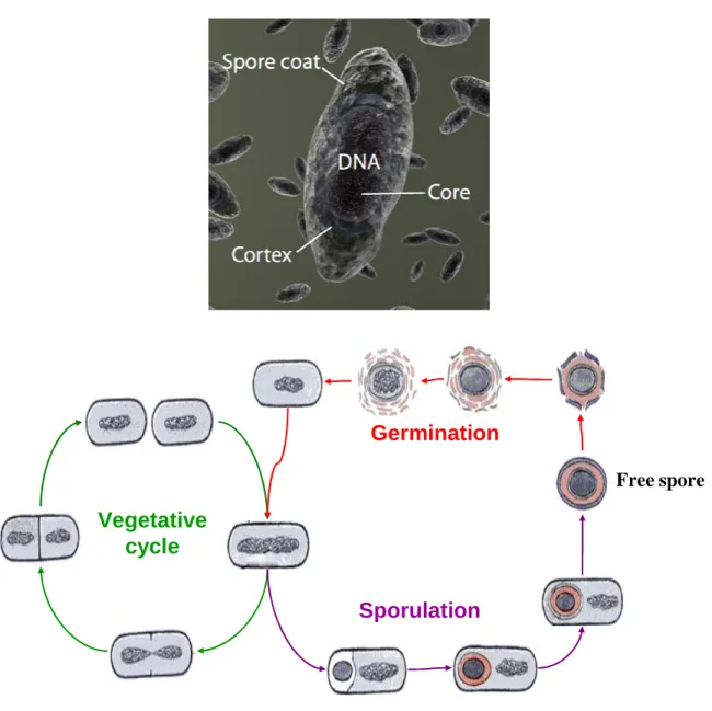
Bacterial spores, also known as endospores, are dormant microbial structures that exhibit remarkable resistance to chemical and physical environmental stresses and are considered to be one of the toughest forms of life on Earth.68 Discovered in 1876, endospores are formed inside the vegetative cells of certain species of Bacillus, Clostridium, and Sporosarcina (hence the ‘endo’ prefix) in a process called
sporulation.69-72 Sporulation is often triggered when the cells are exposed to adverse environmental conditions, such as desiccation or starvation. Bacterial spores house the cell DNA within the spore core, comprised of calcium dipicolinate (CaDPA), and a tough coat composed of protein layers (Figure 1.8). Endospores can remain dormant with no detectable metabolism for potentially millions of years.73-76 When conditions become favorable again, as indicated by the presence of water, nutrients or specific germinants, endospores undergo germination and outgrowth to become vegetative cells, completing the cycle.77, 78
In the dormant spore state, endospores are resistant to a wide variety of chemical and physical stresses, such as UV and gamma radiation, desiccation, temperature and pressure extremes, and attack by a number of toxic agents.79-82 Bacterial spores are 10,000 times more resistant to heat and 100 times more resistant to UV radiation than
their active bacterial cell counterparts.83, 84 As they are resilient to most sterilization procedures, bacterial spores are used in several industries as biological indicators.85, 86 Certain species can even survive the vacuum, extreme temperatures and radiation of space,87, 88 and are the focus of research concerning planetary protection, panspermia (transfer of life from one planetary body to another via meteoritic impacts), and life in extreme environments.89, 90 In addition, detection of bacterial spores became a national priority after the anthrax attacks of 2001, as Bacillus anthracis spore powders are the vectors of the anthrax bioweapon.91-94 Certain species of anaerobic endospores, such as Clostridium botulinum and C. perfringens, are pathogenic and the causative agents of
food poisoning or serious disease.95
Due to its applications in homeland security, sterilization validation and astrobiology, bacterial spore detection has become a rather extensive field. The classical measurement of bacterial spores involves culture-based methods, and is used as the NASA Standard Assay for ascertaining spacecraft sterility.96, 97 This procedure involves swabbing the surface, suspending any collected endospores in water, and then plating this suspension on growth media. Any bacterial spores will germinate in the favorable conditions and produce colonies. However, colony formation requires at least 20 cycles of cell replication, a process that requires 2 to 3 days.97 This method also cannot discriminate between endospores and vegetative cells. Thus, the NASA Standard Assay is not viable as a rapid endospore detection technique.
Direct detection of bacterial spores can be challenging, for the same reasons that make endospores difficult to irradicate. The tough spore coat is impermeable to staining techniques, so most microscopy and flow cytometry methods are unsuccessful.
Endospores are also highly resistant to lysis, meaning DNA extraction protocols are ineffective. The lack of measurable metabolism renders microcalorimetry and cellular respiration techniques inadequate. In order to detect bacterial spores, we must instead consider what makes them unique and take advantage of these factors to produce a highly specific assay.
Bacterial spores contain a unique chemical marker – dipicolinic acid, or DPA.
DPA is present in nearly all bacterial spores and comprises about 10–15 % of a spore’s dry weight, or approximately 108 molecules per spore.84, 98, 99 Some endospore mutants do not contain DPA, but these are rare, and DPA has never been detected in vegetative cells.95 Detection of this chemical marker can therefore serve as a positive signal for the presence of bacterial spores, and the amount of DPA detected can be used to estimate the approximate endospore concentration.95, 100
There are several theories regarding the functionality of such a high concentration of dipicolinate in the endospore. First, this dianion serves in the storage of divalent cations such as calcium and magnesium, which are important for cell function.98 Second, as DPA is an excellent absorber of UV radiation, it appears to play a major role in the UV photochemistry and protection of endospore DNA.101, 102 The high concentration of dipicolinate salts in the core also displaces water and confers additional resistance to desiccation and wet heat.103 Finally, DPA is metabolized in the early stages of germination, and therefore serves as a nutrient source for the growing cell.104, 105
Detection of DPA as an indicator of bacterial spores was first proposed in 1955, in a protocol involving UV absorption of the dipicolinate following acid digestion of the spores, isolation by ether extraction, separation by paper chromatography and finally
elution of the DPA-containing spots.106 A much simpler spectroscopic method of dipicolinate detection was published in 1958, in the form of a colorimetric assay with ferrous iron.107 DPA extracted from endospores was detected via a color change from pale green to red-brown upon complexation with Fe2+ in acidic conditions (pH 4–6).
Following this work, many methods for dipicolinate detection have been published, including fluorescence of CaDPA, UV absorbance spectrophotometry, anti-Stokes Raman spectroscopy, surface-enhanced Raman spectroscopy, gas chromatography, mass spectrometry and high performance liquid chromatography (HPLC).95, 108-111
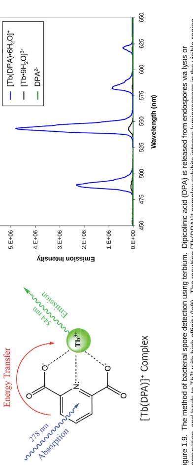
The application of lanthanides to bacterial spore detection began in 1997 with a method using terbium to detect dipicolinate with fluorescence spectrophotometry.112 Addition of terbium chloride to a suspension of lysed endospores causes the formation of [Tb(DPA)n]3-2n complexes, where n varies from 1 to 3, as the Tb3+ displaces the Ca2+ of CaDPA. Dipicolinate is an effective absorber of ultraviolet radiation due to the delocalized π-electrons of the aromatic pyridine ring. The triplet excited state of the DPA anion (26,600 cm-1) is also in the appropriate regime to effectively sensitize the Tb3+
cation via energy transfer to the 5D4 emitting level of the terbium (20,500 cm-1) through the AETE mechanism.113-115 There is some evidence against an intramolecular heavy atom effect in some 4-substituted dipicolinate ligands coordinated to Tb3+, which argues for a singlet-to-metal mechanism for intersystem crossing to the lanthanide.41, 116, 117
However, the contribution of this pathway is most likely very small, so we treat this system in the usual manner and assume the excited dipicolinate singlet state decays to the triplet via internal conversion prior to energy transfer to the lanthanide. The end result is
intense luminescence under UV excitation that is more than three orders of magnitude greater than that of terbium alone (Figure 1.9).95, 118-122
Although the method is rapid and straightforward, there is much room for improvement in the detection of bacterial spores with the Tb-DPA luminescence assay.
The potential for false positives or false negatives through complexation of anionic interferents to the trivalent terbium cation is a serious concern when the method is applied to environmental samples. Previous studies indicate that phosphate in particular can inhibit DPA binding or decrease luminescence intensity.123, 124 Further, in environmental samples with low endospore concentrations where the Tb(DPA)+ species predominates, the six coordinated water molecules can quench the luminescence by nearly an order of magnitude due to radiationless deactivation.125 Finally, the propensity of dipicolinate to form up to three complexes with terbium, namely Tb(DPA)+, Tb(DPA)2- and Tb(DPA)33-, each with different luminescence intensities and lifetimes, precludes a direct correlation between intensity and DPA concentration. This dissertation will focus on methods to mitigate these detrimental factors using a macrocyclic helper ligand to generate a dipicolinate-specific lanthanide-based receptor site.
1.4 Lanthanides and Lanthanide Complexes as Sensors
Lanthanides demonstrate several advantages over traditional organic fluorophores, quantum dots and other fluorescent species commonly used as sensors and switches. They feature significant spectral resolution due to their large Stokes shifts and narrow emission lines, and temporal resolution due to long lifetimes. This allows for the use of time-gated techniques and bandpass filters to reduce interference from native or
auto-fluorescence in the sample, which occurs on the nanosecond timescale.23, 58 Lanthanide complexes also do not suffer from photobleaching as organic dyes do, because lanthanides are effective quenchers of triplet states.126 These qualities allow for the construction of robust sensors for a variety of applications.
Of all the lanthanides, Eu3+, Tb3+ and Gd3+ are the best ions in terms of efficient excited state population, with energy gaps of 12,300 cm-1 (5D0 → 7F6), 14,800 cm-1 (5D4
→ 7F0) and 32,200 cm-1 (6P7/2 → 8S7/2), respectively.12 However, while europium and terbium both emit in the visible region, gadolinium emits in the ultraviolet, making it unfeasible for use in most sensing applications due to significant absorption and emission interference of these high-energy wavelengths. Though not suitable as luminescent sensors, Gd3+ complexes do have a large number of unpaired electrons ([Xe] 4f7) and isotropic magnetic properties. These properties, coupled to a long electron spin relaxation time of 10-9 s, makes this lanthanide highly NMR-active.127 When coordinated to one or more water molecules, Gd3+ enhances the water proton relaxation efficiency by lengthening the rotational correlation time.128 Thus, many gadolinium complexes with one free binding site for solvent coordination, such as Gd(DTPA) and Gd(DOTA), have been developed for use as MRI contrast agents.129-131
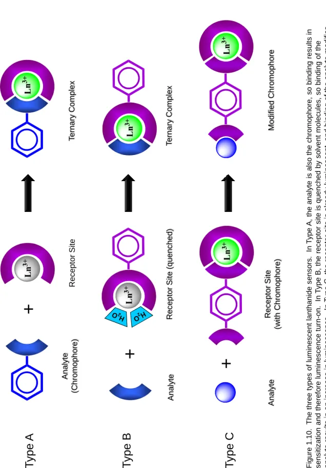
Luminescent lanthanide complexes designed for use as sensors and switches can be classified into three types, based on the position of the chromophore and the state of the lanthanide before and after analyte binding (Figure 1.10). More complex systems have been designed containing two or more lanthanides for multicolor detection in imaging applications, but these are out of the scope of this dissertation and will not be discussed here.132
The type A sensor is the most simple; the receptor site is the lanthanide complex, and the analyte is the chromophore. Binding of the aromatic analyte to the receptor site results in emission due to sensitization. This type of sensor is most effective in cases where the two states – analyte bound or unbound – must be very easily distinguished, as in samples with very low amounts of analyte or where the background noise is high.
Type A sensors have been used with amide-modified DO3A (1,4,7,10-tetraazacyclo- dodecane-1,4,7-trisacetate) ligands bound to Tb3+ to selectively bind the bidentate analytes p-dimethyl amino benzoic acid (DMABA) or salicylic acid (SA).133-135 The binary complex of Tb3+ and ethylenediaminetetraacetic acid (EDTA) can effectively detect salicylic acid, 4-aminosalicylic acid, 5-fluorosalicylic acid.136 The Tb(EDTA) complex has also been used to detect catalysis of hydroxybenzoic acid (HBA) by hemin via formation of a ternary complex with the HBA oxidation product.137 Detection of tetracyclines was realized using sensitized europium luminescence post-column following liquid chromatography; the authors noted a dependence on EDTA concentration but did not hypothesize any ternary complex formation, though this is the likely result.138 Diaza-crown ethers have been utilized with Tb3+ and Eu3+ to detect phthalate, benzoate, dibenzoylmethide and picolinate.139 A pH sensor was developed due to the pH-dependence of a europium ternary complex containing a β-diketonate as the chromophore. When the pH shifted out of physiological range, the chromophore dissociated and sensitization was lost.12 The future of these types of sensors lies in the development of receptor sites with greater analyte affinity, perhaps via stronger host- guest interactions such as π-stacking or modification of receptor site topology to generate a ‘lock and key’ style of hydrophobic pocket.
The type B sensor involves a change in luminescence upon analyte binding. In this case the receptor site is composed of the lanthanide complex with a chromophore already attached, but the complex is unsaturated. This results in labile solvent molecules in the lanthanide coordination sphere, so luminescence is quenched due to nonradiative deactivation. Binding of the analyte displaces these quenchers and produces a change (usually an increase) in emission. This method is most useful in anion sensing, where the anion (acetate, fluoride, etc.) can coordinate well to the lanthanide but is not capable of absorbing UV radiation and sensitizing the lanthanide. For example, several types of Tb3+ and Eu3+ podand complexes have been developed with sensitivity to Cl- and NO3-
, respectively, when detected in acetonitrile.140, 141 A binary complex composed of a bis- bpy-phenyl phosphine ligand chelated to Eu3+ was able to detect NO3-, Cl-, AcO- and F- with varying sensitivities by displacing acetonitrile solvent molecules in the Eu3+
coordination sphere.142, 143 Europium bound to a tri-N-substituted DO3A ligand was able to detect hydrogen carbonate in aqueous solution,144, 145 and when coordinated to a cryptate with a poly-N-methylated flexible arm can act as a sensitive pH sensor over a fairly wide range.146 Conversely, quenching of terbium acetylacetonate (acac) luminescence allowed for a sub-micromolar detection limit of chromate in aqueous solution when coupled to ion chromatography.147 Though sensitive ion detectors have been developed, the selectivity of such receptor sites is still in need of improvement, in particular when discriminating between ions of similar size and/or charge.
The final sensor design motif, type C, also starts with the lanthanide complex and chromophore already bound. In this case, however, the lanthanide coordination sphere is already saturated, so the analyte is not required to chelate the Ln3+ to be detected.
Instead, association of the analyte with the chromophore causes a change in emission due to an alteration of the antenna properties (i.e., change in degree of sensitization, quantum yield, lifetime, etc.). The first examples of these types of sensing complexes were to detect cations such as H+, Na+ and K+ via suppression of luminescence in europium aza- crown ethers.148 Other water-soluble complexes have been developed to detect dO2, halide and hydroxide by quenching of luminescence through various means (protonation, charge transfer, etc.) or decreasing the absorption of the chromophore.39, 149 Detection of Zn2+ using Tb3+ and Eu3+ diethylenetriaminetetraacetic acid (DTPA) bis-amide complexes has also been proposed, where two N,N,N’,N’-tetrakis(2-pyridylmethyl) ethylenediamine (TPEN) groups on the DTPA ligand are brought into closer proximity to the lanthanide upon zinc binding, allowing for sensitized luminescence.150 This sensor demonstrated selectivity over Ca2+ and Mg2+ ions and an apparent dissociation constant for Zn2+ of 2.6 nM, but Cu2+ interfered significantly and luminescence intensity was low due to the large lanthanide-chromophore distance. Sensors based on this strategy are still in their infancy, though many applications can be envisioned following sufficient improvement of stability and sensitivity.
This dissertation summarizes work to improve the Tb-DPA luminescence assay for bacterial spore detection by designing a dipicolinate-specific receptor site in the form of a lanthanide binary complex. Since dipicolinate is both our analyte and chromophore, this assay follows the Type A paradigm, with the receptor site comprising the terbium ion protected by an encapsulating helper ligand. However, dipicolinate is not the only aromatic anion capable of sensitizing lanthanide luminescence (Figure 1.11). We will also investigate receptor site designs for various salicylates and catecholamines with
physiological relevance to generate more robust, rapid sensing technologies for highly sensitive in situ detection.
A number of factors must be considered when designing lanthanide-containing receptor sites. The choice of lanthanide is paramount. The ionic radius varies across the lanthanide series, and the size of this cation can influence the relative binding affinity of the target analyte. The lanthanide excited state must also align correctly with the triplet excited state of the target analyte; if the energy difference is too great, the energy transfer efficiency will be low, but if it is too small, quenching effects such as back-transfer will predominate. The emission properties of the lanthanide, such as luminescence lifetime and quantum yield, must also be considered to produce an adequate signal.
Almost as important as the choice of lanthanide is the choice of helper ligand, which turns the lanthanide into a receptor site. This ligand must bind to the lanthanide with high affinity to prevent solvent coordination, which can severely quench luminescence via nonradiative decay pathways. However, the ligand must not interfere with binding of the target analyte, or the lanthanide loses all functionality as a sensor.
Finally, pH and temperature effects must be considered, such that the receptor site is stable in the pH range where the target analyte is most soluble and/or sufficiently deprotonated to bind effectively.
We will be using a hexadentate macrocyclic ligand, DO2A (1,4,7,10- tetraazacyclododecane-1,7-bisacetate), as our helper ligand, as this chelator meets our initial criteria for designing a terbium-containing dipicolinate receptor site. Macrocyclic ligands are molecules – usually heteroatomic – with a semirigid ring structure as the backbone. These ligands vary in terms of ring diameter and the extent of
functionalization of substituents on or attached to the ring scaffold. Most macrocyclics have a hydrophilic cavity in which an ionic substrate such as a metal ion can nest and be shielded from the environment by its lipophilic envelope.3, 151 Macrocyclic ligands such as the octadentate DOTA (1,4,7,10-tetrakiscarboxymethyl-1,4,7,10-tetraazacyclo- dodecane) have found a convenient niche in the field of bioimaging as magnetic resonance contrast agents due to their tendency to bind gadolinium with high affinity.152,
153 In fact, most macrocyclic ligands seem to exhibit an unprecedented selectivity for lanthanide ions, and the dissociation of these lanthanide-macrocycle complexes appears to be independent of foreign metal ion concentration.154 Lanthanide complexes involving macrocyclic ligands are highly water-soluble, thermodynamically stable, kinetically inert at physiological pH, cell-permeable and nontoxic, making them ideal for use in vivo as bioprobes. The facile derivatization of ligands bound to lanthanides also makes them easily tailored to bind specific biomolecules such as antigens or proteins.
We are therefore interested in constructing receptor sites composed of a lanthanide ion encapsulated by a macrocyclic ligand that confers stability, solubility and high analyte specificity for a target molecule. Binding of the aromatic analyte to this binary complex results in intense luminescence gain upon UV excitation due to the absorbance-energy transfer-emission (AETE) mechanism that is orders of magnitude greater in intensity than the lanthanide alone is capable. We will investigate various binary complexes composed of a luminescent lanthanide (Sm3+, Eu3+, Tb3+ and Dy3+) and a macrocyclic ligand to optimize detection of a particular analyte of interest. These investigations will involve complete analysis of structure, photophysics, stability and resistance to interferents. The optimal receptor site will demonstrate superior
spectroscopic and thermodynamic properties, such as intense luminescence, high quantum yield, long luminescence lifetime, high binding affinity and resistance to a variety of chemical and physical conditions (temperature, pH, ionic strength, etc.). The receptor site with the best performance will be used as a sensor in environmental samples to detect the analyte of interest.
1.5 Outline of Thesis
This dissertation is centered around the design of tailored lanthanide receptor sites for target analytes (Figure 1.12). In Chapter 1, the various unique properties and applications of lanthanides were discussed, including the use of lanthanides and lanthanide complexes as sensors. The importance of bacterial spore detection was also described, and the qualities of an ideal receptor site identified. Chapter 2 covers the complete spectroscopic and structural characterization of the Ln(DO2A)(DPA)- ternary complexes, where Ln = Sm, Eu, Gd, Tb and Dy. Relevant discussions concerning the crystal structures and quantum yields of these complexes are included, as well as lifetime studies to determine the number of coordinated solvent molecules and a brief summary of attempted density functional theory (DFT) calculations. In Chapter 3, the Ln(DO2A)+ complex is investigated as a first-generation bacterial spore receptor site. Experiments involve binding studies, temperature and pH dependence studies, exposure to various cationic and anionic interferents, and examination of DPA analogues to better understand binding geometry and selectivity. The Tb(DO2A)+ complex is chosen as the optimal dipicolinate sensor, and is tested with bacterial spore samples. Chapter 4 discusses recent work on the next generation dipicolinate receptor site, where the DO2A ligand is
modified to append to a solid substrate for future sensor design. This novel Tb(DOAAM)(DPA) complex is characterized and compared to the Tb(DO2A)(DPA)- complex. In Chapter 5, we expand our investigation to include other analytes of interest, such as salicylates and catecholamines. Salicylates, such as salicylic acid (SA) and salicyluric acid (SU), are of medical relevance due to their association with certain metabolic imbalances and disease. Similarly, catecholamines including epinephrine (Epi), norepinephrine (NE) and dopamine (DA) are neurotransmitters of significant importance in normal body function. We apply our sensor design technique in an attempt to improve detection capability for these medically significant analytes. We end this chapter with conclusions drawn during the course of this work, including two important discoveries regarding the effects of helper ligands on lanthanide-based receptor sites.
The various appendices at the end of this dissertation include complete derivations of models used to fit competition experimental data and the crystallographic information on all solved crystal structures.
R
EFERENCES(1) Holden, N. E. In IUPAC General Assembly; US Department of Energy: Brisbane, Australia, 2001.
(2) Haxel, G. B.; Hedrick, J. B.; Orris, G. J.; US Geological Survey, 2002, pp 1-4.
(3) Arnaudneu, F. Chemical Society Reviews 1994, 23, 235-241.
(4) Bünzli, J.-C. G. Journal Of Alloys And Compounds 2006, 408-412, 934-944.
(5) Cotton, S. Lanthanide and Actinide Chemistry; John Wiley & Sons Ltd.: West Sussex, England, 2006.
(6) Galgano, F.; Favati, F.; Caruso, M.; Scarpa, T.; Palma, A. LWT - Food Science and Technology 2008, 41, 1808-1815.
(7) Taylor, S. R.; McLennan, S. M. In Metal Ions in Biological Systems: The Lanthanides and Their Interrelations with Biosystems; Sigel, A., Sigel, H., Eds.; Marcel Dekker, Inc.: New York, 2003; Vol. 40, pp 1-38.
(8) Faulkner, S.; Matthews, J. L. Applications of coordination chemistry: Comprehensive coordination chemistry II, 2nd ed.; Elsevier: Amsterdam, 2003.
(9) Petoud, S.; Cohen, S. M.; Bünzli, J.-C. G.; Raymond, K. N. Journal Of The American Chemical Society 2003, 125, 13324-13325.
(10) Gritmon, T. F.; Goedken, M. P.; Choppin, G. R. Journal of Inorganic and Nuclear Chemistry 1977, 39, 2021-2023.
(11) Gritmon, T. F.; Goedken, M. P.; Choppin, G. R. Journal of Inorganic and Nuclear Chemistry 1977, 39, 2025-2030.
(12) Leonard, J. P.; Nolan, C. B.; Stomeo, F.; Gunnlaugsson, T. Topics in Current Chemistry 2007, 281, 1-43.
(13) Suarez, S.; Mamula, O.; Scopelliti, R.; Donnio, B.; Guillon, D.; Terazzi, E.; Piguet, C.;
Bunzli, J. C. G. New Journal Of Chemistry 2005, 29, 1323-1334.
(14) Horrocks Jr., W. D. Science 1979, 206, 1194-1196.
(15) Bunzli, J. C. G.; Wessner, D. Helvetica Chimica Acta 1981, 64, 582-598.
(16) Bulman, R. A. In Metal Ions in Biological Systems: The Lanthanides and Their
Interrelations with Biosystems; Sigel, A., Sigel, H., Eds.; Marcel Dekker, Inc.: New York, 2003; Vol. 40, pp 39-67.
(17) Cotton, F. A.; Wilkinson, G. Advanced Inorganic Chemistry; John Wiley and Sons: New York, 1988.
(18) van der Ende, B.; Aarts, L.; Meijerink, A. Physical Chemistry Chemical Physics 2009, 11, 11081-11095.
(19) Parker, D.; Williams, J. A. G. In Metal Ions in Biological Systems: The Lanthanides and Their Interrelations with Biosystems; Sigel, A., Sigel, H., Eds.; Marcel Dekker, Inc.: New York, 2003; Vol. 40, pp 233-280.
(20) Dieke, G. H.; Crosswhite, H. M. Applied Optics 1963, 2, 675-686.
(21) Parker, D.; Dickins, R. S.; Puschmann, H.; Crossland, C.; Howard, J. A. K. Chemical Reviews 2002, 102, 1977-2010.
(22) Parker, D.; Williams, J. A. G. Journal Of The Chemical Society-Dalton Transactions 1996, 18, 3613-3628.
(23) Bünzli, J.-C. G. Lanthanide probes in life, chemical and earth sciences: Theory and practice; Elsevier: New York, 1989.
(24) Sabbatini, N.; Guardigli, M.; Lehn, J. M. Coordination Chemistry Reviews 1993, 123, 201- 228.
(25) Carnall, W. T. In Handbook on the Physics and Chemistry of Rare Earths; Gschneider, K.
A., Eyring, L., Eds.; North Holland Publishing Co.: Amsterdam, 1998; Vol. 25, pp 508.
(26) Walsh, B. M. In Advances in Spectroscopy for Lasers and Sensing; Di Bartolo, B., Forte, O., Eds.; Springer: The Netherlands, 2006, pp 403-433.
(27) Bünzli, J.-C. G.; Chopin, G. R. Lanthanide Probes in Life, Chemical and Earth Sciences:
Theory and Practice; Elsevier: New York, 1989.
(28) Richardson, F. S. Chemical Reviews 1982, 82, 541-552.
(29) Bunzli, J. C. G. In Metal Ions in Biological Systems; Sigel, A., Sigel, H., Eds.; Marcel Dekker: Zurich, 2004; Vol. 42, pp 39.
(30) Johnson, D. A. Journal of Chemical Education 1980, 57, 475-477.
(31) El-Sayed, M. A.; Bhaumik, M. L. Journal Of Chemical Physics 1963, 39, 2391-2393.
(32) Bhaumik, M. L.; El-Sayed, M. A. Journal Of Physical Chemistry 1965, 69, 275-280.
(33) Sammes, P. G.; Yahioglu, G. Natural Product Reports 1996, 13, 1-28.
(34) Stryer, L.; Thomas, D. D.; Meares, C. F. Annual Review of Biophysics and Bioengineering 1982, 11, 203-222.
(35) Lehn, J. M. Angewandte Chemie-International Edition 1990, 29, 1304-1319.
(36) de Silva, A. P.; Gunaratne, H. Q. N.; Rice, T. E. Angewandte Chemie-International Edition 1996, 35, 2116-2118.
(37) Sato, S.; Wada, M. Bulletin of the Chemical Society of Japan 1970, 43, 1955-1962.
(38) Leonard, J. P.; Gunnlaugsson, T. Journal Of Fluorescence 2005, 15, 585-595.
(39) Parker, D. Coordination Chemistry Reviews 2000, 205, 109-130.
(40) Hemmila, I.; Laitala, V. Journal Of Fluorescence 2005, 15, 529-542.
(41) Prendergast, F. G.; Lu, J.; Callahan, P. J. Journal of Biological Chemistry 1983, 258, 4075- 4078.
(42) Carnall, W. T. In Handbook on the Physics and Chemistry of Rare Earths; Gschneider, K.
A., Eyring, L., Eds.; North-Holland: Amsterdam, 1979; Vol. 3, pp 171.
(43) Sabbatini, N.; Guardigli, M.; Manet, I. Antenna effect in encapsulation complexes of lanthanide ions; Elsevier: Amsterdam, 1996.
(44) Stein, G.; Würzberg, E. Journal Of Chemical Physics 1975, 62, 208-213.
(45) Horrocks Jr., W. D.; Sudnick, D. R. Journal Of The American Chemical Society 1979, 101, 334-340.
(46) Halverson, F.; Brinen, J. S.; Leto, J. R. Journal Of Chemical Physics 1964, 41, 157-163.
(47) Tsukube, H.; Shinoda, S.; Tamiaki, H. Coordination Chemistry Reviews 2002, 226, 227- 234.
(48) Tsukube, H.; Shinoda, S. Chemical Reviews 2002, 102, 2389-2403.
(49) Rudzinski, C. M.; Hartmann, W. K.; Nocera, D. G. Coordination Chemistry Reviews 1998, 171, 115-123.
(50) Armaroli, N.; Accorsi, G.; Barigelletti, F.; Couchman, S. M.; Fleming, J. S.; Harden, N. C.;
Jeffrey, J. C.; Mann, K. L. V.; McCleverty, J. A.; Rees, L. H.; Starling, S. R.; Ward, M. D.
Inorganic Chemistry 1999, 38, 5769-5776.
(51) Horrocks Jr., W. D.; Wong, C.-P. Journal Of The American Chemical Society 1976, 98, 7157-7162.
(52) Dexter, D. L. Journal Of Chemical Physics 1953, 21, 836-850.
(53) Courrol, L. C.; de Oliveira Silva, F. R.; Gomes, L.; Vieira Junior, N. D. Journal of Luminescence 2007, 122-123, 288-290.
(54) Förster, T. Annalen der Physik 1948, 437, 55-75.
(55) Wu, S. L.; Wu, Y. L.; Yang, Y. S. Journal Of Alloys And Compounds 1992, 180, 399-402.
(56) Brown, T. D.; Shepherd, T. M. Journal Of The Chemical Society-Dalton Transactions 1973, 336-341.
(57) Wang, Q.; Yan, B. Journal Of Materials Chemistry 2004, 14, 2450-2454.
(58) Lakowicz, J. R. Principles of Fluorescence Spectroscopy, 3 ed.; Springer Science+Business Media, LLC: Singapore, 2006.
(59) Lemmetyinen, H.; Vuorimaa, E.; Jutila, A.; Mukkala, V. M.; Takalo, H.; Kankare, J. Journal of Luminescence 2000, 15, 341-350.
(60) Lis, S.; Elbanowski, M.; Makowska, B.; Hnatejko, Z. Journal Of Photochemistry And Photobiology A-Chemistry 2002, 150, 233-247.
(61) Mathis, G. Clinical Chemistry 1993, 39, 1953-1959.
(62) Li, M.; Selvin, P. R. Bioconjugate Chemistry 1997, 8, 127-132.
(63) Soini, A. E.; Kuusisto, A.; Meltola, N. J.; Soini, E.; Seveus, L. Microsc. Res. Technol. 2003, 62, 396-407.
(64) Sjoroos, M.; Ilonen, J.; Reijonen, H.; Lovgren, T. Dis. Markers 1998, 14, 9-19.
(65) Watanabe, K.; Arakawa, H.; Maeda, M. Luminescence 2002, 17, 123-129.
(66) Samiotaki, M.; Kwiatkowski, M.; Ylitalo, N.; Landegren, U. Analytical Biochemistry 1997, 253, 156-161.
(67) Barela, T. D.; Sherry, A. D. Analytical Biochemistry 1976, 71, 351-352.
(68) Roberts, T. A.; Hitchins, A. D. In The Bacterial Spore; Gould, G. W., Hurst, A., Eds.;
Academic Press, 1969; Vol. 1, pp 611-670.
(69) Cohn, F. Beitr. Biol. Pflanz. 1876, 2, 249-276.
(70) Koch, R. Beitr. Biol. Pflanz. 1876, 2, 277-310.
(71) Koch, R. Beitr. Biol. Pflanz. 1877, 2, 399-434.
(72) Tyndall, J. Phil. Trans. Royal Soc. 1877, 167, 149-206.
(73) Church, B. D.; Halvorson, H. Nature 1959, 183, 124-125.
(74) Berg, P. E.; Greez, N. Journal of Bacteriology 1970, 103, 517-519.
(75) Cano, R. J.; Borucki, M. K. Science 1995, 268, 1060-1064.
(76) Vreeland, R. H.; Rosenzweig, W. D.; Powers, D. W. Nature 2000, 407, 897-900.
(77) Johnstone, K. Journal of Applied Bacteriology 1994, 76, 17-24.
(78) Setlow, P. Current Opinion in Microbiology 2003, 6, 550-556.
(79) Byrne, A. F.; Burton, T. H.; Koch, R. B. Journal of Bacteriology 1960, 80, 139-140.
(80) Aronson, A. I.; Fitz-James, P. Bacteriological Reviews 1976, 40, 360-402.
(81) Bisset, K. A. Nature 1950, 166, 431-432.
(82) Driks, A. Proceedings of the National Academy of Sciences of the United States of America 2003, 100, 3007-3009.
(83) Lim, D. V. Microbiology; WCB/McGraw-Hill: Boston, Massachusetts, 1998.
(84) Gould, G. W.; Hurst, A. The Bacterial Spore; Academic Press, 1969.
(85) Albert, H.; Davies, D. J. G.; Woodson, L. P.; Soper, C. J. Journal Of Applied Microbiology 1998, 85, 865-874.
(86) Yung, P. T.; Ponce, A. Applied and Environmental Microbiology 2008, 74, 7669-7674.
(87) NIcholson, W. L.; Munakata, N.; Horneck, G.; Melosh, H. J.; Setlow, P. Microbiol. Mol.
Biol. Rev. 2000, 64, 548-572.
(88) Horneck, G.; Bucker, H.; Reitz, G. Adv. Space Res. 1994, 14, 41-45.
(89) NIcholson, W. L. Orig. Life Evol. Biosph. 2003, 33, 621-631.
(90) Shafaat, H. S.; Ponce, A. Applied and Environmental Microbiology 2006, 72, 6808-6814.
(91) Jernigan, J. A.; Stephens, D. S.; Ashford, D. A.; Omenaca, C.; Topiel, M. S.; Galbraith, M.;
Tapper, M.; Fisk, T. L.; Zaki, S.; Popovic, T.; Meyer, R. F.; Quinn, C. P.; Harper, S. A.;
Fridkin, S. K.; Sejvar, J. J.; Shepard, C. W.; McConnell, M.; Guarner, J.; Shieh, W. J.;
Malecki, J. M.; Gerberding, J. L.; Hughes, J. M.; Perkins, B. A. Emerging Infectious Diseases 2001, 7, 933-944.
(92) Sanderson, W. T.; Stoddard, R. R.; Echt, A. S.; Piacitelli, C. A.; Kim, D.; Horan, J.; Davies, M. M.; McCleery, R. E.; Muller, P.; Schnorr, T. M.; Ward, E. M.; Hales, T. R. Journal of Applied Microbiology 2004, 96, 1048-1056.
(93) Yung, P. T.; Lester, E. D.; Bearman, G.; Ponce, A. Biotech. Bioeng. 2007, 84, 864-871.
(94) Sharp, R. J.; Roberts, A. G. J. Chem. Technol. Biotechnol. 2006, 81, 1612-1625.
(95) Hindle, A. A.; Hall, E. A. H. Analyst 1999, 124, 1599-1604.
(96) Science, O. o. S., Ed.; Jet Propulsion Laboratory, 1980.
(97) Yung, P. T.; Kempf, M. J.; Ponce, A. In IEEE Aerospace Conference; IEEE: Big Sky, Montana, 2006.
(98) Gerhardt, P.; Marquis, R. E. In Regulation of prokaryotic development; Smith, I., Slepecky, R. A., Setlow, P., Eds.; American Society for Microbiology: Washington, D.C., 1989, pp 43-63.
(99) Murrell, W. G. The Bacterial Spore; Academic Press: New York, 1969.
(100) Slepecky, R.; Foster, J. W. Journal of Bacteriology 1959, 78, 117-123.
(101) Douki, T.; Setlow, B.; Setlow, P. Photochemical & Photobiological Sciences 2005, 4, 893- 896.
(102) Setlow, B.; Setlow, P. Applied and Environmental Microbiology 1993, 59, 640-643.
(103) Setlow, B.; Atluri, S.; Kitchel, R.; Koziol-Dube, K.; Setlow, P. Journal of Bacteriology 2006, 188, 3740-3747.
(104) Vary, J. C. Journal of Bacteriology 1973, 116, 797-802.
(105) Prasad, C.; Diesterhaft, M.; Freese, E. Journal of Bacteriology 1972, 110, 321-328.
(106) Perry, J. J.; Foster, J. W. Journal of Bacteriology 1955, 69, 337-346.
(107) Janssen, F. W.; Lund, A. J.; Anderson, L. E. Science 1958, 127, 26-27.
(108) Yung, P. T., California Institute of Technology, Pasadena, 2008.
(109) Raymond Ooi, C. H.; Beadie, G.; Kattawar, G. W.; Reintjes, J. F.; Rostovtsev, Y.; Suhail Zubairy, M.; Scully, M. O. Physical Review A 2005, 72, 023807.
(110) Pestov, D.; Zhi, M. C.; Sariyanni, Z. E.; Kalugin, N. G.; Kolomenskii, A. A.; Murawski, R.;
Paulus, G. G.; Sautenkov, V. A.; Schuessler, H.; Sokolov, A. V.; Welch, G. R.; Rostovtsev, Y.
V.; Siebert, T.; Akimov, D. A.; Graefe, S.; Kiefer, W.; Scully, M. O. Proceedings of the National Academy of Sciences of the United States of America 2005, 102, 14976-14981.
(111) Ghiamati, E.; Manoharan, R.; Nelson, W. H.; Sperry, J. F. Applied Spectroscopy 1992, 46, 357-364.
(112) Rosen, D. L.; Sharpless, C.; McGown, L. B. Reviews in Analytical Chemistry 1999, 18, 1-21.
(113) Arnaud, N.; Vaquer, E.; Georges, J. Analyst 1998, 123, 261-265.
(114) Latva, M.; Takalo, H.; Mukkala, V. M.; Matachescu, C.; RodriguezUbis, J. C.; Kankare, J.
Journal of Luminescence 1997, 75, 149-169.
(115) Carnall, W. T.; Fields, P. R.; Rajnak, K. Journal Of Chemical Physics 1968, 49, 4447-&.
(116) Kleinerman, M. Journal Of Chemical Physics 1969, 51, 2370-2381.
(117) Lamture, J. B.; Hong Zhou, Z.; Kumar, S.; Wensel, T. G. Inorganic Chemistry 1995, 34, 864-869.
(118) Horrocks Jr., W. D.; Sudnick, D. R. Accounts of Chemical Research 1981, 14, 384-392.
(119) Horrocks Jr., W. D.; Albin, M. Progress in Inorganic Chemistry 1984, 31, 1-104.
(120) Balzani, V. Pure and Applied Chemistry 1990, 62, 1099-1102.
(121) Balzani, V.; Decola, L.; Prodi, L.; Scandola, F. Pure and Applied Chemistry 1990, 62, 1457- 1466.
(122) Lehn, J. M. Angewandte Chemie-International Edition In English 1988, 27, 89-112.
(123) Pellegrino, P. M.; Fell, N. F.; Rosen, D. L.; Gillespie, J. B. Analytical Chemistry 1998, 70, 1755-1760.
(124) Jones, G.; Vullev, V. I. Journal of Physical Chemistry A 2002, 106, 8213-8222.
(125) Cable, M. L.; Kirby, J. P.; Levine, D. J.; Manary, M. J.; Gray, H. B.; Ponce, A. Journal Of The American Chemical Society 2009, 131, 9562-9570.
(126) Gassner, A.-L.; Duhot, C.; Bunzli, J. C. G.; Chauvin, A. S. Inorganic Chemistry 2008, 47, 7802-7812.
(127) Bottrill, M.; Kwok, L.; Long, N. J. Chemical Society Reviews 2006, 35, 557-571.
(128) Eisinger, J.; Shulman, R. G.; Blumberg, W. E. Nature 1961, 192, 963-964.
(129) Lauffer, R. B. Chemical Reviews 1987, 87, 901-927.
(130) Sherry, A. D.; Brown, R. D.; Geraldes, C. F. G. C.; Koenig, S. H.; Kuan, K.-T.; Spiller, M.
Inorganic Chemistry 1989, 28, 620-622.
(131) Bousquet, J. C.; Saini, S.; Stark, D. D.; Hahn, P. F.; Nigam, M.; Wittenberg, J.; Ferrucci, J., J. T. Radiology 1988, 166, 693-698.
(132) Brunet, E.; Juanes, O.; Rodriguez-Ubis, J. C. Current Chemical Biology 2007, 1, 11-39.
(133) Gunnlaugsson, T.; Leonard, J. P.; Mulready, S.; Nieuwenhuyzen, M. Tetrahedron 2004, 60, 105-113.
(134) Gunnlaugsson, T.; Harte, A. J.; Leonard, J. P.; Nieuwenhuyzen, M. Supramolecular Chemistry 2003, 15, 505-519.
(135) Gunnlaugsson, T.; Harte, A. J.; Leonard, J. P.; Nieuwenhuyzen, M. Chemical Communications 2002, 2134-2135.
(136) Arnaud, N.; Georges, J. Analyst 1999, 124, 1075-1078.
(137) Zheng, X.-Y.; Lu, J.-Z.; Zhu, Q.-Z.; Xu, J.-G.; Li, Q.-G. Analyst 1997, 122, 455-458.
(138) Wenzel, T. J.; Collette, L. M.; Dahlen, D. T.; Hendrickson, S. M.; Yarmaloff, L. W. Journal of Chromatography 1988, 433, 149-158.
(139) Magennis, S. W.; Craig, J.; Gardner, A.; Fucassi, F.; Cragg, P. J.; Robertson, N.; Parsons, S.;
Pikramenou, Z. Polyhedron 2003, 22, 745-754.
(140) Yamada, T.; Shinoda, S.; Sugimoto, H.; Uenishi, J.-I.; Tsukube, H. Inorganic Chemistry 2003, 42, 7932-7937.
(141) Yamada, T.; Shinoda, S.; Tsukube, H. Chemical Communications 2002, 1218-1219.
(142) Charbonniere, L. J.; Ziessel, R.; Montalti, M.; Prodi, L.; Zaccheroni, N.; Boehme, C.; Wipff, G. Journal Of The American Chemical Society 2002, 124, 7779-7788.
(143) Montalti, M.; Prodi, L.; Zaccheroni, N.; Charbonniere, L.; Douce, L.; Ziessel, R. Journal Of The American Chemical Society 2001, 123, 12694-12695.
(144) Bruce, J. I.; Dickins, R. S.; Govenlock, L. J.; Gunnlaugsson, T.; Lopinski, S.; Lowe, M. P.;
Parker, D.; Peacock, R. D.; Perry, J. J. B.; Aime, S.; Botta, M. Journal of the American Chemical Society 2000, 122, 9674-9684.
(145) Dickins, R. S.; Gunnlaugsson, T.; Parker, D.; Peacock, R. D. Chemical Communications 1998, 1643-1644.
(146) Bazzicalupi, T. C.; Bencini, A.; Bianchi, A.; Giorgi, C.; Fusi, V.; Masotti, A.; Valtancoli, B.;
Roque, A.; Pina, F. Chemical Communications 2000, 561-562.
(147) Schreurs, M.; Somsen, G. W.; Gooijer, C.; Velthorst, N. H.; Frei, R. W. Journal of Chromatography A 1989, 482, 351-359.