Adrian Olivares, who supported me throughout my PhD, during a global pandemic, and always inspired me to pursue whatever career interests I had. And to my in-laws, nieces and nephews for making the holidays feel exciting and fun. Finally, I would like to thank my loving partner, David Brownlee, for inspiring me and helping me be the best person I can be.
Introduction
The importance of ClpP as the peptidase component of a AAA+ machine
Furthermore, in humans ClpP mutants cause Perrault syndrome ( Brodie et al., 2018 ; Jenkinson et al., 2013 ), classified by female infertility and sensorineural hearing loss. The AAA+ motors in these complexes recognize labeled substrates (Burton et al., 2001; Flynn et al., 2003), provide energy through ATP hydrolysis (Olivares et al., 2018), and perform work to unfold protein substrates and threaded them to ClpP for degradation (Sen et al., 2013). Separately, ClpP is captured in three unique structural conformations that also exhibit rearrangement of these loops ( Ye et al., 2013 ; Zhang et al., 2011 ).
Previous evidence of mechanical contributions by ClpP
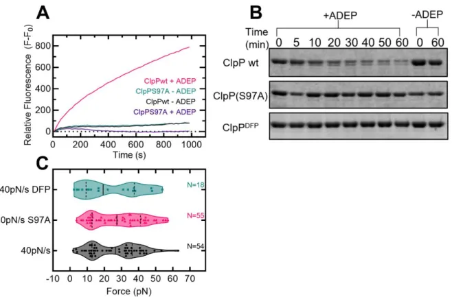
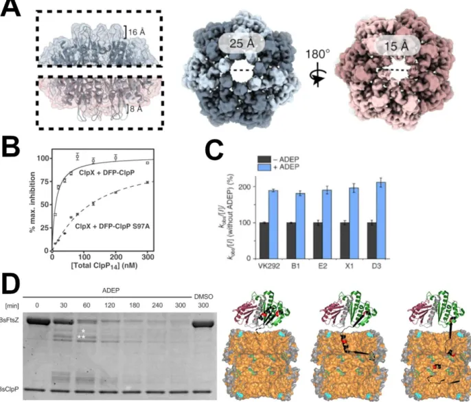
In addition to motor proteins, ClpP is also activated by a class of natural products called acyldepsipeptides (ADEPs) (Brötz-Oesterhelt et al., 2005; Malik & Brötz-Oesterhelt, 2017). In cells, ClpP activation by ADEPs leads to cell death through the indiscriminate proteolysis of nascent polypeptides and metastable protein substrates ( Brötz-Oesterhelt et al., 2005 ). In bacteria, ADEP activation inhibits cell division through the specific degradation of FtsZ ( Sass et al., 2011 ), which forms the Z-ring during cytokinesis.
Structures of the ClpP monomer and tetradecamer
The N-terminal loops form ordered -hairpins in the extension and are disordered in the compact and compressed conformations. Left: Cartoon showing a side view of the ClpP tetradecamer, the color is the same as in A. The N-terminal loops are colored pink and two adjacent monomers are colored cyan and orange for viewing.
ClpP conformational cycling
This is supported by an NMR study of cross-linked EcClpP, which captured substrates when cross-linked and released products when reduced ( Sprangers et al., 2005 ). In addition, chemically inactivated ClpXP released a captured GFP substrate upon addition of ATP and excess substrate ( Kim et al., 2000 ), suggesting that polypeptide egresses outside an opening other than the central pore. The presence of a closed pore suggests a compressed/compact conformation for ClpP; López et al.
The importance of ClpP’s N-terminal loops
However, a higher-resolution crystal structure later found the N-terminal loops in two conformations, named "up" and "down" (Bewley et al., 2006). Later, the crystal structure of ADEP bound to MtClpP also captured the N-terminal loops in an upward conformation similar to EcClpP (Schmitz et al., 2014). In addition, ADEP binding shifts the equilibrium of SaClpP toward an extended conformation in solution (Gersch et al., 2015).
ClpP activation by AAA+ motors
These interactions are highly dynamic and can be overwhelmed by sub-stoichiometric amounts of ADEP (Amor et al., 2016). Motor protein binding only slightly decreases the rate of peptide hydrolysis of ClpP for dipeptide substrates (Thompson et al., 1994). Joshi and colleagues (Joshi et al., 2004) showed this using different competitive and ATPase degradation assays.
ClpP activation by ADEPs
FtsZ forms a Z ring during cytokinesis for bacterial cell division and has many protein partners to regulate its stability and polymerization (Romberg et al., 2017). It remains unclear how ADEP-ClpP unfolds this substrate and why FtsZ can be unfolded but not other protein substrates such as GFP (Kirstein et al., 2009). ADEP-ClpP also degrades - and -tubulin (Sass et al., 2011), suggesting that there may be a conserved structure between bacterial and mammalian cytoskeletal proteins that allows their degradation by ADEP-ClpP.
Implications of ClpP dysregulation in disease
-ClpP also degrades - and -tubulin (Sass et al., 2011), suggesting that this may be a conserved structure between the bacterial and mammalian cytoskeletal proteins that allows it to be degraded by ADEP-ClpP. However, more research is needed to determine the exact mechanism of unfolding and whether it depends on certain structures in substrate proteins, biochemical stability, mechanical stability, or a combination of all these properties. In one family, ClpP mutations were found within the hydrophobic pocket that are thought to affect its ability to bind the ClpX motor protein ( Jenkinson et al., 2013 ). A future study examined two of these mutations, T145P and C147S, and showed that T145P reduced motor binding while C147S bound similar to wild type (Brodie et al., 2018).

History of single molecule studies of ClpXP and ClpAP
Subsequently, the Berkley group showed that both ATP hydrolysis and phosphate release occurred during the power stroke (Rodriguez-Aliaga et al., 2016). Next, the MIT/Vanderbilt group showed that the direction of degradation affected substrate processing ( Olivares et al., 2017 ). Therefore, mechanistic contributions by ClpP to any of these properties will shift the current understanding of degradation in the field and influence our understanding of their biological function as two-component machines.
Possible contributions by ClpP to previous single molecule studies
Aubin-Tam and colleagues (M.-E. Aubin-Tam et al., 2011) looked at ClpXP to degrade a multidomain filamin substrate and compared the translocation from ClpXP to ClpX alone. Similarly, Maillard and colleagues (Maillard et al., 2011) confirmed these findings when studying ClpXP that degrades various multidomain GFP and titin substrates. In solution, ClpA unfolds a dimeric protein substrate more slowly than the ClpAP complex (Baytshtok et al., 2015).
Thesis Overview
These differences are likely caused by the CulpA double-ring architecture, which can increase processivity and decrease stalling when processing substrate (Kotamarthi et al., 2020).
Materials and methods
- Biochemical purification of ClpP and substrate proteins
- Single-molecule optical trapping of ClpP-substrate complexes
- Optical trapping data analysis
- Biochemical degradation assays
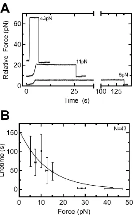
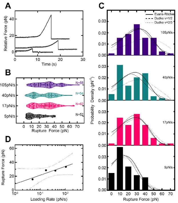
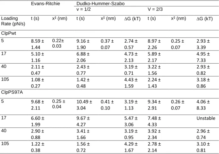
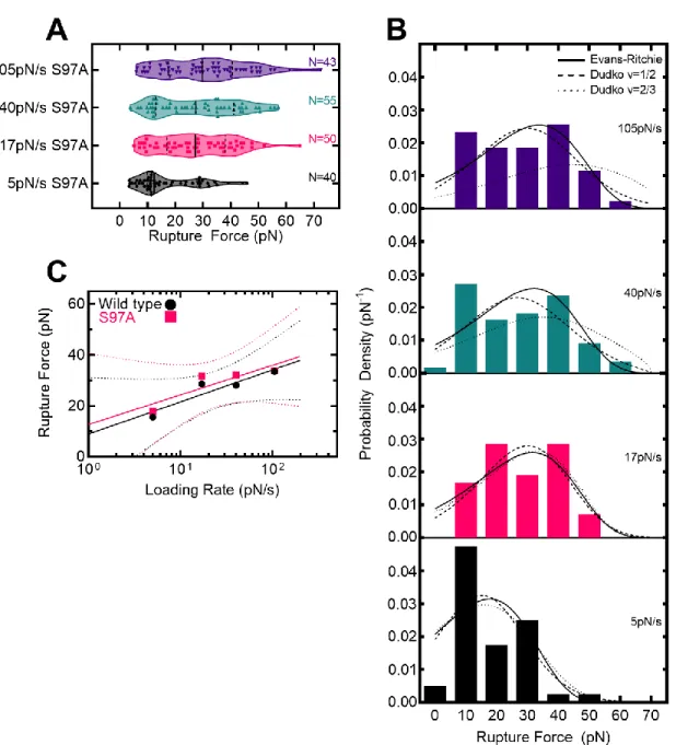
For lifetimes, the end of the force ramp and the terminal fracture were both reported using the first. For lifetimes, data were further downsampled to 5 Hz, which was necessary to automate detection of the end point of the force ramp. Where is the intrinsic lifetime, r is the strain rate, x‡ is the distance to the transition state, and v is a variable representing the shape of the free energy barrier (1/2 for a spike and 2/3 for a linear -cubic) .
The ClpP peptidase forcefully grips a protein substrate
- Abstract and statement of significance
- The role of ClpP as a AAA+ peptidase
- ADEPs activate ClpP for proteolysis
- Single-molecule mechanics of the ClpP peptidase
- Contribution of the ClpP active site serine to substrate grip
- The impact of ClpP grip on degradation mechanics
- Hypotheses for bimodal rupture force distributions
- ClpP possibly contributes to processivity
- Sumo-ClpP control experiments
For example, ClpA unfolds a dimeric substrate more slowly (Baytshtok et al., 2015) and makes slower kinetic steps in the absence of ClpP (Miller et al., 2013; Rajendar & Lucius, 2010). However, a class of natural products called acyldepsipeptides (ADEPs) activates ClpP in the absence of motor proteins (Brötz-Oesterhelt et al., 2005; Malik & Brötz-). ADEPs bind to the same hydrophobic pocket on ClpP that motors bind to (Amor et al.). al., 2016) and are thought to activate ClpP in a similar manner, ie.
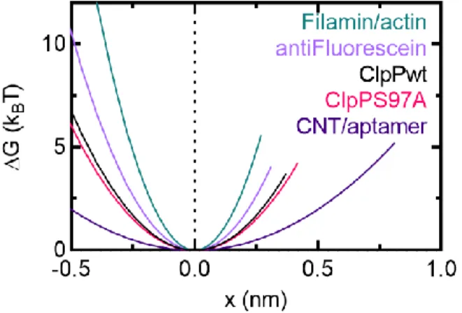
In addition, ADEPs disrupt bacterial cell division by specifically degrading the FtsZ protein with ClpP (Sass et al., 2011). Fits to the Evans-Ritchie model (Evans & Ritchie, 1997) for each load level are shown as solid black lines along with fits to the Dudko-Hummer-Szabo model (Dudko et al., 2006) where v=1/2 (dashed lines ) and v=2/3 (dashed lines). D) The most likely rupture force is plotted as a function of loading level for wild-type ClpP-substrate interactions in the presence of ADEP1. We compared these with other measured protein-protein interactions (M. E. Aubin-Tam et al., 2011) and found that the free energy of activation and the distance to the transition state are comparable to the interaction between fluorescein and an anti-fluorescein antibody.
Free energy diagrams were constructed according to the Dudko-Hummer-Szabo model (Dudko et al., 2006) assuming a rope shape (v=1/2). In addition, we characterized ClpP-substrate disruption forces and fitted them to models of force-dependent protein-ligand interactions based on Kramers theory (Dudko et al., 2006; Evans. & Ritchie, 1997). ClpPs represent another candidate site that mediates substrate adhesion, as they already play a role in substrate binding (Gribun et al., 2005) and modulate the activity of the active site serine (Jennings et al., 2008).
In addition, a ClpX variant with unfolding defects is rescued when complexed with wild-type ClpP, ClpPS97A, and DFP-tagged ClpP ( Joshi et al., 2004 ).
Discussion
Implications of ClpP grip in past studies
The head-to-head distance of the SaClpP tetradecamer changes by 10 angstroms when comparing the extended and compressed conformations (Ye et al., 2013; Zhang et al., 2011, See figure 1.2B). Thus, if ClpP does switch between conformations when bound to a motor, this could add up to 1nm to the distances translocated by ClpXP and ClpAP. For the two single molecule examples above, movement by ClpP may help explain the paradoxes in both observations.
For example, if 4nm bursts involve 1nm movement by ClpP, their frequency would not change with ATP concentration. In addition to step size, ClpP could contribute to the mechanics of ClpXP and ClpAP degradation via substrate adhesion. For example, in solution ClpA unfolds a dimeric protein substrate more slowly than with ClpP present (Baytshtok et al., 2015).
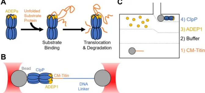
Furthermore, ClpXP unfolds GFP faster with a longer linker between the substrate and the ssrA degron tag ( Martin et al., 2008 ). In this dissertation, I showed that activated ClpP clamped a protein substrate against up to 40 pN of external force. ClpXP or ClpAP, mutations can be made in ClpP that affect substrate binding and then assayed for degradation by ClpAP and/or ClpXP.
Therefore, the direct continuation of my research should be to determine which domains and/or residues in ClpP are responsible for substrate adhesion.
Structural hypotheses for ClpP substrate grip
Furthermore, disrupting the structure of these -hairpins reduces the rate of casein degradation by activated ClpP (Alexopoulos et al., 2013). Overall, the flexibility of the N-terminal loops and their role in substrate permeability make these residues candidates for substrate grip. If true, this mechanism would also provide an explanation by which ADEP-ClpP unfolds and degrades FtsZ in vitro ( Sass et al., 2011 ; Silber et al., 2020 ).
In addition to the N-terminal loops, other residues within the active site may be required and/or cooperate to confer substrate grip. ClpP uses a canonical Ser-His-Asp catalytic triad to carry out peptide hydrolysis (Maurizi, Clark, Katayama, et al., 1990; Maurizi, Clark, Kim, et al., 1990). In bacterial competition assays and in degradation assays in vitro, the mutation of the active site Asp to Ala was less severe for peptidase activity than the mutation of the Ser or His residues (Lin et al., 2020).
Since the catalytic triad residues differentially affect peptidase activity, these residues may also play different roles in substrate binding/grip. Alternatively, it may be necessary to mutate two or more of these residues to significantly affect substrate grip (eg, a Ser/His double mutant). In addition, there are non-catalytic residues within the active site that may be responsible for substrate binding.
These residues may also play a role in substrate grip and would be good candidates to mutate to affect ClpP grip.
ClpP: a model for all AAA+ peptidases?
However, such studies would be more complicated because the AAA+ motor and peptidase components cannot be studied separately, as ClpP and the 20S core particle were. Overall, my work characterizing substrate adhesion by the peptidase ClpP against force raises many questions about past single-molecule studies and offers a new perspective on how the field should view mechanical degradation by AAA+ proteases: the cooperation between AAA+ motors and their peptidase.
ClpP grip informs models of ADEP-mediated degradation
It is possible that, since ADEPs are competitive inhibitors of the natural ClpP-motor interactions, perhaps ADEP-ClpP induces a change that allows ClpP to functionally cooperate with other chaperones to help degrade other substrates in the cell. Highly dynamic interactions maintain the kinetic stability of the ClpXP protease during the ATP-driven mechanical cycle. Effects of protein stability and structure on substrate processing by the ClpXP unfolding and degradation machinery.
Proteomic discovery of cellular substrates of the ClpXP protease reveals five classes of ClpX recognition signals. Roles of the N domains of the ClpA unfoldase in binding substrate proteins and in stable complex formation with the ClpP protease. Sequence and structure of Clp P, the proteolytic component of the ATP-dependent Clp protease from Escherichia coli.
Pool the desired fractions and spin filter concentrate to just over 1 mL (volume can be increased if larger loops are available. Column allows <5 mL of sample). Pellet the resin by spinning at 500xg for 5 minutes and remove most (but not all) of the supernatant. The DNA to be used must not be resuspended in tris buffer (ie, any of the elution buffers).
This dilution may depend on how much material you have lost during each wash cycle. This code contains a function that finds fractions by looking at the first derivative of the data.