OF HeLa MITOCHONDRIA
II. RE PLICATION OF CLOSED CIRCULAR MITOCHONDRIAL DNA IN MOUSE L CELLS
Thesis by
Donald Lewis Robberson
In Partial Fulfillment of the Requirements for the Degree of
Doctor of Philosophy
California Institute of Technology Pasadena, California
1972
(Submitted July 2, 1971)
To Barbara
•
ACKNOWLEDGMENTS
There are many people who have made significant contributions to my graduate career at Caltech. With deepest gratitude I thank the following people, in the order of their appearance in my thesis career:
!1ay Owen-who concerned himself with the difficulties encoun- tered early in my career and encouraged me to continue graduate studies.
I thank him for his faith in me and for his council.
Norman Davidson-who has been a patient research advisor and from whom I have gained much scientific knowledge and self-reliance. I thank him for the high standards of scientific excellence he has imparted to me and for the continued interest and encouragement he has given in my career.
Jerome Vinograd-who has been close to my career and has generously shared his enthusiasm for research. I have been privileged to discuss with him many of the findings that have come from his labora- tory. I am grateful to him for his concern for my scientific understanding and for his continued interest in my career.
Michael Raftery-who has provided sound scientific advice on many occasions and has been a much appreciated friend. I express my gratitude to him for his continued support.
Yosef Aloni and Giuseppe Attardi-who have given me an opportu- nity for my work to be recognized. I have received many benefits from my association with them, as this thesis testifies. I thank them for these collaborations.
Harumi Kasamatsu and Jerome Vinograd-who have provided an
element of excitement in scientific discovery. I thank them for their generosity in collaboration.
James Bonner-who has given moral support and council in my graduate program. I thank him for his involvement.
The Division of Biology at Caltech and USPHS and NSF-whose financial assistance is gratefully acknowledged.
The many friends and colleagues-who have made my stay at Caltech pleasurable at both scientific and social levels over these many years.
I sincerely thank them all.
ABSTRACT
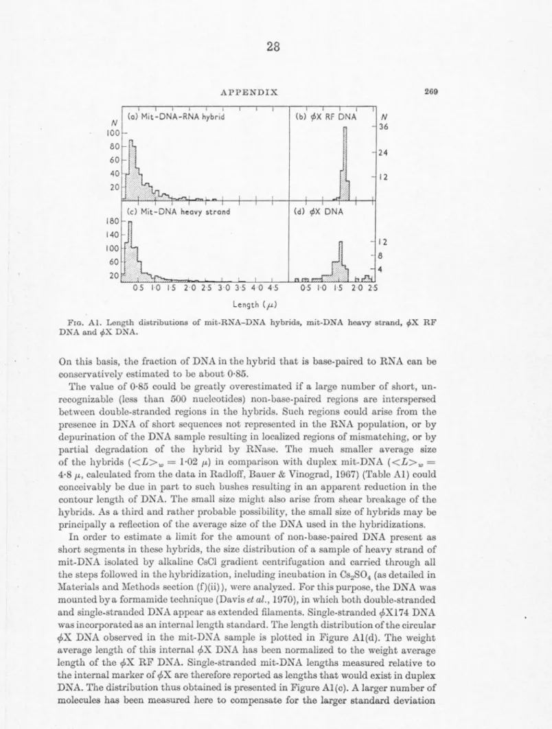
The thesis consists of two parts. In Part I, electron microscope analysis of HeLa mitochondrial RNA-DNA hybrids isolated from CsS04 gradients demonstrates that more than 85% of the heavy strand of HeLa mitochondrial DNA is transcribed in vivo.
A modified basic protein film method of spreading RNA in a strongly denaturing solvent for examination in the electron microscope has been developed and applied to determine the size of the HeLa mito- chondrial specific ribosomal RNA (rRNA) components. Length measure- ments on purified 128 and 168 mitochondrial rRNA, on mixtures of the two, and on mixtures of 128 with 188 cytoplasmic rRNA have given molecular lengths of 0. 27 µ., 0. 42 µ, and 0. 55 µ. for the 128, 168, and 188 rRNAs, respectively. If these molecular lengths are proportional to molecular weight, and if the molecular weight of 188 cytoplasmic
rRNA is taken as 0. 71
x
106 , as determined by sedimentation equilibrium, the molecular weights of the 128 and 168 components are 0. 35 x 106 and 0. 54 x 106, respectively. These molecular weight values are in good agreement with the relative values predicted from sedimentation velocity measurements, but not with the relative values based on gel electrophoresis.Electron microscopy of hybrids between the heavy strand of HeLa mitochondrial DNA and HeLa mitochondrial rRNA demonstrates that the genes for 168 and 12S HeLa mitochondrial rRNAs are situated adjacent, or very close, to each other on mitochondrial DNA. The length of the DNA segment separating the two genes is estimated to correspond to less than 500 nucleotide pairs.
A coupling procedure has been developed for the efficient covalent attachment of periodate oxidized RNA to an insoluble matrix of hydrazide- derived sepharose, which is subsequently available for nucleic acid
hybridization.
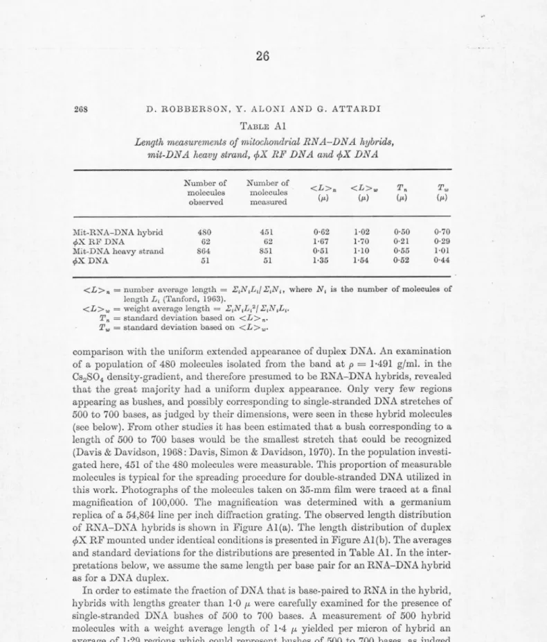
In Part II of the thesis, the properties of a new structure of mito- chondrial DNA are discussed. In two strains of mouse L cells, it was found that approximately one half of the closed mitochondrial DNA mole- cules contain a short three-stranded DNA region in which a short single strand of progeny DNA containing about 350 nucleotides is inserted into the closed circular duplex with the displacement of a corresponding stretch of the still circular parental strand. The three-stranded region is called a D-loop because of its formal shape and because it appears to have been formed by displacement replication. Double-length mitochon- drial DNA molecules often contain two D-loops which are always at
diametrically opposed positions on the 20 megadalton circle. This latter result is taken to indicate that one of the forks in each D-loop marks a unique site for the initiation of replication.
TABLE OF CONTENTS
PART TITLE PAGE
I A STUDY BY ELECTRON MICROSCOPY
OF THE RNA AND RNA-DNA HYBRIDS
OF HeLa MITOCHONDRIA 1
Chapter A General Introduction 2
Publication 3
Chapter B Determination of the Fraction of the Heavy Strand of HeLa Mitochondrial
DNA that is Transcribed 24
Publication 25
Chapter C Size Determination by Electron Micros- copy of HeLa Mitochondrial Ribosomal
RN As 30
Abstract 32
Introduction 33
Materials and Methods 34
Results 41
Discussion 55
References 65
Chapter D Determination of the Relative Positions of the Ribosomal Genes on HeLa Mito-
chondrial DNA by Electron Microscopy 67
Abstract 69
Text 70
References 86
Chapter E A General Approach to the Isolation of
Specified DNA Sequences 87
Abstract 88
Introduction 89
Materials and Methods 89
Results and Discussion 96
References 104
PART TITLE PAGE II REPLICATION OF CLOSED CIRCULAR
MITOCHONDRIAL DNA IN MOUSE L
CELLS 105
Abstract 107
Introduction 108
Materials and Methods 111
Results 117
Discussion 134
R.eferences 138
Appendix A Isolation of E. coli R.ibosomal RNA 139 Appendix B Electron Microscope Techniques 143
Part I
A STUDY BY ELECTRON MICROSCOPY OF THE RNA AND RNA-DNA HYBRIDS
OF HeLa MITOCHONDRIA
A. General Introduction
SPIUXO
Volume XXX \', I 070 Pri.1tted in. U.S.A.
Transcription of Mitochondrial DNA in HeLa Cells
GIUSEPPE ATTA1wr, YosEF ALONI, BARBARA ATTARDI, DEANNA OJALA, LIVIA PICA-?llATTOCCIA, DON L. ROBBERSON, AND BRIAN STORHIE
Biology Division, California Institute of Technology, Pasadena, California
In tho last few years the investigation of the physical properties of mitochondrial DNA in oucaryotic cells has brought to light two remarkable facts. The first is that mitochondrial DNA has undergone an evolution, going from the simpler to the higher cellular organisms, in a direction opposite to that of the nuclear genome. \Vhcrcas the latter has increased in sizo 11nrl complexity, mitochondrial DNA has become smaller aml :simpler. Mitochondrial DNA molecules from lower euco.ryotic cells (protozoa., fungi) and plant cells ho.ve, in fact, been found to be considerably larger (from 3 to 12 times) than those of animal cells (Suyama. and Miura, 1968; Wolstenholme and Gross, 1968; Wood and Luck, 1969; Borst, 1970);
moreover, as far as one can judge from the few available data concerning the sequence length of mitochondrio.l DNA, estimated from renaturntion kinetics (Borst, 1970), the reduction in size of mitochondrial DNA during evolution appears to have been accompanied by reduction in its in- formational content, with the lost functions presumably having been taken over by the nucleus.
The second remarkable fact is that mitochondrial DNA from all animal species examined, from insects to echinoderms to the.highest vertebrates, is in the form of circular molecules about five microns in length (Borst, Van Bruggen, and Ruttcnberg, 1968). Thus, the a.mount of information that animal mitochondrial DNA contains appears to have been preserved over hundreds of millions of years. This suggests that the progressive reduction in informational content of mitochondrial D.N A has reached in animal cells the limit compatible with the survival of the mitochondrial DNA itself. This in turn would imply that whatever the function is which has proven to be advantageous for anima.l cells to keep under the control of cytoplasmic gcnos, it cannot be taken over in any part by nuclear gen rs.
As concerns the possible nature of thi~ function, strong constraints are obviously placed on it by the small size of animal cell mitochondrial DNA itself.
One can think of the information required for the replication and transcription of mitochondrial DXA and their control, ancl for the synthcsi:s of certain components of the intramitochondrial
protein synthesizing machinery which cannot be substituted by their cytoplasmic counterparts for the translation of mitochondrial messages. lt is likely, however, tho.t these represent ancillary functions which would not by themselves confer cvolutiono.ry 11dvantagc upon the persistence of mitochondrial DNA in 11nimal cells. One would like to think tho.t there is 11 unique funct,ion of mitochondrio.l DNA related to the synthesis of some essential mitochondrial constituent. On the basis of various pieces of circumstantial cvidcnco, a good candidate for this unique function is the synthesis of some protein component of mito- chondrial membranes with a structural or or- ganizational role (Attardi and Attardi, 19Ml). It is clear, at any rate, that mitochondria arc vory far from being closed autonomous systems within the cell. The primitive symbiotic bacteria, which 11·cre the probable precursors of mitochondria, adapted to growth in the microchemostat provided by tho ancestral eucaryotic cell, have slowly become, in the course of evolution, domesticated cell organelles constructed according to a nuclear blueprint, though still with some mitochondria.] controlling elements. As a physical corollary of the nuclear control of mitochondrial assembly, mitochondrial membranes, for a long time known to be a site of exchange and transport of small molecules, are thought todo.y to be also an active site of flux inward, and possibly out,rnrd, of macromolecules:
proteins, RNA, and lipids.
The mitochondrial genetic system, because of its relative simplicity and its compo.rtmcntalizcd character, offers unique advantages for the stud,,· of some of the basic problems concerning strncturo, replication and transcription of DNA, and procc-s- sing, transport and translo.tion of RN A 111 cucaryotic cells.
\Ye shall describe in this paper the ,1·ork t.hat \1·e lrnvc co.rriccl out in the last t\1·0 years on the transcription of mitochondrial DNA in a hu1111111 coll line grown in vitro, HcLa cPlls.
RAPIDLY LALIELED Hr:TEHO<:EXEOl'S MITOCJION'DHTAL RNA
\Vhen exponentially growing Hr.Lo. rPlb arc exposed to a lo.bcled RNA precursor, radioact,iYity
ATTARD! et al.
'>-00
0400
,000
0 "'° .E c
~ ";;;
ON 0200 u
"""
ci
40 ml
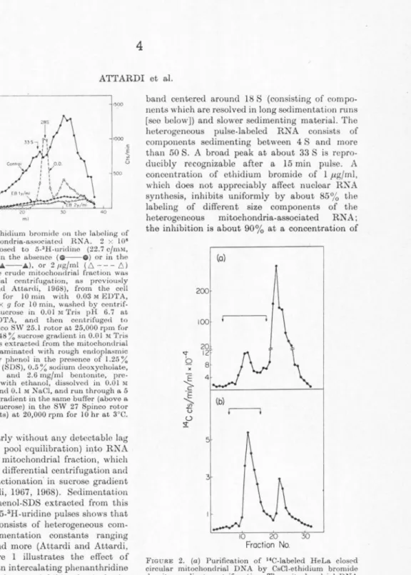
F10u1rn I. Effect of ethidium bromide on tho labeling of
het~rogencous mitochondria-associated RNA. 2 x 10⁸ HoLa cells were exposed to 5-3H-uridino (22.7 c/mM, 1.4 µc/ml) for 15 min in the absence ( 0 - - 0 ) or in tho presence of 1 /tg/ml (.A--.A), or 2 11g/ml ( 6 - - - 6) ethidium bromide. The crude mitochondrial fraction was isolated by differential ccntrifugat.ion, as previously described (Attardi and Attardi, 1968), from the cell homogenate, treated for 10 min with 0.03 M EDTA, centrifuged at I 1,000 X g for 10 min, washed by centrif- ugat.ion with 0.25 M sucrose in 0.01 M Tris pH 6.7 at 25°C and 0.015 M EDTA, and then centrifuged to equilibrium in tho Spinco SW 25.1 rotor at 25,000 rpm for 18 hr at 3°C in a 30 to 48 % sucrose gmdient in 0.01 M Tris buffer pH 6.7. RN"A was extracted from the mitochondrial band (which was contaminated with rough endoplasmic reticulum elements) by phenol in the presence of 1.25 % sodium dodocyl sulfate (SDS), 0.5 ~~sodium dooxycholatc, 0.5 % sodium cholate and 2.6 mg/ml bentonite, pre- cipitated three times with ethanol, dissolved in 0.0 I M
acetate buffer pH 5.0 and 0.1 M NaCl, and run through a 5 to 20 % (w/w) sucrose gradient in the same buffer (above a 4 ml cushion of 6-1 % sucrose) in the SW 27 Spinco rotor (2.54 x 8.83 cm buckets) at 20,000 rpm for 10 hr at 3°C.
is incorporated linearly without any detectable lag (after correction for pool equilibration) into RNA associated with the mitochondrial fraction, which can be separated by differential centrifugation and buoyant density fractionation· in sucrose gradient (Attardi and Attardi, 1967, 1968). Sedimentation analysis of RNA phenol-SDS extracted from this fraction after short 5-3H-uridine pulses shows that the labeled RNA consists of heterogeneous com- ponents with sedimentation constants ranging from 4 S to 50 S and more (Attardi and Attardi, 1967, 1969). Figure 1 illustrates the effect of ethidium bromide, an intercalating phonanthridine dye, which has been shown to inhibit selectively the synthesis of mitochondria-associated RNA (Zylber, Vesco, and Penman, 1969), on the labeling during a 15 min 5-3H-uridine pulse of tho heterogeneous RNA. The mitochondrial fraction had been treated with 3 x 10⁻² M EDTA to remove tho bulk of ribosomes of the rough endoplasmic reticulum which contaminates the mitochondrial fraction.
The OD260 profile shows a prominent 28 S peak (pertaining in its great majority, if not exclusively, to membrane-stuck 50 S subunits not removed by the EDTA-treatment from the contaminating rough endoplasmic reticulum elements), a broad
band centered around 18 S (consisting of compo- nents which are resolved in long sedimentation runs [see below]) and slower sedimenting material. The heterogeneous pulse-labeled RNA consists of components sedimenting between 4 S and more than 50 S. A broad peak at about 33 S is repro- ducibly recognizable after a 15 min pulse. A concentration of cthidium bromide of 1 µg/ml, which does not appreciably affect nuclear RNA synthesis, inhibits uniformly by about 85% the labeling of different size components of the heterogeneous mitochondria-associated RN A;
the inhibition is about 90% at a concentration of
200
100
5
3 (o)
(b)
r---1
10 20
Fraction No.
\
FIGURE 2. (a) Purification of "C-labeled HeLa closed circular mitochondrial DNA by CsCl-ethidium bromide density gradient centrifugation .. Tho mitochondrial DX A was released by SDS, as previously described (Attard i and Attardi, 1969), from the crude mitochondrial fraction of HeLa cells labeled for 60 hr in the presence of 2-"C- thymidine. After addition of CsCl t.o I Mand centrifugation at 15,llOO x a for I 5 min, ethidium hrornide was added to the supernatant to 200 11.g/ml, and the solution was adjw;tcd t,o I .55 g/cm3 with CsCI and centrifuged i11 tho Spinco SW 4 l rotor at 35,000 rpm for 48 hr at :?0°l'.
Fractions were collected dropwise from the bottom of tho tube. (b) After assay of radioactivity, the fractions corre- sponding to the smaller heavy band (designated by arrows in a) were pooled and recentrifuged under the same condi- tions in a CsCl-ethidiurn bromide density gradient. The fractions containing t.lle heavier band, which consi•tcd exclusively of closed circular mitochondrial DXA (Radloff et al., 1967), were pooled and freed of the dye by passage through aDowex 50 W-X8 column (Radloff et al., 1967).
Tl~ANSCRIPTION OF MITOCHONDRIAL DNA
the drug of2 µg/rnl. The almost complete sensitivity to ethidium bromide, in short-term '" periments, of tho labeling of mitochondria-al'··· n,ted hetero- geneous RNA presumably rcflectH a direct effect of this drug on the transcription of a closed circular DNA template: in fact, this intercalating dye is kno"·n to distort the structure of supereoiled circular DNA (Radloff, Bauer, and Vinograd, 1967;
Crawford and Waring, 1967; Bauer and Vinograd, HJGS). These experiments therefore support the idea that mitochondrial DNA is tho template of the fast-labeled heterogeneous mitochondria-associated RNA. In agreement with this interpretation arc the results of RNA-DNA hybridization experiments utilizing closed circular mitochondrial DNA purified by two cycles of cesium chloride density gradient centrifugation in the presence of ethidium bromide (Fig. 2) (Radloff ct al., l!l67; Attardi and Attardi, l!l69). As shown in Table I, after a 7 or 15 min 5-3H-uridine pulse, both the RNA components sedimenting slower than 28 S and tho heavier components hybridize with groat efficiency with mitochondrial DNA and only to a very limited extent with total cell or nuclear DNA.
As shown in Table 2, the nucleotide composition of the mitochondria-associated heterogeneous RNA,
TABLE I. J3A~E SEQUENCE HOMOLOGY TO MITOCHO:>DHIAL D'.liA OF PULSE-LABELED i\IITOCHONDRIA-ASSOCIATED
HETEROOE:>EOUS RXA
% input Expt. Pulse
count/ co_un_t/
mm min 1n
no. (min) Fro.ct ion DKA in hybrid hybrid 10-26 s Mitochondl'ial 194 13.3 J0-26 s Total cell 5 0.3 33-50 s Mitochondrial 284 25.6
33-50 s Nuclear JO 0.9
8-35 s Mitochondrial 30 11.4 2 15 8-35 s Total coll <I <0.4 35-50 s Mitochondrial 5J J4.l 35-50 s Total coll <l <0.3 The mitochondrial fraction was isolated from HcLa cells exposed for 7 or 15 min to 5-'H-uridiue and the RXA was phenol-SOS extracted and run through sucrose gradients as in Fig. I. RNA components from different portions of each grurlient wcro prceipitatccl with ethanol and disAolvod in flSC/ I 0. Tho incubation mixtures, co11taini11g 2 to 3.6 /lg heat-denatured mitochondrial DX A l\nd I 0 to 22 11g HNA in a totl\I vol of 2 ml 2 X SSC, wcro incuLatocl at 70°C for 5 hr. Afto1· cooling, tho mixture"
woro treated with 10 11g/ml heated pancreatic Rl\ase for 60 min l\t 22°C l\ncl the HNA/DNA hybrids isolated Ly Sophadcx chromatography as described in Jeantl'ur l\nd Attardi (1969). (Washing of the nitrocclluloso membranes with 0.5 M KCI, 0.01 M Tris buffer was at ii6°C.) Tho data are correctod for nonspecific background estimated with E.coli DNA.
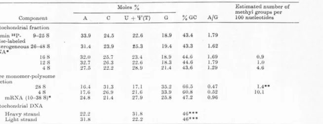
TABLE 2. XucLEOTIDE COMPOSITION 01· R:XA COMPONENTS FROM THE MITOCHONDRIAL FRACTION AND FROM THE FREE 11'.loNoMER-POLYSOME FnACTIO~ OF HELA CELLS
Moles% Estimated number of
methyl groups per
Component A c U + 'Y(T) G %GC A/G 100 nucleotides
Mitochondrial fraction
30 min up_ 9-25 s 33.9 24.5 22.6 18.9 43.4 1.79
pulse-labeled
heterogeneous 26-48 S 31.4 23.9 ~5.3 J9.4 43.3 1.62
RXA•
16 s 32.0 25.7 23.4 18.9 44.6 1.69 0.9
12 s 32.7 26.3 22.6 18.3 44.6 l.i9 1.0
4S 27.5 22.2 28.!) 21.4 43.6 l.29 4.6
Free monomcr-polysomc fraction
28 s 16.4 31.3 17. l 35.2 66.5 0.4 i l ,4••
48 I i.6 :16.!l 21.G 33.9 60.8 0.52 JO.I
mR:XA (10-38 S)• 24.8 21.4 27.9 25.8 47.2 0.96
i\litochondrial DXA
Heavy strand 22.2 31.8 413•••
Light strancl 31.8 22.2 413•••
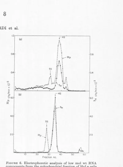
Th<' 16 Sand 12 S R:\ A component> w<'r<' i>olatC'cl by two cycles of sucrose gradiC'nt centrifugal ion from the BX,\ of thl' mitochondrial fraction of H cl.a C!'ll> lab<'lecl for 22 hr with 32 !'-orthophosphate (I. 75 11c/ml) and L-3H-met hyl m<'t hioninP (5.4 mc/J,n1oil•, 2.5 /tc/inl) in modified rrwdiutn containing- 10-3 i\t pho:-:phatC' and 311g/n1l mcthioniuc, in the pn·~t·11ce of 0.04 11g/1nl actinomycin D. To minimi.,,c the labeling of the purinP rings, adPno:-;inc an<l guanosinc were added nt a con- centration of 10-• M to the growth medium for 2~ hr prior to <'xposurc to t.hc isotopes and durin~ th<' lab!'ling period. The 4 fl B'.llA from the mitochondril\I fraction and th<' 28 Sand~ S !{:\,\from the fr<'e rnonorner-polysome fraction WCI'!' isolated from coJI;-; lo.holed with np.orthophosphn.tp u.nd r...aH.mctJlyl 111C'thionino under the 1-10.n1c conditions described nbo\'l' but in t.he absence of act.inornycin D. 'l'he 4 S RXA from the mitochonclrinl fraction and from free ribosomeR were isolatt•d hy polyacryll\mide gel electrophoresis of th<' low mol wt components (up to about 7 S) of the sedimentation pattern (Fig. 6).
32 P-nuclcotido con1po:sition after a.lko.li digestion was carried out as previously de::;cribed (Attardi, Parna~, Hwun~. nnd At tardi, 1966). Tho % of methylated nucleotidPs wa" calculated from the 3H to "P ratios and from the known con lent of methyl groups of cytoplasmic 28 S RXA. 5 S RXA isolated by polyacrylamido gel electrophoresis showed no dPt<'ctal>I<' labeling with L-'H-mcthyl-met-hionine (Fig. 6), thus excluding labeling of the purine rings. The A and T content of th<' heavy and light strunds of mitochondrial DXA was dctcrm incd from the ratio of 2-"C-thymidine incorpornt!'d into each of tho separated strands and from the GC content of human mitochondrial DXA. • Attardi and Attardi, l 96i; •• llrown nnd Attardi, 1965; •••Clayton and Vinograd, 1967.
ATTARDI ct al.
pulse-labeled for 30 min with 32P, is strikingly different (especially for the high A and low G content) from that of 28 S rRNA and from that of free polysome mRNA. Furthermore, this base composition is complementary, as concerns the A and U content, to that of tho heavy strand of mitochondrial DNA; this is in agreement with tho observation that this strand is the transcriptionally active strand of mitochondrial DNA (see below).
DISCRETE RNA COMPONENTS CODED DY
MITOCHONDRIAL DNA
After a 7 min pulse of 5-3H-uridine, and more clearly after a 15 min pulse, discrete labeled components sedimenting at about 16 S, 12 S, and
d 0 0200
0800
(
\~0.500 0.300
. I
i I
I I \ 0200' \
0.100
185
~ 165 I
'
185
I
:..
125
Froclion no 45
I
90min (a)
90 min (b) +act. D 45
90min (c) + eth. brom.
45
I
60
40
2.0
.,.
40 ~ E '
·~
2.0 -;;;
u
3.0
20
10
Fro URE 3. S~dirncntation pattern of RN A component.- sedimenting slowPr than 28 S RNA from the mitochondrial fraction of HeLa cells exposed to 5-3H-uridinc for 90 min in the absence or in the presence of actinomycin Dor cthidium bromide.
2.4 x 10⁸ HeLa cells were labeled for 90 min with 5-3H-uridine (22.8 c/mM. 1.4 µc/ml) in the absence (a) or in t.he presence of 0.04 µg/ml actinomycin D (b) or I µg/ml cthidium bromide (c). R~A was extracted from tho F:DTA-treated and isopycnically separated mitochondrial fraction, and run through a 15 to 30 % (w/w) sucrose gradient in SDS buffer (0.5 % dodecyl SO,, 0.1 M NaCl, 0.01 M TRIS buffer pH 7.0, 0.001 M EDTA) in the SW 25.3 Spinco rotor at 25,000 rpm for 28 hr at 20°C.
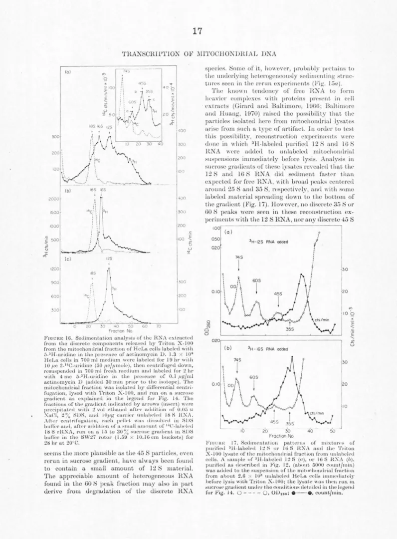
4 S can bo seen to emerge over the background of hctorogenoous RNA (Attardi et al., 1969). These labeled components can be easily recognized in long so<limcntation runs which fully display the com- ponents sedimenting slower than 28 S RNA.
Figure 3a shows the labeling pattern of these components from the EDTA-troated mitochondrial fraction of cells exposed to a 90 min 5-3H-uridine pulse. The OD260 profile shows near the bottom of the tube the prominent peak (partially pelleted) of 28 S RNA, near the center of the gradient an 18 S RNA peak with a shoulder at about 16 S, and t\\·o clear peaks at 12 S and 4 S. The labeling profile shows peaks at 12 S and 4 S; in contrast, the considerable labeling of the 18 S RNA species after a 90 min pulse masks in part the 16 S component, which is recognizable only as a shoulder. However, if the labeling is carried out in the presence of 0.04 µg/ml actinomycin D to block selectively rRNA synthesis (Dubin, 1967; Penman, Vesco, ancl Penman, 1968), the labeling of 18 S RNA is abolished and the 16 S component stands out clearly (Fig. 3b). Under the same conditions, tho labeling of the 12 S and 4 S peaks is also not affected to any appreciable extent. Ethidium bromide, at a concentration of 1 µg/ml, besides strongly inhibiting the labeling of the heterogeneous RNA, abolishes that of the 16 S, 12 S, and to a great extent (about 80%) that of the 4 S com- ponents (Fig. 3c). After 4 hr labeling with 5-3H- uridine in the presence of 0.04 µg/ml actinomycin D the 16 S, 12 S, and 4 S components appear as very prominent peaks (Fig. 4). The ethidium bromide sensitivity and actinomycin D resistance of the synthesis of these components suggests that they are coded by mitochondrial DNA. This in tcrpretation has been fully corroborated by RNA-DNA hybridization tests in which the sedimentation distribution of long-term labeled RNA from an EDTA-treatcd mitochondrial frac- tion of HeLa cells was scanned for capacity to hybridize with mitochondrial DNA (Attardi and Attardi, 1969). In these experiments, in fact, the sedimentation profile of RNA components homol- ogous to mitochondrial DNA has revealed a broad band between 9 and 15 to 16 S, with a peak at 12 S and a p~ominent 4 S peak; the shoulder of RNA homologous to mitochondrial DNA at 15 to 16 S presumably corresponds to the 16 S ethidium bromide-sensitive species which was not resolved in that analysis.
An analysis by polyacrylamide gel electro- phoresis of individual components isolated by sucrose gradient centrifugation (Fig. 5) shows that the RNA species sedimenting at 12 S moves through the gel, with respect to the 18 S and 28 S rRNA markers, as would be expected from its
0 "'
"'
::::i ::::i
TRANSCRIPTION OF MITOCHONDRIAL DNA 3600~
2.600t~· '·
1.400 '1 I
::~f
1~
(a) 4 hr
0.500
4.000 2.000
1.
0500
0.500 265 I
I
~
cP"",
265\
11
I I I I I I
~ I I I I
\ I I I I
~
10
165
l
Fraction No.
(b)
4 hr + ac1inamycin D
(c) 4 hr + ethidium bromide
60 10
20
10
15 10
4.0
2.0
"' Q
... E c:
... E
"' u
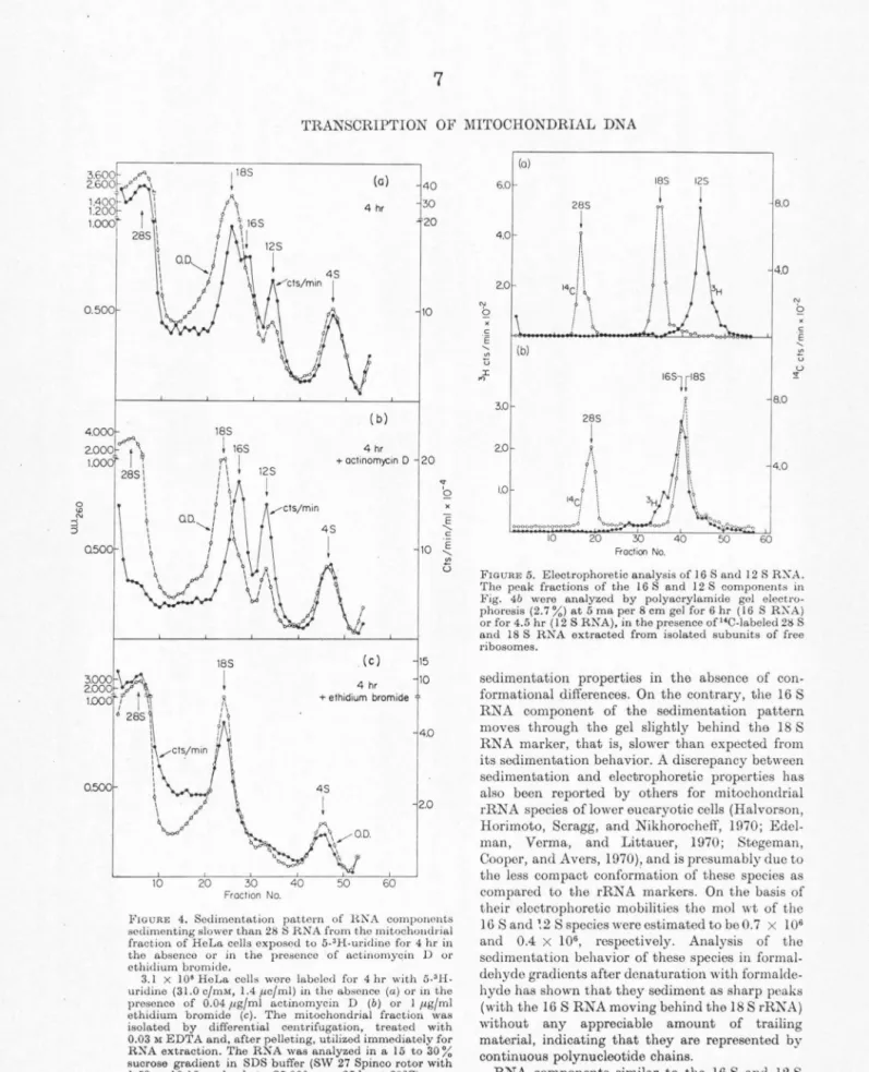
FIGURE 4. Sedimentation pattern of HKA components sedimenting slower than 28 SH.NA from tho mitochoudrial fraction of HeLa cells exposed to 5-'H-uridine for 4 hr iu tho absence or in the presonco of actinomyqin D or othidium bromide.
3.1 x I 08 He La cell• were labeled for 4 hr with 5-' H.
uridine (31.0 c/mM, 1.4 µc/ml) in tho absence (a) or in the prosonco of 0.04 µg/ml actinomycin D (b) or I µg/ml ethidium bromide (c). The mitochondrial fraction was isolated by differential centrifugation, treated with 0.03 M EDTA and, after pelleting, utilized immediately for RNA extraction. The RNA was analyzed in a 15 to 30%
sucrose gradient in SDS buffer (SW 27 Spinco rotor with l.59 x 10.16 cm buckets, 25,000 rpm, 25 hr at 20°0)
(a)
6.0 165 125
285 8.0
4.0 1\ ~
.. , I~
4.02.0
~ "' Q
x i x
c c
e ~
' "' u (b) "' u
I '-'
165lt85 ~
"'
3.0 8.0
265 2.0
I
"
I \
'. 4.0
! I
1.0 !
14ci '
!
60 Fraction No .
FIGURE 5. Electrophoretic analysis of I 6 Sand 12 S RX.A.
The peak fractions of the 16 S and 12 S components in Fig. 4b were analyzed by polyn.crylamide gal electro- phoresis (2.7 %) at 5 ma per 8 cm gel for 6 hr ( 16 S RKA) or for 4.5 hr ( 12 S RN A}, in the presence of "C-labeled 28 S and 18 S RNA extracted from isolated subunits of free ribosomes.
sedimentation properties in the absence of con- formational differences. On the contrary, the 16 S RNA component of the sedimentation pattern moves through the gel slightly behind the 18 S RNA marker, that is, slower than expected from its sedimentation behavior. A discrepancy between sedimentation and electrophoretic properties has also been reported by others for mitochondrial rRNA species of lower eucaryotic cells (Halvorson, Horimoto, Scragg, and Nikhorochcff, 1970; Edel- man, Verma, and Littauer, 1970; Stegeman, Cooper, and Avers, 1970), and is presumably due to the less compact conformation of these species as compared to the rRNA markers. On the basis of their electrophoretic mobilities the mo! wt of the 16 Sand ~2 S species were estimated to be 0.7 x I 06 and 0.4 x 10⁶, respectively. Analysis of tho sedimentation behavior of these species in formal- dehyde gradients after donaturation with formalde- hyde has shown that they sediment as sharp peaks (with the 16 S RNA moving behind the 18 S rRKA) without any appreciable amount of trailing material, indicating that they are represented by continuous polynucleotide chains.
RNA components similar to the 16 S and 12 S
ATTARD! et al.
RNA described horo have been detected in the mitochondrial fraction from hamster cells treated for a long time with actinomycin D (Dubin and l\fontenecourt, 1970). Likewise, the 16 S and 12 S RNA appear to correspond to the "21 S" and
"12 S-13 S" electrophoretic components previously described in the mitochondrial fraction of HeLa cells (Vesco and Penman, 1969) and Xenopus cells (Dawid, 1969).
An analysis by polyacrylamide gel electrophoresis of low molecular weight components from the mito- chondrial fraction of coils labeled with 32P-ortho- phosphate and 3H-mothyl-methionine has shown a prominent methylated 4 S peak and a small non- methylated 5 S component, which migrated in the gel similarly to the 4 Sand 5 S components isolated from free ribosomes (Fig. 6). As shown in Table 2, the 16 Sand 12 S components have a base composi- tion which is very similar to that of the hetero- geneous RN A. The mitochondria-associated 4 S RNA has a nucleotide composition characterized by relatively high A and U content; it therefore resembles that of tho JG S, 12 S and 32P-pulso- labolnd het,orogonoous ltNA in its high A conLent, but diffcrn from it sig11ific1u1tly in its high U con- tent, and is strikingly different from 4 S RNA associated with cytoplasmic ribosomes which is of high GC content. The 16 Sand 12 S components are methylated, though to a lesser extent than the 28 S rRNA. Tho mitochondria-associated 4 S RNA is also methylated and appears to have less than one- half as many methyl groups as cytoplasmic 4 S RNA; this represents probably an overestimate of tho mothylation level of mitochondria-associated 4 S RNA since it is likely that this is contaminated by about 20% cytoplasmic 4 S RNA from endoplasmic reticulum bound ribosomes, as estimated from the incomplete othidium bromide sensitivity of its labeling. The othidium bromide-sensitive 4 S RNA present in tho mitochondrial fract,ion presumably contains at least some of tho mitochondria-specific transfer RNA (tRNA) species which have boon dcscri bed in animal cells, including HeLa cells (Buck and Nass, Hl68, 1969; Nass and Buck, 1969;
Galper and Darnell, 1969; Smith and Marckcr, l!lG9).
Tho labeling of tho 16 S, 12 S and othidium bromide-sensitive 4 S RNA proceeds in time in a proportional fashion and extrapolates back. to the same initial time (5 to 8 min); this is in keeping with the idea of a coordinated synthesis and accumulation of these components in the mito- chondrial fraction and, furthermore, argues against any precursor to product relationship between them.
The 16 S and 12 S components appear to be unstable, with an estimated half-life of less
l.2r--(o-) ---.--,4"'S---~
"' Q
x c E
o.e
0.4
~ (b) 06.o
a._
f:j
4.0
5S
o.e
0.4
c E
~ ;:;
6.0 ,..,I
4.0
FIGURE 6. Eloctrophoretic o.no.lysis of low mol wt RNA components from the mitochondria.I fraction of HoLo. cells lo.boled for 22 hr with 3H-methyl-methionino and "P- orthophospho.to.
(a) HeLo. cells (which had been grown for 24 hr in the presonce of 10-• M o.denosine and Jo-• M guo.nosine) wero exposed for 22 hr to 32P-orthophospho.te (1.75 µc/ml) and
L- 3H-methyl methionine (5.4 mc/1<molo, 2.5 J.<e/ml) in modified medium containing 10-3 M phosphate and 3 11g/mi methionine, in the presonce of 10-• M o.denosine and Jo-• M guo.nosino. The RN A Wll.S extracted from the EDTA-treo.ted and isopycnico.lly separated mitochondrial fraction and o.no.lyzed in sucrose gradient o.s explained in Fig. 3. Tho components sedimonting in the J to 7 S region woro collected by ethanol precipitation and centrifugation and ano.lyzod by polyo.crylo.mido gel oloctrophorosis (IO%
ncrylo.mido gol o.t 5 mo.) for :i hr.
(b) HNA was extracted from the freo monomor-polysomo fraction from tho so.me oxporimont and o.no.lyzod in sucroso gradient o.s in Fig. 3. The components sodimonting in the I to 7 S region of the gradient wero collocted by ethanol precipitation and centrifugation and o.no.lyzcd by polyo.crylo.mide gel electrophoresis o.s in (a).
t,han 4 hr, if tho synthesis of mitochondrial DNA-coded RNA is blocked by othidium bromide (Fig. 4c). That this instability is in part a con- sequence of tho abnormality induced by the drug treatment is suggested by the observation that the specific activity of the l 2 S RNA after 22 hr labeling with 32P-orthophosphate, in tho presence of sufficient unlabeled phosphate to allow normal growth, is only slightly (IO to 15%) higher than that of tho stable 28 Sand 18 S rRNA components.
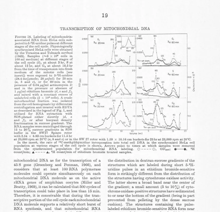
However, it is likely that the 16 S and 12 S RNA