A NOVEL ROLE FOR SCLEROSTIN IN AORTIC VALVE STENOSIS By
Jeffery Ethan Joll II
Dissertation
Submitted to the Faculty of the Graduate School of Vanderbilt University
in partial fulfillment of the requirements for the degree of
DOCTOR OF PHILOSOPHY in
Biomedical Engineering March 31, 2022 Nashville, Tennessee
Approved:
W. David Merryman, PhD Antonis K. Hatzopoulos, PhD Brian R. Lindman, MD, MSCI
Jeffry S. Nyman, PhD Cynthia Reinhart-King, PhD
ii
Copyright © 2022 Jeffery Ethan Joll II All Rights Reserved
iii
Dedicated to my grandparents.
iv
ACKNOWLEDGEMENTS
Kami Walters, Stephanie Joll, Jeff Joll, Sophie Joll, Judy Joll, Jim Fassinger, Bill Joll, Donald Midkiff, Karen Midkiff, Chris Midkiff-Brandt, Aimee Walters, Brent Walters, Michael Raddatz, Natalie Noll, Tessa Huffstater, Caleb Snider, Mark Vander Roest, Cami Johnson, Meghan Bowler, Nathan Bloodworth, Alison Vander Roest, Matt Bersi, Cyndi Clark, Erin Booton, Lance Riley, Mike Valentine, Olu Ogungbesan, Dave Armstrong, Dave Merryman, Cyndi Reinhart-King, Jeff Nyman, Brian Lindman, Antonis Hatzopoulous, Lin Zhong, Todd Giorgio, Heather Fedesco, Cynthia Brame, Leah Roberts, Evan Glass, Nick Goodell, Kelly Swope, Derek Price, Max Hamilton, Mike Miga, Brandi Gilbert, Jason Organ, Sungsoo Na, Rowdy, Buster, Vanderbilt Graduate Workers United, the Vandy Pickup Soccer Facebook group, Squad, Salt, Bernie Sanders, Three Brothers Coffee, Kay Bob’s, 51st Street Deli, the Nashville Public Library, the Belcourt Theater, and Montgomery Bell State Park.
Thank you.
v
TABLE OF CONTENTS
Page
DEDICATION...iii
ACKNOWLEDGEMENTS...iv
LIST OF TABLES...vi
LIST OF FIGURES...vii
GLOSSARY OF TERMS AND ABBREVIATIONS...ix
CHAPTER 1. Dissertation Overview...1
2. Aortic Valve Disease...5
3. Sclerostin...18
4. Sclerostin Ablation Prevents Aortic Valve Stenosis in Mice...37
5. Evaluation of Early Bilateral Ovariectomy in Mice as a Model of Aortic Valve Stenosis or Left Ventricle Hypertrophy...65
6. Improving Programming Content Delivery in an Introductory Biomechanics Course Using a Blended Classroom Approach...85
7. Impact and Future Directions...106
BIBLIOGRAPHY...120
APPENDIX...138
vi
LIST OF TABLES
Table Page
3.1. Summary of cardiovascular side-effects in ARCH trial...35
6.1. Traditional and blended course structure comparison...88
6.2. Data collection strategy...92
A.4.1. Primer sequences used for RT-qPCR analysis...138
A.4.2. Raw echo data separated by genotype and sex...139
A.6.1. Module completion statistics and survey response rate...142
A.6.2. Blended course module effectiveness survey...143
A.6.3. MATLAB coding confidence survey...144
vii
LIST OF FIGURES
Figure Page 2.1. Overview of AV cellular and molecular pathophysiology...13 2.2. Valve disease prevalence increases exponentially with age...15 3.1. Sclerostin is a potent Wnt-signaling inhibitor whose activity is blocked by
Romosozumab...24 3.2. Genetic mutation of the sclerostin gene and regulating elements causes a severe bone
overgrowth phenotype...28 3.3. Romosozumab is a more effective post-menopausal osteoporosis treatment than
Alendronate...33 4.1. The effects of genetic ablation of the Sost gene, aging, and high cholesterol diet were
assessed using in vivo and in vitro models...42 4.2. Genetic ablation of Sost results in a bone overgrowth and prevention of AVS...52 4.3. Sost knockout prevents early myofibroblast related AVS phenotype...54 4.4. RNA sequencing of aortic roots shows over 1000 differentially regulated genes and an
increase in pan-HOX gene expression in NULL groups...56 4.5. Sost null AVIC have altered HOX genes and decreased contractility...58 5.1. The ovariectomy surgical model of post-menopausal osteoporosis, combined with aging and
high-cholesterol diet, was evaluated as a potential model for pre-clinical study of aortic valve stenosis...74 5.2. Ovariectomy results in increased left ventricular mass and an inconclusive aortic valve
stenosis phenotype...76
viii
5.3. Left ventricle hypertrophy was noted in OVX hearts via ex vivo analysis, but valves were
absent of any significant thickening, collagen alteration, or calcification...78
5.4. Ovariectomy did not induce expression of markers of dystrophic or osteogenic calcification...80
6.1. A sample of how the online modules were structured (A) and the new video-based project delivery (B)...90
6.2. Module effectiveness survey results...95
6.3. Blended course pre- and post-MATLAB project coding comfort...97
6.4. Traditional vs. blended course post-MATLAB project coding comfort...99
6.5. Traditional vs. blended course MATLAB project coding quality (A) and characteristics (B)...101
A.4.1. 12-month whole body mass of experimental mice...145
A.4.2. Peak velocity normalized to left ventricular outflow tract diameter...146
A.6.1. Blended course module contents...149
A.6.2. Traditional course assignment...151
A.6.3. Blended course assignment video link...152
A.6.4. MATLAB coding ability rubric...153
ix
GLOSSARY OF TERMS AND ABBREVIATIONS
Abbreviation Term
ARCH Study to Determine the Efficacy and Safety of Romosozumab in the Treatment of Postmenopausal Women with Osteoporosis
ARS Alizarin Red S
aSMA alpha smooth muscle actin AVIC aortic valve interstitial cell
AV aortic valve
AVEC aortic valve endothelial cell
AVS aortic valve stenosis
BMP bone morphogenetic protein
BMD bone mineral density
CDH11 cadherin-11
CAVD calcific aortic valve disease
CV cardiovascular
DXA dual-energy x-ray absorptiometry
EF ejection fraction
EndMT endothelial-to-mesenchymal transition
ECM extracellular matrix
FRAME Fracture Study in Postmenopausal Women with Osteoporosis
HF heart failure
HOX homeobox
IF immunofluorescence
IACUC Institutional Animal Care and Use Committee IRB Institutional Review Board
LV left ventricle
LRP low density lipoprotein receptor related protein
MTC Masson’s Trichrome
MG mean pressure gradient
x
mAb monoclonal antibody
OCT optimal cutting temperature
OVX ovariectomy
PV peak velocity
PSR Picrosirius Red
PMO post-menopausal osteoporosis
qPCR quantitative real-time polymerase chain reaction RANKL receptor activator of nuclear factor kappa-B ligand
RNA ribonucleic acid
STRUCTURE
Study Evaluating Effects of Romosozumab Compared with Teriparatide in Postmenopausal Women with Osteoporosis at High Risk for Fracture Previously Treated with Bisphosphonate Therapy
TAVR transcatheter aortic valve replacement TGF-β transforming growth factor beta
VCPC Vanderbilt Cardiovascular Physiology Core VANTAGE Vanderbilt Technologies for Advanced Genomics
WT wild-type
1 CHAPTER 1 Dissertation Overview
2
There are many shared mechanisms of osteogenesis and bone growth and the ectopic development of calcific lesions often found in cardiovascular (CV) diseases, including aortic valve stenosis (AVS) [1]. Research performed as an undergraduate focused on diseases of the musculoskeletal system, including osteoporosis [2–6]. This work was related to research that discovered the fundamental role of sclerostin in the bone remodeling axis. Closely following the clinical trial results of sclerostin neutralizing antibodies in the treatment of skeletal disease led to interesting questions in the field cardiovascular pathophysiology. Specifically, an outcome of a Phase III clinical trial evaluating the sclerostin neutralizing antibody romosozumab was of great interest. Unexpected CV side-effects of the drug were discovered when compared to current treatments [7]. This provided a fruitful area to study the linkages between skeletal signaling and CV ectopic calcification, thus fusing previous research experience with a new area of focus on which to base the graduate studies found within this document. The first aim of this dissertation is focused on understanding the role of sclerostin on AVS development and how targeting this protein in osteoporosis may inadvertently affect the CV system.
Technical questions arising from this first aim lead to the discovery of the extent of understudy of female CV disease in pre-clinical studies. Only 10% of studies focus on female- only populations compared to 60% in that of men [8]. It is necessary to address this understudy problem head-on in order to accurately study the links between osteoporosis and valve disease.
Therefore, the second aim of this dissertation is studying how AVS progresses in a common pre- clinical model of post-menopausal osteoporosis (PMO) – bilateral ovariectomy (OVX). The goal for this aim is to determine if a commonly used female-specific model for pre-clinical PMO can double as a pre-clinical model of female-centric CV disease in order to better understand the relation between bone and CV disease as well as to provide a tool to address understudy of
3
female CV disease. This topic is closely related to the first in that sclerostin neutralizing antibodies have become a first-in-class drug therapy for treating PMO but questions remain about the off-target side effects [9]. Developing a strong pre-clinical model to study the PMO patient population will be very valuable in understanding the potential CV side effects.
The final arm this dissertation steps out of the laboratory and evaluates biomedical engineering classroom instruction. Approximately 40% of the work in this document was completed during the COVID-19 pandemic. Performing research in a hybrid style led to fundamental questions about the culture of knowledge production and transmission in the biomedical engineering field. In order to systematically evaluate this question, a study was performed to understand how the undergraduate learning experience is affected by a hybrid approach. In an introductory biomechanics course, the effect of hybrid and traditional classrooms was assessed and compared. This study is crucial for evaluating how the biomedical engineering education field develops post-COVID19. While many are eager to return to pre-pandemic teaching habits, there may be something to be learned the necessary shift to virtual and hybrid classrooms.
In summary, this dissertation is organized as follows: first, the foundation of the field of AVS pathophysiology is detailed in how the system works and what can potentially go wrong.
Next, the linkages between diseases of the skeleton and aortic valve (AV) are explained. Then, novel evidence of the role of sclerostin in AVS is presented. Next, the sex differences of AVS diagnosis, disease progression, and treatment are introduced. Then, evidence of a potential new pre-clinical model for the evaluation of AV and left ventricle (LV) disease in PMO is described.
Finally, evaluation of how a biomedical engineering classroom can be reformatted into a hybrid style and improve student confidence and performance is detailed. The dissertation is concluded
4
with a look at the impact of the novel research as well as a discussion on the potential future paths new research could follow.
It is the hope of the author that the work contained in this document will allow for new avenues of evaluation of AVS, understanding female specific AVS, and optimal methods of teaching biomedical engineering content in the modern college classroom.
5 CHAPTER 2 Aortic Valve Disease
6 Aortic valve development
The heart is the first functional organ in the developing fetus. The heart tissue arises from the mesoderm germ layer 18 to 19 days after fertilization and the primitive AV region begins to form a few days later from a substance known as cardiac jelly. This substance acts as an
extracellular matrix (ECM) and lies between the inner and outer tubes of the myocardial heart consisting of collagens, glycoproteins and polysaccharides [10]. The region of the jelly at the outflow tract lying in the atrioventricular canal develops into a region called the endocardial cushions. Endocardial cells begin to undergo an important process called endothelial-to-
mesenchymal transition (EndMT) whereby endothelial cells take on a mesenchymal phenotype akin to a fibroblast or smooth muscle cell [11,12]. This transition process is crucial to CV development and disease.
The signaling cytokine Transforming Growth Factor Beta (TGF-β) and protein Bone Morphogenetic Protein (BMP) from the myocardium and Notch signaling from the endothelium work together to spur EndMT and ECM maturation in the cardiac jelly [11]. Sox9 transcription factor is involved in maturation and proliferation of the new mesenchymal stem cells which use integrin receptor signaling and secretion of metalloproteinases to infiltrate and reconfigure the ECM tissue [13,14]. These mesenchymal cells begin to form the stratification of the AV leaflets, maintaining the inner region of the structure with endothelial cells forming the outer barrier.
Mechanical forces are crucial in developing the structure to allow unidirectional blood flow in the valves. Endothelium cells physically rearrange their cytoskeletons to align parallel to blood flow [15]. In 2017, a major component of the mechanobiological mechanism of EndMT was discovered. A shear responsive transcription factor called KLF2 induces WNT9B paracrine signaling which spurs EndMT. Without these elements, the AV does not develop properly [16].
7 Aortic valve functional anatomy
The AV is one of four heart valves. It is a tri-leaflet structure situated between the LV and aorta. The three leaflets meet along a ridged area known as the commissures. Each leaflet has a cusp which joins the aorta and myocardium at a transitional area known as the annulus.
Endothelial cells cover the outer blood contacting regions of the leaflets. In healthy humans, each leaflet has a trilayer interior with distinct regions determined by their primary ECM composition.
The region nearest the aorta (the outflow surface) is termed the fibrosa and contains densely packed collagen (particularly type I, III, and V fiber types) to provide support and provide continuous structure with surrounding tissues [17,18]. The area nearest the ventricle (inflow) is termed the ventricularis and is rich in elastin and is thought to maintain collagen configuration and provide structure in relaxed states [19]. Central to these regions is an area termed the spongiosa which is rich in glycosaminoglycans and allows structure flexibility and rearrangement of structural elements due to the mechanically strenuous heart cycle [17].
The AV experiences the highest mean pressure gradient (MG) and peak velocity (PV) magnitude and the largest amount of volumetric flow [20]. In diastole, the ventricle fills with blood and the AV is responsible for preventing regurgitant flow. During systole, the ventricle contracts and blood is expelled from the heart into systemic circulation. During this time, the AV must deform readily to allow the easy and unobstructed flow to the body. This cycle occurs over three billion times over the course of a lifetime [21].
In diastole, the AV is tightly closed in order to prevent regurgitation of blood. The anisotropic nature of the valve anatomy allows for different mechanical properties dependent on the direction on which stresses and strains are applied. Radially, the AV leaflets are extremely compliant in that they deform readily with low strains. This allows for a strong coapting under
8
high pressures which prevents collapse [22]. Collagen provides stress bearing capabilities and the arrangement of collagen fibrils changes in the systolic and diastolic phases. Collagen achieves a flat and ordered shape during diastole but undergoes a crimping action in order to contract and decrease surface area in response to blood being expelled through the outflow area.
With the return of diastole, the collagen resumes its ordered formation to maintain the mechanical barrier between aorta and ventricle. Elastin fibers contract during systole, further reducing the area of the valve [20,21].
During disease, the ordered conformation of the valve subregions can become disorganized, disturbing the hemodynamics found in the healthy valve. Understanding the complex form-function relationship presented here makes clear why disturbances in valve microarchitecture can have drastic effects on the flow characteristics of the region.
Aortic valve cell biology
There are two primary resident cell populations of the AV: valve endothelial cells (AVEC) and valve interstitial cells (AVICs) [23]. The AVECs form an external barrier to the valve, regulating the infiltration of various factors and cells as well as sensing the fluid flow dynamics. AVECs interact with the blood and are thus the first line of interaction of the valve with circulating factors. Indeed, AVECs are involved in response to thrombotic stimuli,
inflammation, and are engaged in cross-talk with AVICs in order to regulate valvular physiology [24]. AVECs are unique in that they maintain the ability to naturally undergo EndMT. This drastic phenotypic shift is an important stage of embryological development of the valve and also may be a method by which AVECs can replenish the AVIC population during adulthood [25].
9
This mechanism therefore is being investigated in its role in calcific aortic valve disease (CAVD) pathophysiology [26].
AVICs are present in all three layers of the AV [27,28]. These quiescent cells have relatively low basal activity but can be activated in response to injury, mechanical perturbation, disease/infection, and other biomolecular stimulations [21]. The activated form of AVICs is generally termed “myofibrobolast” and is characterized by the cells ability to synthesize fibrous matrix and contract its local environment as part of the natural repair process [23,29]. Only a small portion of AVICs are activated in the healthy heart, providing further evidence of their predominantly quiescent state with chronic activation only occurring during disease [21,30,31].
Evidence shows that AVICs re-gain their quiescent phenotype after return to equilibrium [32].
AVIC phenotype is relatively fluid with a number of phenotypes represented in explanted cells. AVICs are also sensitive to their microenvironment, local stiffness, and mechanical strain imposed during the cardiac cycle. These all contribute to AVIC homeostasis, and changes in these factors are seen in many disease states, including CAVD[23,27]. AVIC biology is associated with age-related changes in valve morphology where AVIC numbers drastically decrease with age with a concurrent reduction in the dynamism of the collagen architecture. This leads to a less compliant valve with increasing age [33–35].
Aortic valve disease
CV disease is the most common cause of death in the United States. Much of the focus of societal and research focuses on the more prevalent and acute CV diseases such as coronary artery disease (the most common CV disease) and myocardial infarction (carrying the highest
10
mortality rate). As a result, heart valve disease is often overlooked. In particular, AVS is the third most common CV disease, responsible for around 16,000 deaths per year [36].
AS is defined as the narrowing of the LV outflow area in such a way that valve
hemodynamic properties are drastically altered. The AV acts as a one-way valve controlling the flow of oxygen-rich blood from the LV (where it has arrived from the lungs) into systemic circulation. The delivery of vital oxygen and nutrients to the many tissues of the body requires regular and unobstructed flow between the LV and the rest of the body. Therefore, stenosis of the AV results in less efficient pumping between these two bodies. This is predominantly caused by a congenital defect called bicuspid AV where patients have two AV leaflets instead of three. AS is also seen in older individuals that have anatomically normal AVs that have accrued ectopic calcification over time, altering the structure-function relationship of the healthy valve anatomy [37].
Calcified nodules build up on the outflow surfaces of the valve via dystrophic (cell-death mediated) or osteogenic processes. Dystrophic calcification has found to be more prevalent in explanted valves [38]. In vitro studies have shown that AVICs are capable of undergoing both dystrophic and osteogenic mechanisms of calcification and are a likely source of calcification from either mechanism [23]. Numerous cell interactions and biological stimuli can augment or prevent the calcific potential of AVICs in vitro. These lesions cause a material stiffening (causing more than a doubling in young’s modulus of leaflet stiffness) and the once pliable leaflets no longer fully open and close to their full capacity, resulting in lower efficiency of oxygenated blood delivery [39]. In response to the increased resistance of the diseased valve, the LV will enlarge itself with muscle to try and overcome the resistance. This feedback loop can
11
lead to congestive heart failure (HF) which results in a decrease in quality of life and potentially death if left untreated.
AS is generally preceded by a thickening of the valve without disturbed hemodynamics – termed sclerosis [40]. The gradual fibrosis and accumulation of calcific nodules within the valve structure decreases the compliance and reduces the outflow area, producing extremely high exit velocities and pressure gradients between the aorta and the LV during systole [21,37]. This leads to progressive cardiac hypertrophy which can lead to congestive HF. Patients generally do not present until severe CAVD symptoms occur such as angina (chest pain) or syncope (loss of consciousness). These patients require immediate surgical replacement of the valve to remove the obstruction [41]. There are currently no treatments beside surgical intervention which is problematic due to around half of patients being ineligible for surgery due to high risk factors such as obesity, hypertension, and kidney disease [21].
The past decade has marked a shift in the core understanding of CAVD pathophysiology.
The disease was originally postulated to be degenerative and passive accumulation of
calcification and wear on the valve structure, through which local cells may respond in a way that furthered the disease phenotype. Instead, recent work has shown the disease is actively regulated and cell driven with AVIC disease activation, phenotypic shifts, mineralization, inflammation, matrix dysregulation, and other cellular functional mechanisms [42]. Further, many of these pathological mechanisms seemed to be regulated by AVICs [21,23].
Dystrophic calcification is dependent on programmed cell death, cell membrane damage, oxidative stress, inflammation, osteogenic phenotype switch, and lipid infiltration, among others [42]. Transforming growth factor, fibroblast growth factor, bone-morphogenetic protein,
lipoprotein, and Wnt signaling have shown to be major molecular drivers of CAVD disease
12
phenotype in cell and animal models [23,43–45]. AVIC phenotype shift to osteogenic and chondrogenic lineage has also shown to be associated with CAVD [46]. Major lineage regulators of Runx2, Sox9, Msx, among others also have varying roles in the prevention or causation of CAVD symptoms in various pre-clinical studies. Osteogenic, dystrophic, chondrogenic, and atherosclerotic regions have all been identified in explants of disease human valves [45,47–49].
The role of inflammatory cell infiltration is a rapidly growing area of CAVD research but results are by no means definitive. The most likely immune cell contributor to CAVD progress are cells of the macrophage/monocyte lineage which are able to interact with AVECs and enter the interior of the valve where release of pro-inflammatory cytokines can contribute to chronic activation of AVICs in disease [21,50].
AVICs activated to become myofibroblasts dysregulate the AV architecture by
dysregulated production of ECM factors such as collagen and fibronectin [51]. Activated AVICs also disturb the valvular structure through expression of matrix remodeling factors. AVIC activation is shown to be regulated by TGF-β, FGF, nitric oxide, Wnt signaling, and mechanical strains [21,23]. Hypercontractile AVICs become enriched in cell junctional proteins such as cadherin-11 which allow for tight attachment leading to damaging of neighboring cell
membranes where cell materials become the nucleation point for dystrophic calcifications [52–
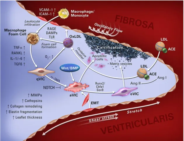
55]. Chronic activation of these cellular processes can lead to CAVD symptoms. The cell biology and molecular pathophysiology of the AV is summarized in Fig 2.1.
13
Figure 2.1. Overview of AV cellular and molecular pathophysiology. The aortic valve controls blood flow from the left ventricle into systemic circulation. AVIC and AVEC cells populate the tissue. Quiescent AVICs can become activate through a number of different factors
leading to dystrophic or osteogenic calcification. This figure summarizes many of the primary factors in the disease progression of the AV. Reprinted with permission from © 2014 Yutzey et.
al. Originally printed in ATVB [56].
14 Aortic valve disease burden and treatment
CV disease is an increasing public health concern in our aging population. Furthermore, poor diet and sedentary lifestyle contribute to poor overall CV health leading to a myriad of diseases [57]. Valvular diseases contribute to approximately 25,000 deaths per year, of which most are associated with AS and mitral regurgitation [58]. 25% of patients over the age of 65 present with aortic sclerosis – thickening of the AV. While this does not always progress to more advanced AS or CAVD, the risk of CV event increases by 50% in sclerotic patients. Further, this correlates to an 80% chance of HF, necessity of surgical valve replacement, or death over the next 5 years of life [59].
AS affects 3% of individuals over the age of 75, around 1.5 million patients in total, and accounts for 17,000 deaths per year in the United States. The number of individuals perishing from this disease is expected to double over the next 25 years [21]. Valvular stenosis lead to global changes in heart and peripheral blood vessels, and other organ systems in the body. Death by this disease usually occurs from cardiac hypertrophy and failure stemming from LV overload [37]. There are, in fact, no drug treatments known to slow, stop, or reverse the damage that eventually leads to AVS. Currently the only effective treatment for CAVD is surgical
replacement of the diseased valve with a synthetic replacement. This is because most clinical presentations of the disease are already in the severe, symptomatic stages, which require
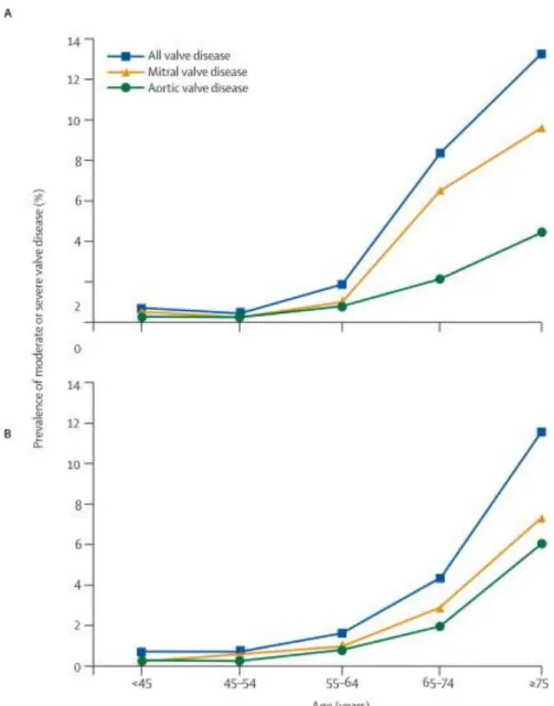
immediate relief. However, only about half of severe CAVD patients are candidates for surgical replacement due to high surgical risk [21,41]. This is problematic for several reasons. AVS primarily affects the extremely elderly, and complications due to age can disqualify the elderly from complete valve replacements (Fig. 2.2). Further, AVS is associated with a few other risk factors that make the potential patient population extremely vulnerable to surgery.
15
Figure 2.2. Valve disease prevalence increases exponentially with age. Results from two clinical trials indicating the prevalence of all valve disease (as well as mitral and aortic valve
specific) prevalence. The rate of disease increases drastically as patients age past 55-64, demonstrating how the disease primarily afflicts the elderly presenting treatment challenges.
Reprinted with permission from © 2006 Nkomo et. al. Originally printed in The Lancet [60].
16
Open-heart surgical replacement of the valve was long the standard for treatment. For appropriate patients, it can greatly improve quality of life and life expectancy. However, there are many potential operative and post-operative complications. The transcatheter aortic valve replacement (TAVR) technique has recently presented a percutaneous solution to this problem – avoiding the risks of open surgery. TAVR, which involves threading a catheter through the vasculature and remotely deploying a prosthetic with no need for heart resection, may be as effective at reducing symptoms and restoring healthy blood flow. Additionally, TAVR is safer for those patients at highest risk for open surgery. Early results show that TAVR is an effective treatment for people with CAVD at high surgical risk [61]. However, the TAVR placed
prosthetic is notorious for earlier failure and may necessitate multiple replacement surgeries which can be costly and time-consuming. Additionally, there are still no treatments that target the disease non-surgically. This is due to an unclear understanding of the mechanism of disease combined with an unclear effective therapeutic window due to the long-time course of disease progression.
AV disease is also not responsive to drugs which are effective in treating related
pathologies. Lipid lower drugs, such as statins, were evaluated in clinical trials of AVS but were shown to have no effect on the progression of severe AV disease [62]. Anti-hypertensive drugs have shown confounding results from exploratory studies [63]. Drugs which affect the
osteoclastogenesis pathways such as receptor activator of nuclear factor kappa-B ligand (RANKL) inhibitors have also shown inconclusive results [64]. It is therefore necessary to attempt to better understand the pathobiological mechanisms of AV disease so that new
pharmaceuticals can be developed to avoid the necessity for costly and risky AV replacement.
17
Clinical trials investigating the use of statins were conducted as the pathology of CAVD shares some similar aspects with that of atherosclerosis. However this trial failed to affect the progression of CAVD [65]. This is likely due to the differences in disease mechanism as well as the stage at which the intervention occurred [66]. Thus, there is theorized to be a point of
irreversibility where pharmacological targeting can no longer halt or reverse the progression of CAVD [67].
Currently, trials for RANKL inhibitors in slowing CAVD are in progression. This drug is used to slow bone resorption in osteoporosis by targeting the activation and activity of
osteoclasts [68]. Interestingly, RANKL has shown to be a pro-disease factor in CAVD and it is currently undergoing studies to see if RANKL targeting may have the dual effect of reducing CV calcium burden while improving skeletal BMD parameters [69,70]. While RANKL contributes to osteoclastogenesis in the bone, it is involved with myofibroblast activation and calcification in AVICs [71]. Our lab has produced robust pre-clinical justification for CDH11 blocking
monoclonal antibody (mAb) as a potential therapeutic option for treating CAVD. This mAb reduces dystrophic calcification and upregulates protective Sox9 signaling in a Notch1
haploinsufficiency model of CAVD [72]. Further research needs to be performed in this area but could represent an exciting new avenue for CAVD treatment.
Therapeutic options for treating CAVD are severely lacking and there is great potential benefit in elucidating the molecular mechanism of disease and factors that may be targeted to prevent, slow, or even reverse disease progression. This will improve outcomes for patients who may experience early stages of disease but would not be candidates for surgical replacement later down the road. Identification and early intervention with a pharmacological strategy could prevent the high mortality rates associated with CAVD.
18 CHAPTER 3
Sclerostin
19 Aortic valve disease and osteoporosis
A link between CAVD and osteoporosis has been proposed since the late 1990’s [73].
Recent studies using molecular imaging techniques have identified there is concomitant valvular calcium burden inversely correlated with BMD in osteoporosis [67,74]. Clinical studies have shown osteoporosis is independently associated with CV mortality and CV calcification is associated with increased incidence of fracture due to low BMD [75]. High-fat diet induced CV disease can also cause obesity-induced osteoporosis in small animals [76].
The paradoxical nature between CV calcification and low-bone mass has raised
interesting possibilities in treatment of these diseases. Factors that have disparate effects on bone and CV cells may prove fruitful areas of multi-scale treatment. The most common example is RANKL. In bone, RANKL activates osteoclasts and upregulates resorption which leads to osteoporosis [77]. The mAb denosumab targets this factor to slow the breakdown of bone tissue.
RANKL also acts on AVICs to drive inflammatory signaling in the AV, leading to CAVD. It stands to reason that targeting this protein may have the dual effect of decreasing bone resorption as well as calcific deposition on the AV. Unfortunately, clinical trials for denosumab in the treatment of AVS have shown little effect [78].
Sclerostin in cardiovascular disease
Sclerostin activity was originally thought to be confined to the skeletal system. However recent work shows expression in the protein of a number of organ systems and in various
physiological and disease processes. Sclerostin has shown to be present in the CV system, which is of great interest due to the apparent CV effects observed when targeting the protein in
osteoporosis patients.
20
Wnt signaling itself is known to play a role in a number of CV systems and diseases. The pathway is involved in cardiac and vascular development. It is particularly important in
atherosclerosis and CAVD [79]. Canonical Wnt signaling is a driver of CAVD in
hypercholesteremia models and is involved in the osteogenic shift of AVICs in a substrate stiffness dependent manner [47,80–82]. Low density lipoprotein receptor related protein (LRP) 5 is a major contributor to CAVD [80]. LRP6 genetic mutations result in coronary artery disease, osteoporosis, and hypertension indicating a dual role in low bone mass disease and CV disease in a paradoxical nature [83,84]. Warfarin induces vascular calcification by up-regulation of Wnt signaling in vascular smooth muscle cells [85].
While the role of Wnt signaling is fairly well characterized in CV development and disease, the role of sclerostin is relatively unclear and very few studies investigate the molecule specifically. As previously described, the specific CV status of patients with genetic disease of sclerostin is unclear. Many sclerosteosis patients perish due to brain herniation at a relatively young age when CV disease is quite uncommon. Elderly patients with sclerosteosis and van Buchem disease do not appear to have a propensity to CV disease but these patients come from relatively homogenous groups and may not be indicative of the effects sclerostin deficiency on the population overall [86]. Small animal models of this disease have not been thoroughly investigated for CV phenotype.
Sclerostin has recently been shown to be associated with calcific nodules on diseased AVs at the ribonucleic acid (RNA) and protein level [87,88]. Upregulation has also shown to be present in atherosclerotic lesions [89]. In vitro experiments focusing on vascular smooth muscle cell lines show that these cells upregulate sclerostin expression in osteogenic conditions which is recapitulated at the in vivo level [90]. Further, sclerostin has shown to be present in the
21
extracellular proteome of the aorta [91]. Sclerostin was shown to be associated with vascular calcifications at the in vivo level as well. The most thorough investigation of the sclerostin mutant phenotype in CV disease showed that sclerostin overexpression and excess protein treatment was able to ameliorate atherosclerosis and aortic aneurysm in an angiotensin-II mouse infusion model. This was associated with decreased immune factor circulation and macrophage infiltration. These effects were associated with downregulation of the Wnt signaling pathway [92]. This study paints a potentially protective role for sclerostin in the vascular disease and may begin to explain the negative effects on stroke and heart attack in clinical trials targeting the factor.
Many studies have been performed investigating circulating levels of sclerostin and association with CV disease progression and mortality. Results have been heterogeneous:
circulating sclerostin has shown positive associations with CAVD and arterial calcification in postmenopausal women [87,88,93]. There was no association between sclerostin and coronary artery calcification [94]. In African-American patient cohorts, a negative association between serum sclerostin and vascular calcification [95]. Studies also show that sclerostin and aortic calcification are inversely proportional in CKD patients [96]. Meta-analysis of these disparate findings asserts there is no overall association with serum sclerostin and CV mortality [97].
These complex and sometimes contradictory results do little to resolve the specific mechanism of action of sclerostin in the CV system. Signs point to a complex and potentially multifactorial role across a number of different CV tissues and diseases. Further investigation of these effects is crucial to better understand the off-target effects of targeting this protein in the treatment of osteoporosis.
22 Molecular biology of sclerostin signaling
Sclerostin is a secreted glycoprotein comprised of 190 amino acids. While originally described as an antagonist of BMP signaling, it was found to primarily be an antagonist of Wnt signaling through direct inhibitory effectors on the LRP family (Fig 3.1) [98,99]. LRP5 and 6 are the primary receptors of canonical Wnt signaling and sclerostin has been found to interact
directly with their extracellular signaling regions.
Normal Wnt signaling is activated via the binding of Wnt ligands to the LRP5/6 co- receptors and the interaction with the Frizzled co-receptors. The activation of this unit sequesters a multi-protein destruction complex to the cell membrane allowing for the stabilization of
cytoplasmic β-catenin. Β-catenin is then translocated to the nucleus where it activates Wnt gene transcription in concert with the TCF/LEF transcription factor family [100]. Additionally, sclerostin has also shown to interact with LRP4 which helps facilitate its negative effect on the Wnt signaling
pathway [101–103].
Wnt signaling is involved in a number of important biological processes in development and homeostasis as well as diseases such as CV calcification disorders, cancer, and many others [104]. Sclerostin is one of the most well studied Wnt signaling antagonists and is particularly active in regulating bone remodeling in the skeletal system. Sclerostin is secreted by osteocytes under varying stimuli. It travels through the canalicular network to the surface of bone where it interacts with the first or second EGF propeller domain of the LRP proteins [98]. Sclerostin blocks the binding of canonical Wnt signaling proteins to these sites and blocks the activation of this protein [105]. Through this mechanism, sclerostin is involved in the osteocyte-controlled inhibition of Wnt signaling driven bone formation.
23
Mechanical strain is a well characterized factor in increased bone formation. Increases in activity induce mechanical strains on the bone matrix which are sensed by embedded osteocytes [106,107]. These strains translate to downstream activation of pathways which cause the
deposition of new matrix in an effort to better resist these mechanical forces. This phenomenon is known as Wolff’s Law [108]. Experiments show that sclerostin is a major regulator of this mechanodependent bone remodeling. Specifically, expression is enhanced during unloading and downregulated under loading exercise by osteocytes [109]. Abundant sclerostin production in the absence of mechanical strain in the hindlimbs of mice inhibits Wnt-dependent bone deposition by osteoblasts and also contributes to the production of RANKL which acts to cause the
differentiation and activation of osteoclast cells to begin resorption [110,111]. Sclerostin mutant mice do not experience disuses osteopenia whereas sclerostin overexpression mice are resistant to loading induced bone formation [112,113]. In this way, sclerostin is a unique regulator that affects new bone deposition and resorption of existing matrix.
24
Figure 3.1. Sclerostin is a potent Wnt-signaling inhibitor whose activity is blocked by romosozumab. The Sclerostin protein primarily binds to the canonical surface receptor LRP5
and LRP6 to inhibit Wnt signaling (left). This results in sequestration, phosophorylation, and degradation of the β-catenin protein. Romosozumab inactivates the sclerostin protein (right).
This allows the Wnt ligand to facilitate formation of a dimer between LRP and Frizzled surface receptors. This prevents degradation of β-catenin which translocates to the nucleus and affects downstream gene activation. Reprinted under the Creative Commons license © 2016 Suen and
Qin. Originally printed in the Journal of Orthopaedic Translation [114].
25 Sclerostin associated genetic diseases
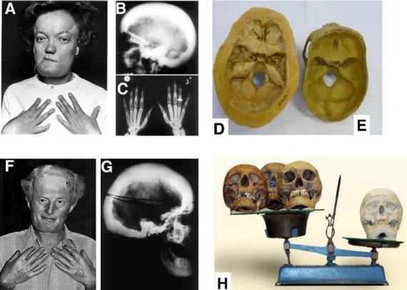
Sclerosteosis is a genetic disorder first characterized in the 1950’s which predominantly affects the Afrikaner population of South Africa [115]. There were approximately 100 identified cases in the 20th century [116]. The hallmark of the disease is progressive skeletal overgrowth and a number of symptoms stem from this phenomenon. Bone overgrowth affects both the axial and appendicular skeleton. Patients are highly resistant to fracture with the only case arising from a vertebral fracture due to increased weight of the skull [116]. Sclerosteosis patients are generally well above average height and weight [86]. Facial overgrowth results in deformity characterized by prominent jaw due to overgrown mandible and enlarged and protruding forehead [116,117]. Facial overgrowth results in loss of function of nerves involved in hearing and vision, which commonly result in impairment [86,118]. The first symptom most commonly observed is syndactyly and deformities of the digits such as fusion or webbing [86,116]. Skull overgrowth leads to compression of the intracranial space resulting in high pressure leading to persistent headaches, nausea, and dizziness which often requires surgical intervention [119,120].
Most patients die in early adulthood (~30 years) from brain herniation due to increased pressure or complications from the craniotomy procedure [116]. Notably, patients who survive into adulthood generally experience stabilization of symptoms [86,117,121]. While two patients to date have died due to heart disease, elderly patients with sclerosteosis do not show signs of impaired cardiac or vascular function or disease [116].
Van Buchem disease is another rare disorder affecting residents of a genetically isolated northern Netherlands community [122]. There were about 30 cases identified in the 20th century [116]. First described in 1955, it presents with symptoms similar to sclerosteosis – skeletal overgrowth, facial deformity, deafness, facial palsy, and increased intracranial pressure. The
26
phenotype of van Buchem patients is milder than sclerosteosis and only one case required craniotomy and carries a normal life expectancy [116,123,124]. Further, patients are of normal height, do not display digital deformities, and have only mildly impaired hearing. Fractures are infrequent in this population but occur at a higher rate than in sclerosteosis patients [123].
Clinical presentation of these diseases is shown in Fig. 3.2.
Similarities between sclerosteosis and van Buchem disease were identified in the 1980’s and genetic analysis revealed mutations throughout chromosome location 17q12-q21
[119,125,126]. Patients with sclerosteosis shared a number of mutations in the coding region of the gene SOST while van Buchem patients shared a deletion in the spacer region between SOST and neighbor MEOX1 [127–130]. Later studies revealed SOST regulatory elements within this spacer region. This reduced (but not eliminated) sclerostin expression is thought to explain the intermediate disease phenotype found in van Buchem patients [131]. Patients heterozygous for sclerostin coding deletion found in sclerosteosis also exhibit an intermediate phenotype with a high BMD and lower sclerostin expression but are free of the severe symptoms found in homozygous deletion [121].
In order to better understand the various mechanisms through which sclerostin acts on the skeletal system, numerous small animal models were generated to study the phenotype in more detail. Germ-line knockouts of sclerostin mimicking the sclerosteosis phenotype exhibit a very high bone mass phenotype as well as increased bone formation rates and mechanical strength.
This model does not recapitulate the digital or facial deformations commonly found in human patients and does not carry a decreased life expectancy compared to wild-type (WT) controls.
However osteoblast activity appears to be upregulated and circulating markers of bone formation are increased with deletion of the sclerostin gene [132]. Conversely, mice overexpressing human
27
sclerostin showed opposite effects and displayed an osteopetrotic phenotype with decreased mineralization in the axial and appendicular skeleton. Deposited bone was less organized and strength testing displayed higher fragility and susceptibility to fracture. There was also a corresponding decrease in metrics of osteoblast activity [131].
28
Figure 3.2. Genetic mutation of the sclerostin gene and regulating elements causes a severe bone overgrowth phenotype. A patient (A) with a full frame deletion of the SOST gene exhibits skeletal overgrowth in the skull (B, D, E) and digits (C). A patient (F) with a mutation in a SOST
enhancing regulation region also displays skull overgrowth (G) but at a lower severity. These diseases are called Sclerosteosis (full SOST) deletion and Van Buchem Disease (enhancer element deletion). Reprinted under the Creative Commons license © 2018 Sebastian and Loots.
Originally printed in Metabolism [133].
29
Preclinical studies for sclerostin in osteoporosis treatment
Sclerostin’s effects were long thought to be isolated to the skeleton. Due to its negative effect on bone formation and positive effect on resorption, its pharmacological targeting seemed to be a promising strategy for diseases of low bone mass such as osteoporosis, disuses
osteopenia, and osteogenesis imperfecta. A mAb was developed and tested in a number of pre- clinical animal models with the expectation that blocking the protein would increase bone mass and reduce fracture incidence with minimal off-target effects.
Sclerostin mAb was shown to be effective in a rat model of disuse osteopenia. Rats were normally mobilized or subject to underloading of the hindlimb bones. Vehicle and sclerostin mAb were administered and the trabecular and cortical anatomy of the tibial metaphysis and shaft were analyzed. Immobilized rats with vehicle treatment had increased bone resorption and decrease of bone formation. In normally loaded and unloaded mice, sclerostin neutralizing mAb increased bone mass and decreased resorption [134,135]. This study showed potential efficacy for sclerostin mAb treatment in patients with disuse osteopenia such as those confined to bed rest.
Osteoporosis affects a large number of women in post-menopause due to the loss of the protective effect of estrogen [136]. Sclerostin neutralizing mAb was potentially thought to be a mechanism of reducing loss of bone mass in these patients. To test the hypothesis, the OVX procedure was performed in rats. This model induces rapid development of PMO symptoms included signatures of osteoporosis. One year after OVX, rats were treated with sclerostin neutralizing mAb. The PMO phenotype was reversed and OVX rats actually displayed greater metrics of bone formation, mass, and strength when compared to non-OVX control mice [137].
This study was a strong endorsement for sclerostin mAb treatment in management of PMO.
30
Decrease in bone mass and incidence of osteoporosis is prevalent in aging. Reducing bone mass, mineral content, and susceptibility to fracture are major factors associated with increasing age and neutralization of sclerostin is thought to be a potential mechanism of reducing these negative events [138,139]. Pre-clinical studies in aged rats show improvements in bone mineral density (BMD) in vertebrae and long bones and improved bone structure in vehicle controls. This correlated with bone strength and increased metrics of bone formation [140].
Simultaneously, aged female rats were examined in a similar fashion at 10 months of age. One month of sclerostin mAb treatment resulted in improved bone formation metrics and trabecular volume. Bone resorption was also decreased [141]. Sclerostin mAb seems promising for treatment of age-related low-bone mass in both men and women.
In addition to efficacy in common low bone mass disease, sclerostin mAb treatment has also shown to be effective in fracture healing in numerous studies in rats as well as improvement of healing after surgical osteotomy in a non-human primate study [142–145].
To prove efficacy in a non-human primate model, sclerostin neutralizing mAb was administered to female cynomolgus monkeys with no alteration of hormonal state. Only two treatments displayed dose-dependent increases in bone anabolism in all studied compartments.
This was also seen in bone mineral studies and bone strength and structure in comparison to vehicle controls [146]. This study greatly improved the rationale for study of sclerostin neutralization in humans suffering from low-bone mass disorders.
31 Clinical trials for sclerostin in osteoporosis treatment
Long theorized to be a potential target in treating low bone mass disease in humans, positive results from pre-clinical animal studies justified beginning clinical trials in a number of patient populations.
Phase I trials were first published in 2010. 72 individuals comprised of healthy men and postmenopausal women were administered sclerostin blocking mAb or placebo in ascending dosage via subcutaneous or intravenous injection. After three months of treatment there were minimal adverse effects with only one case of hepatitis and zero deaths or treatment
discontinuations. Markers of bone formation increased in a dose-dependent fashion and BMD improved in treated patients compared to placebo in the spine and hip [147]. A follow up phase I trial was then performed and published in 2014 focusing on men and women who presented with low bone mass phenotype. 48 patients received ascending dosage of sclerostin mAb or placebo and bone formation metrics were assayed along with BMD assessed by imaging. Markers of bone formation increased drastically with blocking of sclerostin with a concurrent increase in BMD. Adverse effects were once again minimal and balanced between groups [148].
Following the promise of phase I trials, phase II trials were published in 2014 and were focused on comparing the efficacy of sclerostin mAb treatment in comparison to current standards of treatment – bisphosphonates. These drugs take advantage of the affinity of the bisphosphonate structure to bind to hydroxyapatite – a primary component in actively remodeled bone. When osteoclasts encounter bone matrix with embedded bisphosphonate molecules they cease resorption and undergo apoptosis – thus slowing bone loss [149]. These drugs therefore slow bone loss but do little to encourage new bone formation. Over 400 postmenopausal women with low bone mass phenotype received sclerostin neutralizing mAb, vehicle, or a
32
bisphosphonate. Treatment occurred for one year and BMD and markers of bone remodeling were assessed. Adverse effects were balanced amongst groups and sclerostin blocking improved metrics of bone formation compared to bisphosphonate treatment, indicating potential for a new gold standard in osteoporosis care [150].
Large-scale phase III trials were conducted to assess long-term effects of sclerostin neutralization treatment and effects on fracture reduction and improved bone metrics. Results from the Fracture Study in Postmenopausal Women with Osteoporosis (FRAME) trial were published in 2016. Patients were treated for one year with sclerostin mAb or placebo followed by treatment with denosumab. This drug is a RANKL inhibitor acting to downregulate activation of osteoclasts and reduce new bone resorption. Patients treated with sclerostin mAb had a 0.27 risk ratio of vertebral fracture when compared to placebo with no significant decrease in nonvertebral fractures. After denosumab treatment there was a 0.25 risk ratio of vertebral fracture in sclerostin mAb treated individuals and 0.75 hazard ratio for non-vertebral fractures however this effect had a non-significant adjusted p-value [151].
The Active Controlled Fracture Study in Postmenopausal Women with Osteoporosis at High Risk (ARCH) trial was published in 2017 and included over 4000 patients (Fig. 3.3).
Patients were given sclerostin blocking or bisphosphonate treatment for one year followed by one year of further bisphosphonate treatment in each group. Patients with initial treatment of sclerostin blocking were half as likely to experience vertebral fracture as patients only treated with bisphosphonates. Overall fracture risk reduced by 27% when patients were treated with combination therapy as opposed to bisphosphonates alone [7].
33
Figure 3.3. Romosozumab is a more effective post-menopausal osteoporosis treatment than alendronate. Results from the ARCH trial were acquired based on BMD and bone remodeling
agent levels. BMD at the spine (A) was nearly tripled compared to alendronate after one year.
BMD approximately doubled at the hip (B). P1NP (C) and β-CTX (D) circulating levels were similarly diminished between the two drugs. Reproduced with permission from 2017 Saag et. al.
© Massachusetts Medical Society. Originally printed in NEJM [7].
34
Finally, the Study Evaluating Effects of Romosozumab Compared with Teriparatide in Postmenopausal Women with Osteoporosis at High Risk for Fracture Previously Treated with Bisphosphonate Therapy (STRUCTURE) trial analyzed over 400 women who had been treated for over three years with bisphosphonate therapy. Half of patients were continued on
bisphosphonate therapy while the others were transitioned to a sclerostin blocking therapy and analyzed after one year. Results showed a 2.6% increase in BMD in sclerostin mAb patients compared a 0.6% decrease in bisphosphonates. These changes were consistent in other skeletal sites and indicate an advantage of sclerostin blocking even when bisphosphonates have become ineffective in patients at severely high fracture risk [152].
While the skeletal effects of sclerostin neutralization are extremely positive in treating patients with a number of low bone mass afflictions, results of the ARCH trial indicated troubling CV side effects (Table 3.1). Specifically, serious CV events were more frequent in patients treated with sclerostin blocking mAb than bisphosphonates. Digging into the reported differences, cardiac ischemic and cerebrovascular events were more commonly observed in sclerostin treated individuals with odds ratios of 2.65 and 2.27, respectively. Death after
sclerostin treatment was numerically higher than bisphosphonate and became level after groups switched to bisphosphonates alone. Interestingly, incidence of noncoronary revascularization and HF were lower in sclerostin treated groups and remained lower after reverting back to
bisphosphonate treatment [7]. These effects resulted in a safety flag by the U.S. Food and Drug Administration and delayed approval for treatment [153].
35
Table 3.1. Summary of cardiovascular side-effects in ARCH trial. Results of cardiovascular side-effects after one-year of treatment of alendronate or romosozumab. There were significant increases in cardiac ischemic and cerebrovascular events. Adapted with permission from 2017
Saag et. al. © Massachusetts Medical Society. Originally printed in NEJM [7].
36
Data gathered from robust clinical trials indicates a strong effect of sclerostin blocking therapy in improving the effects of low bone mass in old age and particularly in PMO. However, the myriad of CV side effects is obviously a cause for concern. The safety flags even resulted in postponement of FDA approval of the drug for treatment. However, approval was first gained in Japan and was nearly unanimously approved in the U.S.A. In April of 2019 for postmenopausal women at very high risk of fracture. This took place after pooling of data from multiple trials to try to determine overall CV risk. The drug is marketed under the brand name Evenity, carries a warning label, and is not recommended for patients at high risk for CV diseases or those with a history of CV disease. This obviously limits the clinical utility of the drug, as the patients most likely being treated are elderly and at higher risk for CV disease the general population.
Therefore, it is important to develop a thorough understanding of sclerostin in the CV system in order to fully understand the effects of targeting this drug for low bone mass diseases.
37 CHAPTER 4
Sclerostin Ablation Prevents Aortic Valve Stenosis in Mice
38 Introduction
Sclerostin (human gene: SOST, mouse gene: Sost) is a secreted glycoprotein that has shown to be a potent regulator of bone remodeling [154]. It was first discovered to be the causative root of excessive bone overgrowth in Sclerosteosis (full SOST deletion) and van
Buchem diseases (deletion of a genetic regulator) [125,126,130,155]. Further studies clarified the mechanisms whereby sclerostin regulates ossification, resorption, and mechanotransduction in bone by altering the Wnt and RANKL pathways of the skeletal system [110,111,156,157].
Due to the protein’s involvement in bone remodeling and its apparent localization to the skeletal system, sclerostin became a target of great interest for the treatment of low bone mass diseases such as osteoporosis. Clinical trials of a mAb targeting sclerostin - known as
romosozumab - substantially improved bone quality metrics and reduced hospitalization in PMO patients [7,151,152,158,159]. However, the stage III ARCH trial noted an imbalance in CV effects compared to the current standard bisphosponate medication, alendronate. The authors specifically noted a significant increase in cardiac and cerebrovascular ischemic events and a numerical decrease in HF [7]. This resulted in the drug receiving a warning of CV side effects which must be weighed when considering prescribing romosozumab to patients who may have, or be at risk for, CV disease.
Discovery of off-target effects in the CV system prompted a review of existing literature as well as new studies directly interrogating the role of sclerostin in various disease models.
While the body of literature for sclerostin’s role in the AV is quite limited, there are some studies of interest. AVs excised from human patients with AVS showed that sclerostin protein and mRNA are upregulated in diseased tissue near sites of ectopic calcification [88]. A study from the same year found that serum sclerostin levels are associated with valve calcification in
39
individuals undergoing hemodialysis. This was also confirmed at the tissue level using
immunohistochemistry and quantitative real-time polymerase chain reaction (qPCR) [94]. While these studies show correlation between valve disease and sclerostin expression, it is unclear if the increase is a direct contributor to AVS.
The pathways associated with sclerostin signaling in the skeletal system also contribute to the progression of AVS which augment the necessity for direct investigation. Activated Wnt and RANKL signaling have shown to be involved in ectopic calcification of the AV, indicating there may be shared processes sclerostin acts on in the skeletal and CV systems [49,160].
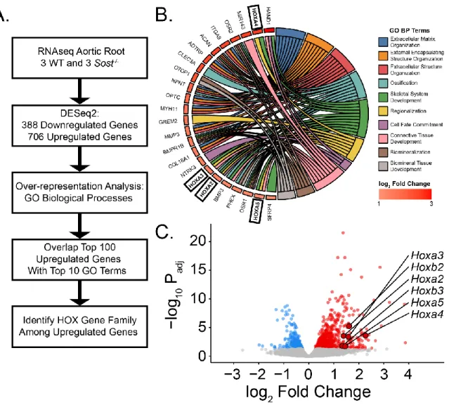
AVS is a complicated and poorly understood disease process and it is unclear whether sclerostin plays a significant role in its progression. To this end, we hypothesized, based on the CV events in the stage III ARCH trial, long-term loss of sclerostin would worsen AVS in a mouse model. Surprisingly, we found Sost ablation had a protective effect in the progression of AVS via reduced myofibroblast activation and AVIC contractility potentially due to upregulation of protective Homeobox (Hox) gene expression.
40 Materials and Methods
Animals
All mouse experiments were carried out under appropriate approval and supervision from the Vanderbilt University Institutional Animal Care and Use Committee (IACUC). Sost
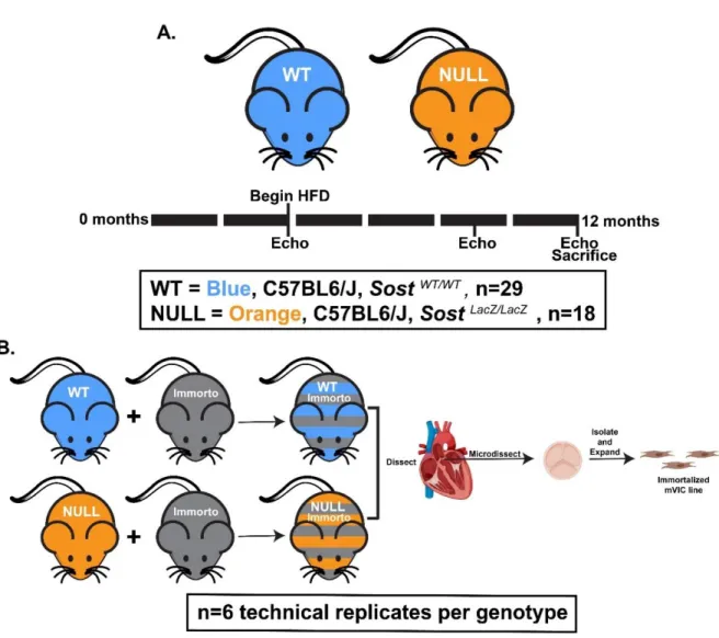
knockout mice (NULL) were provided by Dr. Gabriela Loots and bred as previously described [161]. Littermates homozygous for the WT Sost gene were used as controls. Equal amounts of male and female mice were used. At 4 months of age, mice transitioned from standard chow to a 1% cholesterol Western diet (TestDiet 5TJT). Food and water were provided ad libitum. Mice were aged for 8 additional months on the Western diet to 12 months of age, euthanized by carbon dioxide inhalation followed by cervical dislocation, and AV tissue was harvested for processing and analysis (Fig 4.1A).
Cells
Mouse AVICs were isolated as described previously [53]. Briefly, WT mice were crossed with the “Immortomouse” line (Charles River, 237 HO, 238 HE) in order to generate cell lines capable of undergoing prolonged growth under specific culture conditions. Mice were euthanized and the heart carefully excised under sterile conditions. Using a dissection microscope, the aortic root was isolated and the individual leaflets removed, soaked in 600 U/mL collagenase II
(Worthington Biochemical Corp, Lakewood, NJ) for 30 minutes, and centrifuged. Leaflets were then placed on 0.1% gelatin coated tissue culture dishes in immortalized media (10% fetal bovine serum, 1% penicillin/streptomycin antibiotic, 10 U/mL interferon-Ɣ in Dulbecco’s Modified Eagles Medium), with environmental conditions of 33° C and 5% CO2. AVICs were allowed to adhere to the culture dish, migrate from the leaflet tissue, and expand for about one
41
week until confluence was reached (Fig 4.1B). To verify the isolation of a myofibroblast cell line, cell morphology was confirmed visually and the presence of the contractile protein alpha- smooth muscle actin (aSMA) was confirmed using immunostaining. Prior to all experiments, cells were maintained for 24 hours at 37° C and 5% CO2,in complete media without interferon-Ɣ (10% FBS, 1% pen/strep antibiotic in DMEM) to inactivate the immortalization element.
42
Figure 4.1. The effects of genetic ablation of the Sost gene, aging, and high-cholesterol diet were assessed using in vivo and in vitro models. A. Mice were given high-cholesterol diet beginning at 4 months and aged to 12 months. The development of AVS was tracked using
echocardiography at 4, 9, and 12 months of age. Black bar = 2 months. B. In parallel, immortalized mouse lines were generated for WT and NULL groups. Aortic valve interstitial
cells were isolated and expanded for in vitro analysis. Image partially created with Biorender.com.
43 Echocardiography
Echocardiographic assessment was performed at 4, 9, and 12 months of age to identify changes in cardiac structure and function. All imaging was performed by skilled technicians in the Vanderbilt Cardiovascular Physiology Core (VCPC) using the Vevo 2100 small animal imaging system. Mice were lightly anesthetized (mean: 492 bpm) using isoflurane and laid supine on a heated platform. Transthoracic aortic pulsed wave Doppler imaging was used to generate AV velocity profiles. Parasternal short-axis M-mode imaging was performed to measure LV performance and cardiac function as indicated by changes in strain throughout the cardiac cycle.
For AV-specific measurements, a custom MATLAB script was used to automatically trace pulsed wave Doppler waveforms and determine cardiac-gated hemodynamic metrics such as PV and MG [162]. At each time point, three Doppler measurements were gathered for each mouse resulting in 50 to 100 independent cardiac cycles being averaged to produce
representative metrics. LV thickness and motion were manually measured over each cardiac cycle. Three cycles were averaged for each M-Mode image for a total of 9 cycles analyzed per mouse per time point.
Dual Energy X-Ray Absorptiometry
Femur bone metrics were measured using the Hologic UltraFocus Dual Energy X-Ray Absorptiometry and associated software. Samples were placed on the stage at 2X magnification and images were acquired in a series of 4 captures at 40 kV followed by 4 captures at 80 kV captures. Using a custom Region of Interest analysis in the system software (Version 3.1), the bone mineral content, BMD, and femur length were measured.
44 Histological Staining
Excised aortic roots were embedded in Optimal Cutting Temperature (OCT) compound and flash frozen. Using a -20° C cryostat, aortic root samples were serially sectioned at 10 μm.
For all staining analysis, 3 sections per aortic root were analyzed for a representative metric.
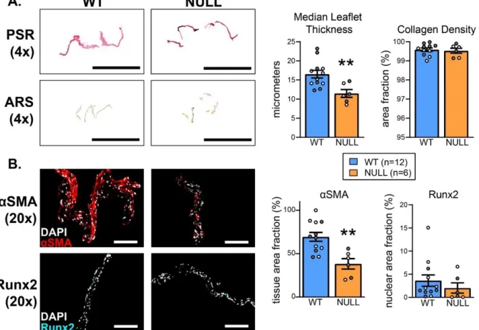
Picrosirius Red (PSR) (FisherScientific) staining was performed to assess collagen
characteristics and morphology of the aortic roots. Alizarin Red S (ARS) (Sigma-Aldrich) staining was performed to assess the extent of valve calcification. For both stains, standard manufacturer protocols were followed without deviation. After staining, slides were dehydrated in progressively concentrated alcohol baths (70%, 90%, 100%), cleared in xylene, mounted in organic mounting media, coverslipped, dried overnight, and sealed prior to imaging. Brightfield images were captured at a 4X magnification objective using a Nikon Eclipse E800 microscope equipped with an Olympus DP74 digital polychromatic camera. Representative images were shown to best illustrate the quantitatively determined phenotype.
Immunohistochemical Staining
Fluorescent immunostaining was used to characterize the prevalence and localization of aSMA (contractile) and Runx2 (osteogenic) proteins - both of which are common markers of AV disease. Slides were washed in PBS prior to fixation/permeabilization for 10 minutes in a 4%
paraformaldehyde/0.1% Triton solution in PBS. Sections were then blocked for non-specific binding using 10% bovine serum albumin in PBS for 1 hour at room temperature. Primary antibodies were added to a 10% diluted blocking solution at the following concentrations: 1:100 rabbit polyclonal anti-aSMA (Abcam ab5694), 1:100 rabbit monoclonal anti-Runx2 (Cell
Signaling Technology 12556) and incubated overnight at 4° C. The following day, sections were
45
washed in PBS prior to incubation in 1:1000 goat anti-rabbit IgG Alexa Fluor 647 (ThermoFisher A-21245) for 1 hour at room temperature and covered from light. After
secondary staining, slides were once again washed in PBS prior to being mounted in ProLong Gold Antifade with DAPI (Cell Signaling Technology 8961), coverslipped, allowed to dry overnight at room temperature, then sealed. Fluorescent images were captured at 20X
magnification objective using an Olympus BX53 microscope and Qimaging Retiga 3000 digital monochromatic camera.
Quantitative Image Analysis
For the analysis of PSR staining, brightfield images of the stained aortic root were acquired and cropped to contain only the AV leaflets. Total leaflet area was calculated from brightfield images and collagen composition within the leaflet area was determined as previously described [163,164]. Expression of contractile (aSMA) and osteogenic (Runx2) proteins of interest in the AV was determined from analysis of immunofluorescence (IF) images, as
previously described [165]. Briefly, AV sections fluorescently labeled for either aSMA or Runx2 were stained with DAPI to indicate leaflet nuclei and provide an estimate of leaflet area based on a manually defined boundary. Segmentation of individual nuclei was performed using a modified watershed transform and concave object separation algorithm [163,166]. aSMA area fractions were then computed as the ratio of positive pixels to total pixels within the leaflet boundary.
Runx2 positive nuclei were defined based on the ratio of positive pixels to total pixels within the identified nuclear boundary.