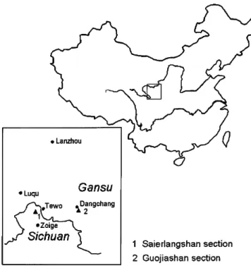
This report extends knowledge of the biogeographic distribution of gastropods during the recovery from the Permian mass extinction. Triassic gastropods of the southern Qinling Mountains, China / Jinnan Tong and Douglas H. Smithsonian contributions to paleobiology ; no. 92) Includes bibliographical references. Guojiashan is part of the northern area while Saierlangshan lies in the southern area (Figure 2).
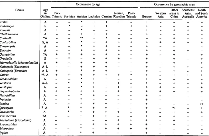
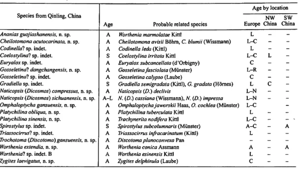
The Saierlangshan section contains two beds containing gastropods from the Zalishan Formation, but only four specimens from the Maresongduo Formation. Most of the fossils discussed in this paper are from the Anisian Guojiashan Formation of Saierlangshan, Sichuan Province and Guojiashan, Gansu Province. Naticopsis {Dicosmos) eyerichi was previously described from the Muschelkalk and Coelostylina ahlburgi was described from the upper part of the Wellenkalk to Muschelkalk in Europe.
Slugs of Ladinian age were found only in the lower part of the Guanggaishan Formation in the Saierlangshan section. The coexisting Naticopsis (Vernelia) sublimneiformis and Neritaria plicatilis were described in the Ladinian and Carnian of the Southern Alps. More than half of the Triassic snail species from the South Qinling Stratigraphic Province were first described in the European Alps or Polish Upper Silesia.
The new information presented here suggests that many Triassic lineages originated in the Early Triassic and Early Anisian (Table 4).
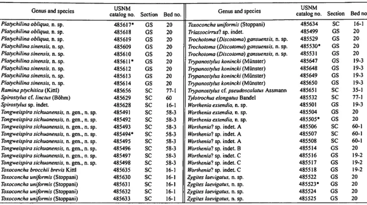
USNM 485500, 485503, 485509
17 leya. Although the aperture is broken and the columellar lip is
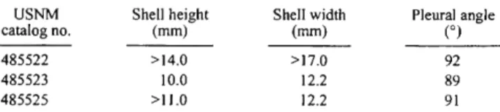
However, most species of Amberleya have more ornamentation than this specimen, although they are usually not as elaborate as Eunemopsis. DISCUSSION.-This Triassic genus, which probably arose from Paleozoic murchisoniids, is characterized by a peripheral keel in the middle of the crest that forms the lower limit of the selenison. These easily identified specimens exhibit extensive morphological variation in pleural angle at a single locality.
Glyptospira from the Permian is quite similar, and a single specimen of Eunemopsis from this collection may represent a member of this clade. MATERIAL EXAMINED.—One partial specimen with the last two of the six original whorls; other whorls broken during preparation. A relatively large pleural angle and a stronger upper row of nodules distinguish this shell from other species of this genus (e.g. Eunemopsis epaphus (Laube) and E. praecurrens Kittl).
DIAGNOSIS. - Sharply conical shell with a pleural angle of 45°-65° and with four to six strongly convex whorls divided by deep sutural grooves.
USNM 485621-485625, 485627
Internal resorption of early whorls was considered a key feature distinguishing Neritopsidae from Neritidae. Teeth or growths on the inductura of the inner lip together with resorption of the internal gyrus have been used as the most important markers of the separation of genera. In living neritoids, folds on the inductura of the inner lip improve the position of the operculum within the opening and are related to the ecology of the animal.
DISCUSSION.-The Neritopsidae are distinguished from the Neritidae by the neritopsids' resorption of the inner wall during ontogeny. MATERIAL EXAMINED.-Six specimens, most complete, from the middle to upper part of the Guojiashan Formation of Guojiashan. MATERIAL EXAMINED.—Six specimens from the middle to upper part of the Guojiashan Formation: USNM.
MATERIAL STUDYED. Five specimens, three of which are well preserved, from the lower part of the Guojiashan Formation of Saierlangshan: USNM. MATERIAL STUDYED. Thirty specimens from the lower and middle parts of the Guojiashan Formation of Saierlangshan and two from the lower part of the Guanggaishan Formation of Lagecaimo, Zoige Xian, Sichuan Province: USNM. The upper edge of the opening in the Chinese forms also extends further abaxially than obliquely as in the two European species.
MATERIAL EXAMINED.—Five specimens, including a very small one, from the middle and upper parts of the Guojiashan Formation of Guojiashan and two specimens from the lower part of the Guanggaishan Formation of Lagecaimo, Sichuan Province. DISCUSSION.—The ornamentation of this neritopsid easily distinguishes members of the genus from the smooth naticopsids. MATERIAL EXAMINED.—One well-preserved specimen and one with the apex broken, from the middle part of the Guojiashan Formation: USNM.
MATERIAL EXAMINED.—A single well-preserved specimen from the middle part of the Guojiashan Formation at Guojiashan. DISCUSSION.—The taxa assigned to this genus show no significant differences from the unornamented neritopsids in external form, but the resorption of the inner wall of the early whorls is the standard character that distinguishes this taxon from other groups in the clade. MATERIAL EXAMINED.—Six deformed specimens from the middle and upper part of the Guojiashan Formation of Guojiashan and additional, fractured specimens from other beds at the same locality: USNM.

USNM 485593-485598
25 Neritaria ingrandita (Kittl, 1894)
The inner lip is covered with a thin, wide, smooth and distinctly concave inductura, which covers the umbilicus but forms a wide depression in the umbilical region. MATERIAL EXAMINED.—Six specimens from the lower and middle part of the Guojiashan Formation, Guojiashan, and one from the lower part of the Guojiashan Formation, Saierlang-shan. DISCUSSION.—These specimens are identical to Neritaria ingrandita (Kittl) in the broad naticiform form, fewer whorls, and the deepened suture near the aperture.
MATERIAL EXAMINED.—A single specimen from the lower part of the Guanggaishan Formation, Lagecaimo, Sichuan Province: USNM 485608. DISCUSSION.—The shell shape and prominent spiral of this specimen support its assignment to Nerititaria, anplitaria between N . Inner lip curved with smooth induration, moderately wide and slightly convex, mainly on the parietal lip.
MATERIAL STUDYED. Seventeen generally well-preserved specimens from the middle and upper part of the Guojiashan Formation, Guojiashan.
DISCUSSION.—The spherical shape, flat upper surface of fi-
FIGURE 31
29 nament except for faint sigmoidal growth lines. Aperture evi-
Whorls flat to slightly convex in middle to lower part; ratio of whorl height to width slowly and gradually increasing from 1:2 to 2:3. MATERIAL EXAMINED.—Six specimens from the middle part of the Guojiashan Formation at Guojiashan: USNM. DISCUSSION.—Kittl (1894a: 194) listed six key features that help identify this species; our specimens agree with these, although they are not as tall as the figure given by Kittl (1894b, fig. 39).
DESCRIPTION.—Medium-sized, slender, high-pointed cochlea with deeply incised sutures and eight weakly convex whorls. MATERIAL EXAMINED.—A single deformed steinkern from the upper part of the Zalishan Formation, Saierlangshan: USNM 485651. Confusion arose when Cossmann (1909:119) attributed Kittle's figures of the type species Macrochilia ptychitica to this genus.
Kittl's figures were quoted by Bohm (1895) when he founded Rama, but Bohm reversed the order of the text figures to those of Kittl. Wenz (1938) did not notice the problem when he renamed Bohm's genus, resulting in an incorrect view of the type species and an incorrect generic diagnosis. The bottom two figures by Bohm (1895) have a larger end whorl and differ greatly from this example.
31 DISCUSSION.—Only the type species of Spirocyclina had
USNM 485654
47 Wenz, W
Manuscripts intended for serial publication receive substantive review (performed by their original Smithsonian museums or offices) and are submitted to the Smithsonian Institution Press with Form SI-36, which must show the approval of the appropriate authority assigned by the sponsoring organization— unit is designated. Requests for special treatment—use of color, fold-outs, case-bound covers, etc.—require, on the same form, the additional approval of the sponsoring authority. Review of manuscripts and art by the Press for requirements of serial format and style, completeness and clarity of copy, and arrangement of all materials, as set forth below, will, at the Press's discretion, govern acceptance or rejection of manuscripts and art.
On the first page of the text, the title and author should be at the top of the page; the second page should contain only the author's name and work mailing address, which are used as an unnumbered footnote on the first page of the printed text. Central headings, regardless of level, should be capitalized for larger words, with extra space above and below the heading, but without other preparations (such as all caps or underscores, except for the underscore required for general and specific epithets). Official tables (numbered, with captions, frames, stubs, rules) should be submitted as a carefully typed copy, double spaced separately from the text; they will be typed unless otherwise requested.
Synonymy in zoology should use the short form (taxon, author, year: page), with the full reference at the end of the paper below. Footnotes, when few, whether notes or bibliography, should be printed on separate sheets and inserted immediately after the pages of the text on which the references are found. Extensive notes should be collected together and placed at the end of the text in a notes section.
For book and article titles, sentence style capitalizes according to the rules of the language used (exception: capitalize all main words in English). Legends for illustrations should be submitted at the end of the manuscript, with as many legends typed, double-spaced, on a page as is convenient. They should be called figures and should be numbered consecutively as they will appear in the monograph.
34;Figure 9b." Illustrations intended to follow the printed text may be called plates, and all components should have the same letters and for reference: "Plate 9b." Keys to symbols within an illustration should appear on the image in instead of in the legend. Use of the metric system of measurement is preferred. If use of the English system is unavoidable, indicate metric equivalents in brackets. Index copies may be submitted at the page proof stage, but plans for an index should be indicated when the manuscript is ready. submitted.