M E E T I N G A B S T R A C T S Open Access
Sepsis 2014
Paris, France. 3-5 December 2014
Published: 3 December 2014
These abstracts are available online at http://ccforum.com/supplements/18/S2
POSTER PRESENTATIONS
P1
TLR-independent activation of NK cells during systemic inflammation.
O Rasid*, J-M Cavaillon
Cytokines and Inflammation, Institut Pasteur, Paris, France Critical Care2014,18(Suppl 2):P1; doi:10.1186/cc14004
Introduction:During the course of systemic inflammation, most of the immune cell types get activated to a certain degree as part of, or contributing to, the cascade of physiopathological events. Whether for some cells, classically phagocytes of the innate immune system, it is clear that direct sensing of pathogen-associated molecular patterns leads to activation initiating systemic inflammation, the picture is not so clear for natural killer (NK) cells. While NK cells have been shown to express toll-like receptors (TLR), the role of these receptors on NKs during systemic inflammation has not been directly addressed.
Methods:To directly assess the role of TLR expression on NK cells we used an adoptive transfer model in which NKs purified from the spleens of WT, TLR4KO and TLR2/4DKO mice were transferred intravenously to RAG2-/-gc-/-(devoid of T, B and NK cells). Five days after reconstitution the mice were challenged intraperitoneally with conventional or TLR-grade lipopolysaccharide (LPS). Immune cell activation and production of IFNg by NK cells was determined after 6 hours by FACS analysis.
Results:We observed no differences in reconstitution of the recipient mice with NK cells from different backgrounds suggesting no difference in trafficking and survival of the transferred cells. At 6 hours after LPS challenge, WT, TLR4KO or TLR2/4DKO NK cells recovered from the spleen and lungs of RAG2-/-gc-/- mice showed comparable levels of CD69 activation marker expression. Intracellular labeling for IFNgin NK cells also revealed no significant differences.
Conclusion:Whether there is a role for direct TLR signaling on NK cells remains the objective of further investigations; however, our data show that in the course of a systemic inflammatory process, like endotoxinemia, the expression of TLR2 and TLR4 by NK cells makes no difference in terms of their activation and secretion of IFNg
P2
Role of 6-hour, 12-hour, and 24-hour lactate clearance in mortality of severe sepsis and septic shock patients.
V Herwanto1*, KC Lie2, S Suwarto2, CM Rumende3
1Department of Internal Medicine, University of Indonesia, Jakarta, Indonesia;
2Division of Tropical Medicine and Infectious Diseases, Department of Internal Medicine, University of Indonesia, Jakarta, Indonesia;3Division of Respirology and Critical Care, Department of Internal Medicine, University of Indonesia, Jakarta, Indonesia
Critical Care2014,18(Suppl 2):P2; doi:10.1186/cc14005
Introduction:Lactate is one of biomarkers used for risk stratification, resuscitation target, and death prediction in sepsis [1,2]. Interpretation of lactate clearance was proven more superior than single measurement to
evaluate resuscitation adequacy and to determine prognosis [3,4]. This study aimed to find out whether mean differences of 6-hour, 12-hour, and 24-hour lactate clearance were observed between nonsurvivors and survivors of acute phase mortality in severe sepsis and septic shock patients.
Methods:The study design was prospective cohort. Subjects were collected by consecutive sampling from the emergency department, hospital ward, and ICU at Cipto Mangunkusumo Hospital, Jakarta. Lactate levels were measured at 6, 12, and 24 hours, and subjects were subsequently followed to evaluate 3-day mortality. To determine their association with mortality, we used mean difference analysis of those three lactate clearance periods between nonsurvivors and survivors. In addition, to determine the cutoff value, we used receiver operator curve analysis.
Results:Eighty-one subjects were included in this study. Eighty of 81 were followed until 12 hours, and 72 out of 80 were followed until 24 hours. Twenty-five subjects (31%) did not survive within 3 days of hospitalization. Only 24-hour lactate clearance had significant median difference (-17.0% in nonsurvivor vs. 15.2% in survivor group;P= 0.034).
The best cutoff value for 24-hour lactate clearance was -6.0% (AUC 0.744, sensitivity 62.5% and specificity 87.5%, positive predictive value 58.8%
and negative predictive value 89.1%, relative risk 5.39). From multivariate analysis, 24-hour lactate clearance was proven to be an independent predictor of mortality.
Conclusion:Median of 24-hour lactate clearance was significantly lower in nonsurvivors of severe sepsis and septic shock patients. Its cutoff value was -6.0%.
Acknowledgements:Gratitude to Dr Imam Subekti, head of Internal Medicine Department, Faculty of Medicine University of Indonesia, and Dr Aida Lydia, head of Internal Medicine study program, Faculty of Medicine University of Indonesia for their guidance and useful critiques of this research work. Thanks also to Dr Kuntjoro Harimurti, Dr Esthika Dewiasty, and Ms Utami for their advice and assistance in doing methodology and statistic of this study
References
1. Okorie ON, Dellinger P:Lactate: biomarker and potential therapeutic target.Crit Care Clin2011,27:299-326.
2. Schuetz P, Haubitz S, Mueller B:Do sepsis biomarkers in the emergency room allow transition from bundled sepsis care to personalized patient care?Curr Opin Crit Care2012,18:341-349.
3. Nguyen HB, Loomba M, Yang JJ, Jacobsen G, Shah K, Otero RM:Early lactate clearance is associated with improved outcome in severe sepis and septic shock.Crit Care Med2004,32:1637-1642.
4. Nichol A, Bailey M, Egi M, Pettila V, French C, Stachowski E:Dynamic lactate indices as predictors of outcome in critically ill patients.Crit Care 2011,15:R242.
© 2014 various authors, licensee BioMed Central Ltd. All articles published in this supplement are distributed under the terms of the Creative Commons Attribution License (http://creativecommons.org/licenses/by/4.0), which permits unrestricted use, distribution, and reproduction in any medium, provided the original work is properly cited.
P3
High frequency of myeloid-derived suppressor cells in sepsis patients, with the granulocytic subtype dominating in Gram-positive cases.
H Janols1,2*, C Bergenfelz2, R Allaoui2, A-M Larsson3, L Rydén4, S Björnsson5, S Janciauskiene6, M Wullt1, A Bredberg7, K Leandersson2
1Department of Infectious Diseases, Skane University Hospital, Lund University, Malmo, Sweden;2Center for Molecular Pathology, Skane University Hospital, Lund University, Malmo, Sweden;3Department of Laboratory Medicine, TCR, MV, Lund University, Lund, Sweden;4Department of Surgery, Lund University Hospital, SUS, Lund, Sweden;5Cytometry Laboratory and Department of Laboratory Medicine, Skane University Hospital, Lund University, Malmo, Sweden;6Department of Pulmonology, Hannover Medical School, Hannover, Germany;7Department of Medical Microbiology, Skane University Hospital, Lund University, Malmo, Sweden Critical Care2014,18(Suppl 2):P3; doi:10.1186/cc14006
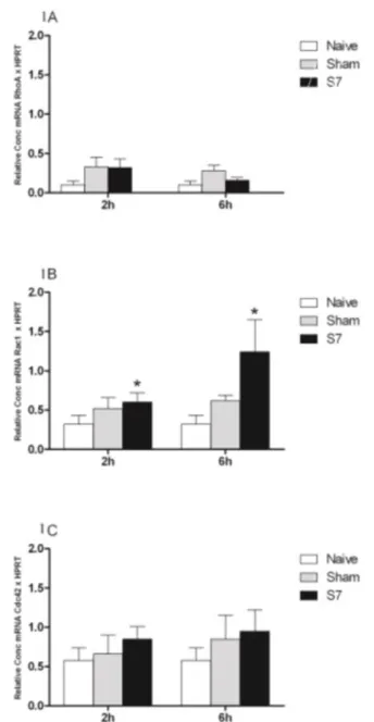
Introduction:Myeloid-derived suppressor cells (MDSCs) constitute a heterogeneous population of immature myeloid cells that potently suppress immune responses. They were originally identified in cancer patients and have since been reported to occur also in chronic inflammation, autoimmunity and even bacterial infections. Human MDSCs are commonly divided into monocytic (Mo-MDSCs) and granulocytic (PMN-MDSCs) subtypes. To what extent thebona fidecancer MDSCs are representative of the proposed MDSCs found in other diseases is not well known. PMN-MDSCs have previously been found to be enriched among low-density granulocytes (LDGs) in density gradient centrifuged blood.
Methods:In this study we analyzed potential MDSCs in sepsis patients with different causative microorganisms, using total peripheral blood as compared to density gradient centrifuged blood.
Results:We found a high frequency of typical CD14+HLADRlow Mo- MDSCs in all sepsis patients, whereas the typical PMN-MDSCs as well as a prominent CD14lowPMN-MDSC-like population appeared preferentially in Gram-positive cases (Figures 1 to 3). The CD14lowPMN-MDSC variant was demonstrated to suppress T-cell proliferationin vitrovia a ROS-dependent
mechanism, to display an increased IL-10:TNFaratio, and to present with signs of immaturity: blast morphology and low cytokine levels (Figures 4 and 5).
Conclusion:We conclude that a spectrum of cells with MDSC features are enriched in sepsis, and that microbial origin of sepsis contributes to the substantial interindividual patient variation in MDSC pattern.
P4
Selective decontamination using antibiotics in ICU patients:
counterfactual protection versus contextual hazard toward bacteremia incidences.
JC Hurley
Melbourne Medical School, University of Melbourne and Ballarat Health Services, Ballarat, Australia
Critical Care2014,18(Suppl 2):P4; doi:10.1186/cc14007
Introduction:Among methods for preventing pneumonia and possibly also bacteremia in ICU patients, selective digestive decontamination (SDD;
topical with or without protocolized parenteral antibiotic) appears most effective within randomized concurrent controlled trials (RCCTs) [1].
However, whether parenteral antibiotic is required, and whether SDD actually increases pneumonia incidences in SDD RCCTs versus the broader ICU pneumonia evidence base, remain unresolved [2,3]. The purpose of this analysis is to test for counterfactual and contextual effects of the topical and parenteral SDD components on the bacteremia incidence versus the broader evidence base related to the patient group at risk of VAP.
Methods:Bacteremia incidence proportion data were extracted from component (control and intervention) groups from studies investigating antibiotic (SDD) or nonantibiotic methods of VAP prevention. Both the counterfactual and the contextual effects of SDD factorized as topical or protocolized parenteral exposures were estimated using random-effects meta-analysis of study and group level data. Studies without any prevention methods under study constituted the reference category (benchmark groups).
Results:As a counterfactual within RCCTs, SDD when given as combined topical and parenteral antibiotic appears to halve the bacteremia incidence (odds ratio (OR) 0.59; 0.48 to 0.73;n= 18 studies). As a contextual however, the mean bacteremia incidence among 27 control groups (17.1%; 13.1 to 22.1%) and 12 intervention groups receiving topical antibiotic alone (16.2%; 9.1 to 27.3%) from SDD RCCTs is double that of 36 benchmark groups (8.3; 7.0 to 10.8%) and 19 control groups from studies of nonantibiotic methods (7.7%; 5.2 to 11.1). The upward dispersion in bacteremia incidence among component groups from SDD RCCTs away from this benchmark is striking with all but two of the 27 control groups and all but two of 12 SDD intervention groups that did not receive PPAP being above this benchmark.
Conclusion:The major contextual hazard of SDD toward bacteremia among ICU patients is inapparent within individual studies. The apparent protection in SDD RCCTs is spurious as the SDD counterfactual is conflated by the strong contextual effect with partial mitigation by SDD Figure 1(abstract P3)
Figure 2(abstract P3)
Figure 3(abstract P3)
Figure 4(abstract P3)
Figure 5(abstract P3)
protocolized parenteral antibiotic. Not only is the safety of SDD within the ICU environment unclear, but this SDD contextual effect may conflate the apparent SDD counterfactual effect on the incidence of bacteremia, as with VAP.
References
1. Hurley JC:Prophylaxis with enteral antibiotics in ventilated patients:
selective decontamination or selective cross-infection?Antimicrob Agents Chemother1995,39:941-947.
2. Hurley JC:Ventilator associated pneumonia prevention methods using topical antibiotics: herd protection or herd peril?Chest2014 in press.
3. Hurley JC:The perfidious effect of topical placebo: Calibration of Staphylococcus aureus ventilator-associated pneumonia incidence within selective digestive decontamination studies versus the broader evidence base.Antimicrob Agents Chemother2013,57:4524-4531.
P5
Thalidomide exerts protective immunomodulatory action during Klebsiella pneumoniaeB5055-induced acute lung infection in BALB/c mice.
V Kumar*, S Chhibber
Department of Microbiology, Panjab University, Chandigarh, India Critical Care2014,18(Suppl 2):P5; doi:10.1186/cc14008
Introduction:Thalidomide (a-naphthylimidoglutarimide), a psychoactive drug that readily crosses the blood-brain barrier, has been shown to exhibit anti-inflammatory, anti-angiogenic, immunomodulatory properties through a mechanism that is not fully established. Keeping these properties in mind, we have tried to find out the anti-inflammatory and immunomodulatory properties of thalidomide in mouse model of acute inflammation by introducingKlebsiella pneumoniaeB5055 in BALB/c mice via the intranasal route.
Methods:Acute lung infection (ALI) or pneumonia in BALB/c mice was induced via instillation of selected dose (104CFU/ml) of bacteria (that is, K. pneumoniaeB5055) intranasally. Mice were observed for 7 days and lungs were isolated on designated days for studying difference in bacterial load and other proinflammatory mediators using standard biochemical methods and ELISA.
Results:The intranasal instillation of bacteria in this mouse model of acute pneumonia-induced inflammation led to significant increase in neutrophil infiltration into the lungs. This was further accompanied by an increased production of proinflammatory cytokines (that is, TNFaand IL-1a) and other mediators of inflammation (that is, malondialdehyde (MDA), myeloperoxidase (MPO) and nitric oxide (NO)) in the lung tissue.
The animals, which received thalidomide alone orally or in combination with augmentin, 30 minutes prior to bacterial instillation into the lungs
via intranasal route, showed significant (P≤0.05) decrease in neutrophil influx into the lungs. A significant (P≤0.05) decrease in the production of proinflammatory cytokines (that is, TNFa and IL-1a) and other biochemical mediators of acute inflammation (that is, MDA, MPO, and NO) was also observed in this group. But the augmentin treatment alone did not decrease these proinflammatory mediators significantly (P≥0.05) as compared to the control group.
Conclusion:We therefore conclude that thalidomide ameliorates lung inflammation induced byK. pneumoniaeB5055 without significantly (P≤0.05) decreasing the bacterial load in the lung tissue whereas augmentin takes care of bacterial proliferation. Hence, it can be used as an adjunct therapy along with antibiotics as an anti-inflammatory or an immunomodulatory agent in case of acute lung infection or pneumonia.
P6
Impact of purulent complications and sepsis on cardiovascular system in patients with type 2 diabetes.
E Shalaeva*, B Babadjanov, U Pulatov, N Dadabayeva, A Shalaeva Republican Center of Purulent Surgery and Complications of Diabetes, Tashkent Medical Academy, Tashkent, Uzbekistan
Critical Care2014,18(Suppl 2):P6; doi:10.1186/cc14009
Introduction:Purulent complications in patients with type 2 diabetes are usually severe, often complicated by sepsis and require emergency surgery. Noncardiac surgery is associated with a 7 to 11% complication rate and mortality of 0.8 to 1.5% [1], up to 42% are cardiac reasons [2].
After surgery, 2% of patients suffer major cardiac complications [3], and 8% show evidence of significant myocardial injury [2]. The aim of this study was to identify the impact of purulent complications and sepsis on cardiovascular system in patients with type 2 diabetes.
Methods:We analyzed 112 consecutive patients (54 men and 58 women) aged 57.2 ± 8.4 years with purulent-necrotic complications (gangrene, phlegmon, and abscess) of type 2 diabetes and sepsis in 2013. We compared laboratory and instrumental data (blood tests, ECG, echocardiography and others), which were previously obtained in the same patients receiving inpatient treatment before sepsis (2011 to 2012).
Results:Gangrene of lower extremities in 59 (52.7%) prevailed among purulent complications. After the development of sepsis we detected in all patients significantly increased heart rate, respiratory rate per minute, leukocytosis, anemia, worse glucose metabolism and renal function (Table 1).
Congestive heart failure became more severe. This was confirmed by decrease of left ventricle ejection fraction (55.2 ± 5.1% before sepsis vs.
49.3 ± 4.1% after) and increase brain natriuretic peptide (291.4 ± 34.5 ng/ml vs. 395.2 ± 28.1 ng/ml,P< 0.001). Prior sepsis in 66 (58.9%) of patients with arterial hypertension was observed, after in 88 (78.6%). After admission to
Table 1(abstract P6) Hemodynamic parameters and blood tests in patients with purulent complications of type 2 diabetes and sepsis
Parameter Before sepsis (n= 112) After sepsis (n= 112) Pvalue
Heart rate (beats/minute) 78.4 ± 15.2 112.5 ± 18.9 < 0.001
Respiratory rate (breaths/minute) 18.0 ± 2.0 29.5 ± 5.5 < 0.001
Systolic BP (mmHg) 155.7 ± 35.4 154.2 ± 58.5 n.s
Diastolic BP (mmHg) 90.4 ± 10.3 91.9 ± 8.6 n.s
Left ventricle ejection fraction (%) 55.2 ± 5.1 49.3 ± 4.1 0.033
Fasting plasma glucose (mmol/l) 8.4 ± 2.5 15.4 ± 4.8 < 0.001
Two-hour plasma glucose (mmol/l) 10.2 ± 2,8 19.9 ± 3.3 < 0.001
HbA1c (%) 8.4 ± 0.5 12.1 ± 0.5 < 0.001
Hemoglobin (g/l) 121.5 ± 12.5 105.4 ± 11.7 0.04
White count (103) 6.7 ± 1.2 14.4 ± 2.1 < 0.001
Fibrinogen (mg%) 411.6 ± 103.6 715.4 ± 215.5 < 0.001
Blood urea (mmol/l) 6.1 ± 2.9 8.8 ± 2.5 0.011
Blood creatinine (mmol/l) 88.4 ± 18.5 105.6 ± 17.3 0.02
Brain natriuretic peptide (ng/ml) 291.4 ± 34.5 395.2 ± 28.1 < 0.001
BP, blood pressure; HbA1c, glycosylated hemoglobin A1c
the centre, patients had no signs of septic shock. In 13 (11.6%) patients, the perioperative period was complicated by acute myocardial infarction, which was accompanied by a fall in blood pressure. We detected an increase of the functional class of stable angina, congestive heart failure, 4.2 times increased incidence of unstable angina, 2.6 times ventricular and four times supraventricular extra systole after septic complications (Table 2).
Conclusion:After the development of purulent complications and sepsis in patients with type 2 diabetes, we observed increased incidence of arterial hypertension, arrhythmias, worsened severity of coronary artery disease and congestive heart failure. Perioperative risk of acute myocardial infarction amounted to 11.6%.
References
1. Haynes AB, Weiser TG,et al:A surgical safety checklist to reduce morbidity and mortality in a global population.N Engl J Med2009, 360:491-499.
2. Devereaux PJ, Chan MT,et al:Association between post-operative troponin levels and 30-day mortality among patients undergoing noncardiac surgery.JAMA2012,307:2295-2304.
3. Devereaux PJ, Goldman L,et al:Perioperative cardiac events in patients undergoing noncardiac surgery: a review of the magnitude of the problem, the pathophysiology of the events and methods to estimate and communicate risk.CMAJ2005,173:627-634.
P7
Severity of sepsis in patients with acute purulent destructive pulmonary disease depending on the presence of type 2 diabetes:
impact on the forecast.
A Babobekov*, B Babadjanov, S Atakov, E Shalaeva
Republican Center of Purulent Surgery and Complications of Diabetes, Tashkent Medical Academy, Tashkent, Uzbekistan
Critical Care2014,18(Suppl 2):P7; doi:10.1186/cc14010
Introduction:Lung abscesses and gangrene are the most severe clinical manifestation and outcome among acute purulent destructive pulmonary disease (APDPD). Mortality ranges from 10 to 35%, and in the presence of diabetes increases up to 30 to 90% [1]. The main reason for this is the generalization of infection (sepsis), leading to the development of multiple organ failure [2,3]. The aim of this study was to identify the severity of sepsis in patients with APDPD depending on the presence of type 2 diabetes, and the impact on the forecast.
Methods:During the period 2012 to 2013, we examined 408 patients aged 48.5 ± 12.5 years (258 men/150 women) who underwent surgical treatment for APDPD. The patients were divided into two groups: 144 patients with type 2 diabetes, and controls (n = 246). We carried out computed tomography, ECG, echocardiography, laboratory biochemical testing, and bacteriological analysis of pathologic material and blood samples.
Results:Patients with type 2 diabetes had much more complications and cases of severe sepsis and septic shock (Table 1). Bacteriological analysis of the pathologic material showed Gram-positive bacteria in 35 to 45%, anaerobic association in 55 to 65%, pathological fungi in 50 to 60%. The patients with type 2 diabetes had much more time from the onset of the first symptoms of lung disease prior to admission (12.5 ± 3.5 vs. 7.5 ± 2.5 days,P= 0.002), and the duration of inpatient treatment was significantly longer (13.8 ± 5.5 vs. 7.1 ± 3.4 days,P= 0.001). Only 53 (36.8%) of patients with type 2 diabetes and 68 (29.5%) without it had bacteriological positive blood culture. The analysis of the distribution of pathogens in groups is presented in Figure 1. Patients with diabetes had moreCandidaspp.
Table 2(abstract P6) Cardiovascular comorbidity in patients with type 2 diabetes before and after purulent- necrotic complications and sepsis
Parameter Before
sepsis (n= 112)
After sepsis (n= 112)
Insulin dependence 42 (37.5) 112 (100)
Normal blood pressure (110 to 139 mmHg)
46 (41.1) 11 (9.8)
Arterial hypertension 66 (58.9) 88 (78.6)
First degree (140 to 159 mmHg) 33 (29.5) 21 (8.9) Second degree (160 to 179 mmHg) 21 (18.6) 43 (38.4) Third degree (>180 mmHg) 12 (10.7) 24 (21.4)
Arterial hypotension (<90 mmHg) - 13 (11.6)
CAD, stable angina 108 (94.6) 82 (73.2)
FC I 18 (16.1) -
FC II 29 (25.9) 18 (16.1)
FC III 52 (46.4) 45 (40.2)
FC IV 9 (8.0) 19 (17.0)
CAD, unstable angina 4 (3.6) 17 (15.2)
Acute myocardial infarction - 13 (11.6)
Postinfarction cardiosclerosis 7 (6.3) 7 (6.3)
Atrial fibrillation 7 (6.3) 7 (6.3)
Supraventricular arrhythmia 3 (2.7) 12 (10.7)
Ventricular arrhythmia 14 (12.5) 36 (32.1)
Congestive heart failure 112 (100) 112 (100)
FC II (NYHA) 76 (67.8) 26 (23.2)
FC III (NYHA) 36 (32.1) 65 (58)
FC IV (NYHA) - 21 (18.7)
Abscesses of the lower extremity - 22 (19.6)
Phlegmon of the lower extremity - 31 (27.7)
Gangrene of lower extremity - 59 (52.7)
Data presented asn(%). CAD, coronary artery disease; FC, functional class;
NYHA, New York Heart Association
Table 1(abstract P7) Clinical symptoms and severity of sepsis in patients with acute purulent destructive pulmonary disease depending on the presence of type 2 diabetes
Data Type 2
diabetes (n= 144)
Control (n= 264)
Acute lung abscess 59 (40.9) 122 (46.2)
Necrotizing pneumonia 47 (32.6) 98 (37.1)
Lung gangrene 38 (26.4) 44 (16.7)
Empyema 88 (61.1) 81 (30.7)
Pyopneumothorax 16 (11.1) 9 (3.4)
Mediastinitis 34 (23.6) 16 (6.1)
Body temperature >38°C/<36°C 98 (68.1)/21 (14.6)
261 (98.9)/3 (1.1) Respiratory rate >20/minute 144 (100) 264 (100) Heart rate >90 beats/minute 138 (95.8) 242 (91.7)
PaCO2<32 mmHg 144 (100) 264 (100)
Leukocytes >12,000/<4,000 cells/
mm3
111 (77.1)/13 (9.1)
202 (76.5)/11 (4.2)
Renal failure, oliguria 42 (29.2) 34 (12.9)
Increase liver enzymes 34 (23.6) 45 (17.1)
Systolic blood pressure <90 mmHg 33 (22.9) 51 (19.3)
Sepsis 101 (70.1) 223 (84.5)
Severe sepsis 25 (17.4) 29 (11.0)
Septic shock 18 (12.5) 12 (4.5)
Data presented asn(%)
(Figure 1). Figures 2, Figure 3 and Figure 4 present the X-ray dynamics of a 42-year-old man with lung abscess. Clinical recovery in patients with type 2 diabetes was significantly worthy compared to controls (45 (31.2%) vs.
153 (57.9%)), mortality rate 48 (33.3%) versus 39 (14.7%), respectively.
Conclusion:In patients with acute purulent destructive pulmonary disease and type 2 diabetes, severe sepsis and septic shock more often prevailed, inpatient mortality rate was 2.27 times higher, compared to patients with normal glucose metabolism.
References
1. Defraigne JO,et al:Cavernostomy: an old but effective technique in the treatment of pulmonary abscess.Rev Med Liege2007,52:498-501.
2. Refaely J, Weissberg D:Gangrene of the lung: treatment in two stages.
Ann Thorac Surg1997,64:970-973.
3. Rice TW, Ginsberg PJ, Todd TR:Tube drainage of lung abscesses.Ann Thorac Surg1987,44:356-359.
P8
Risk factors and incidence of mediastinitis in patients with lung abscess and sepsis.
E Shalaeva*, B Babadjanov, B Janabaev, A Shalaeva
Republican Center of Purulent Surgery and Complications of Diabetes, Tashkent Medical Academy, Tashkent, Uzbekistan
Critical Care2014,18(Suppl 2):P8; doi:10.1186/cc14011
Introduction:Mediastinitis is a life-threatening condition, which is accompanied by high rates of mortality in cases of delayed diagnosis and inadequate treatment. The aim of the study was to identify the risk factors and incidence of mediastinitis in patients with lung abscess and sepsis.
Methods:In 2013, 218 consecutive patients (83 women and 135 men) with lung abscess and sepsis aged 45.8 ± 13.2 years were operated. They had a full range of laboratory and instrumental examinations, including echocardiography and computed tomography.
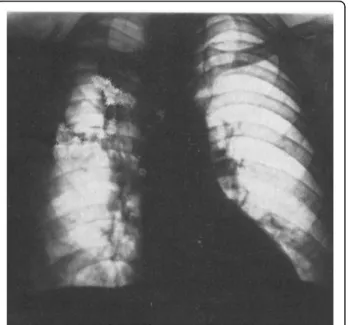
Results:Aerobic-anaerobic association in sputum was revealed in all patients with lung abscess and sepsis,Candidaspp. in 34 (15.6%). Blood culture was positive only in 59 (27%) patients, which had not previously received antibacterial therapy (polymicrobial flora includingStaphylococcus andStreptococcusspecimen). Empyema was diagnosed in 123 patients (56.4%), 31 (14.2%) of them were complicated by mediastinitis. The main clinical symptoms of mediastinitis were hyperthermia (100%), dysphagia (83.8%), dyspnea (80.6%), chest pain (61.3%), orthopnea Figure 1(abstract P7)
Figure 2(abstract P7) Man, 42 years old, with right lung abscess after admission to the centre
Figure 3(abstract P7) The same patient on the fourth day after drainage of the abscess
Figure 4(abstract P7) The same patient before discharge, 14 days in dynamics
(61.3%), and tachycardia (87.1%). The computer tomography revealed an increase in mediastinum size with accumulation of fluids and fluid in the pleural cavities (100%), free gas in the mediastinum (45.1%), enlarged mediastinal lymph nodes (45.1%), and fluid in the pericardium cavity (48.4%). To analyze the risk factors, we include 31 patients with lung abscess and sepsis complicated by mediastinitis in the first group, and 187 patients without mediastenitis in the second group. Groups were similar in age (46.1 ± 8.2 years vs. 45.8 ± 13.2 years). A total 77.4% of patients with mediastinitis were women who suffered from type 2 diabetes (HbA1c = 9.7 ± 1.4%), congestive heart failure and anemia. Significant differences in the groups according to the data of laboratory and instrumental studies are presented in Table 1.
Conclusion:In total, 14.2% of patients presented with lung abscess and sepsis complicated by mediastinitis, more commonly in women with diabetes mellitus, obesity, anemia and reduced ejection fraction of the left ventricle.
P9
Impact of KDO in biological activity of Re-LPS.
I Prokhorenko*, S Zubova, D Kabanov, S Grachev
Institute of Basic Biological Problems, Pushchino, Moscow Region, Russia Critical Care2014,18(Suppl 2):P9; doi:10.1186/cc14012
Introduction:The minimal biological active structure of endotoxins (lipopolysaccharides (LPS)) is Re-LPS (KDO2-lipid A), which consist of lipid A and two (or three) molecules of 3-deoxy-D-manno-2-octulosonic acid (KDO) [1,2]. Biological activity of endotoxins is defined in general by the number and distribution of acyl residues on the lipid A backbone [3].
Recently it has been reported that KDO-treated RAW 264.7 cells exhibited a gene expression pattern similar to that in LPS-treated cells. These authors revealed that free KDO participated in crosstalk between Toll-like receptors (TLR) and G protein-coupled receptors and so that regulated activators and repressors of immune signaling [4]. LPS-dependent TLR4-triggered activation of target cells leads to specific changes in the levels of surface receptors and induces synthesis of proinflammatory cytokines [5]. However, the dependence of these processes on the structural composition of LPS is not well understood. To extend our knowledge in this field, the effects of free KDO as well as KDO as covalently linked to lipid A constituent of Re-LPS on expression of TLR4, CD11b and CD14 receptors and TNFasynthesis in whole human blood have been investigated.
Methods:Human blood was incubated with Re-LPS fromEscherichia coli JM103 orSalmonella enterica sv TyphimuriumSL1181 (100 ng/ml) or with lipids A fromE. coliF583 orS. entericasv Minnesota R595 (80 ng/ml) or with ammonium salt of KDO (20 ng/ml) at 37°C in 5% CO2-humidified atmosphere for 2 or 6 hours to determine receptor expression or TNFa release, respectively. Receptor expression was monitored by EPICS XL-MCL flow cytometer using Alexa Fluor 488 anti-TLR4 (HTA125), anti-CD11b (ICRF44) and anti-CD14 (HCD14) antibodies. Human TNF-a ELISA Kit II was exploited to TNFadetermination.
Results:Re-LPSE. colior Re-LPSS. entericadifferentially affected receptor expression in comparison to their respective lipids A. Free KDO in the equimolar concentration as it exists in KDO2-lipid A (Re-LPS) did not influence the level of CD14 but downregulated the expression of TLR4 and CD11b (Figure 1). Tenfold increased KDO concentration did not affect further the receptor expression. The addition of KDO2 to lipid AE. coli- that
is, applying KDO as covalently linked constituent of Re-LPS - led to upregulation of CD14 and TLR4 but downregulated CD11b expression. The expression of TLR4 was most pronounced upregulated by Re-LPSS. enterica but in the case of CD14 and CD11b this Re-LPS had an opposite effect in comparison toE. coliendotoxins (table in Figure 1). Lipid AS. entericawas a less potent TNFainductor than that fromE. coli(Figure 2). This may be explained by the differences in lipid A composition determining lipid A affinity to target receptor(s). LPSE. coli, as had been shown early, caused MyD88-dependent fast NF-B degradation (rapid TNFaresponse) whereas LPSS. entericainduced MyD88-independent signaling (delayed TNFa response) [5]. In our study, free KDO did not stimulate TNFarelease. KDO2 as a constituent of Re-LPSS. entericaincreased significantly the TNFa- inducing activity of lipid AS. entericabut this effect was not so distinguished between Re-LPSE. coliand lipid AE. coli(Figure 2).
Conclusion: Free KDO in the used concentration was inactive in regulation of TLR4, CD11b and CD14 expression and did not induce TNFa release but its impact in biological activity was detected when KDO was applied as constituent of Re-LPS. This may be explained by the effect of KDO on the spatial conformation of Re-LPS.
Acknowledgements:The work was supported by Grant 16.N08.12.1014 established by the Russian Ministry of Education and Science
References
1. Olsthoorn M, Peterson B, Schlecht S, Haverkamp J, Bock K, Thomas-Oates J, Holst O:Identification of a novel core type in Salmonella
lipopolysaccharide.J Biol Chem1998,273:3817-3829.
2. Fregolino E, Fugazza G, Galano E, Gargiulo V, Landini P, Lanzetta R, Lindner B, Pagani L, Parrilli M, Holst O,et al:Complete lipooligosaccharide structure of the clinical isolate Acinetobacter baumannii, strain SMAL.
Eur J Org Chem2010,7:1345-1352.
3. Rietschel E, Kirikae T, Schade U, Mamat U, Schmidt G, Loppnow H, Ulmer A, Zahringer U, Seydel U, Di Padova F,et al:Bacterial endotoxin: molecular Table 1(abstract P8) Risk factors for mediastinitis in patients with lung abscess and sepsis
With mediastinitis (n= 31) Without mediastinitis (n= 187) Pvalue
Gender, male/female,n(%) 7 (22.6)/24 (77.4) 128 (68.5)/59 (31.5) 0.001
Type 2 diabetes mellitus,n(%) 26 (83.9) 42 (22.5) 0.001
Body mass index 32.3 ± 5.3 27.8 ± 6.1 0.031
Hemoglobin (g/l) 87.9 ± 9.2 128.4 ± 18.4 < 0.001
Fibrinogen (mg%) 800 ± 200 533 ± 166 < 0.001
End-diastolic volume of the left ventricle (ml) 139 ± 27 108 ± 28 0.024
End-systolic volume of the left ventricle (ml) 66.1 ± 8.2 46.8 ± 10.4 < 0.001
Left ventricular ejection fraction (%) 52.4 ± 2.2 57.2 ± 3.4 0.002
Figure 1(abstract P9) Expression of TLR4, CD11b and CD14 on monocytes after incubation of whole blood with Re-LPS, lipid A or KDO. Presented are the results of six independent experiments.
Alteration in receptor expression was calculated according to the control level that had been expressed as 100%. *Changes in receptor expression were calculated as %MnIX [KDO2-lipid A] - %MnIX [lipid A]
relationships of structure to activity and function.FASEB J1994, 8:217-225.
4. Krishnan J, Choi S:Systems biological approaches reveal non-additive responses and multiple crosstalk mechanisms between TLR and GPCR signaling.Genomics Inform2012,10:153-166.
5. Zughaier S, Zimmer S, Datta A, Carlson R, Stephens D:Differential induction of Toll-like receptor 4-MyD88-dependent and -independent signaling pathways by endotoxins.Infect Immun2005,73:2940-2950.
P10
Two novel formulae are superior to procalcitonin for prediction of sepsis in trauma patients.
H-p Liang*, H Jin, Y Xiao, Z Liu
State Key Laboratory of Trauma, Burns and Combined Injury, Research Institute of Surgery, The Third Military Medical University, Chongqing, People’s Republic of China
Critical Care2014,18(Suppl 2):P10; doi:10.1186/cc14013
Introduction:The purpose of this study was to verify the predictive value of two novel formulae and compare them with that of procalcitonin (PCT) for predicting sepsis in trauma patients.
Methods:We performed a retrospective study of trauma patients treated at Daping Hospital in Chongqing, China and Affiliated Hospital of Zunyi Medical College between 2010 and 2013. Patients≥16 years old, admitted to hospital after injury within 24 hours and with length of hospital stay≥48 hours were included. Predictive ability of two formulae based on LD50values of the Injury Severity Score (ISS) and New Injury Severity Score (NISS) were verified: ISS/LD50ISS+SIRS score and NISS/LD50NISS+SIRS score, and then were compared with the most common used biomarker PCT. LD50values for different age groups and genders were obtained in our former study. The statistical performance of the two formulae and PCT to predict sepsis was evaluated using receiver operating characteristic (ROC) curve analysis.
Results:Two hundred and twenty-one trauma patients’data were enrolled in the study, including 44 females and 177 males. The average age of the patients was 44.77 ± 15.01 years. The performance of the ISS/LD50ISS+SIRS score and the NISS/LD50NISS+SIRS score was equivalent (area under the ROC curve (AUC) = 0.816 vs. 0.819,P>0.05) and both performed better than PCT (AUC = 0.592,P< 0.05) in predicting posttraumatic sepsis. For the ISS/
LD50ISS+SIRS score, the cutoff value was 2.38, with a positive predictive value of 78.08%, a negative predictive value of 81.33%, a sensitivity of 89.06%, a specificity of 65.59%, a positive likelihood ratio of 2.59, a negative likelihood ratio of 0.17, a Youden index of 0.5465, an odds ratio of 15.52, and an accuracy of 79.19%. For the NISS/LD50NISS+SIRS score, the cutoff value was 2.4677, with a positive predictive value of 79.70%, a negative predictive value of 75.00%, a sensitivity of 82.81%, a specificity of 70.97%, a positive likelihood ratio of 2.85, a negative likelihood ratio of 0.24, a Youden index of 0.5378, an odds ratio of 11.78, and an accuracy of 77.83%.
Conclusion:The two novel formulae ISS/LD50ISS+SIRS score and NISS/
LD50NISS+SIRS score performed well and were both better than PCT in predicting sepsis post trauma. The value of the two formulae can be easily calculated in real time and can identify the high-risk patients susceptible to sepsis. This method may become an effective way to guide the early assessment and treatment in trauma patients.
P11
Inhibitory effects of evodiamine on zymosan-induced inflammation:
inactivation of NF-B by inhibiting IBaphosphorylation.
X Fan*, J-y Zhu, Z Liu, J Yan, Q Ma, H-p Liang
State Key Laboratory of Trauma, Burns and Combined Injury, Research Institute of Surgery, The Third Military Medical University, Chongqing, People’s Republic of China
Critical Care2014,18(Suppl 2):P11; doi:10.1186/cc14014
Introduction:Inflammation is a host defense reaction against pathogenic infection. In this process, inflammatory cytokines contribute to combat against infection, but excess cytokines will lead to tissue damage.
Nonsteroidal anti-inflammatory drugs and corticosteroids are commonly used for regulating these inflammatory mediators and treatment of inflammatory disorders. But these drugs are not sufficient for clinical practice due to their adverse effects, so new anti-inflammatory drugs are still needed. Evodiamine (EVO), an important alkaloidal component extracted from the fruit of Evodiae fructus, possesses the property of analgesia, antiemesis, and vascular dilatation. Its anti-inflammatory effect and underlying mechanism were investigated using a zymosan-induced inflammatory model.
Methods:In vitro, RAW264.7 cells and primary peritoneal macrophages were treated with different doses of EVO (25, 50 or 100μM) for 1 hour prior to incubation with zymosan (100μg/ml), and the effects of EVO on protein and mRNA levels of proinflammatory cytokines were determined by ELISA and qRT-PCR, respectively.In vivo, peritonitis was induced in C57BL/6 mice by zymosan (500 mg/kg) injection, and the effects of EVO (10 mg/kg) on plasma cytokine levels and organ injury were evaluated.
Activation of the NF-B signal pathway was investigated by ELISA-based Trans-AM transcription factor NF-B p65 kit, immunocytochemistry and western blotting.
Results:EVO effectively suppressed the expression of IL-1b, IL-6 as well as TNFain both protein and mRNA levelin vitro. It can also attenuate zymosan-induced DNA-binding activity of NF-B, which was achieved through the inhibitory effects on the phosphorylation of inhibitory kappa Ba(IBa) and p65 nuclear translocation, but there was little association with mitogen-activated protein kinase activation.In vivo, treatment with EVO could ameliorate inflammatory cell infiltration and vascular ectasia induced by zymosan in both lung and intestine tissues. EVO can markedly decrease the level of TNFaand IL-6 in plasma, and effectively downregulate expression of IL-6, TNFaand myeloperoxidase in both lung and intestine. Moreover, cell apoptosis in organs was also attenuated by treatment of EVO. The underlying mechanism that a decrease in the phosphorylation of IBaand the subsequent transcription activity of NF-B was also confirmed.
Conclusion:Taken together, our data demonstrate that EVO displays anti- inflammatory actionsin vitroandin vivoby suppressing the phosphorylation of IBaand inactivating NF-B, which suggests that EVO is a potential therapeutic agent against inflammatory disorder.
P12
Imaging in severe sepsis and septic shock: is early radiological identification of occult sources of infection needed?.
A Creamer*, J Keep
Emergency Department, Kings College Hospital, London, UK Critical Care2014,18(Suppl 2):P12; doi:10.1186/cc14015
Introduction:The importance of imaging in establishing the focus of infection is recognised in current guidelines for the management of severe sepsis [1], with decisions regarding timing and modality of imaging left to the physicians’clinical judgement. In the emergency department (ED), clinical assessment combined with bedside investigations of chest X-ray Figure 2(abstract P9) Production of TNFaafter incubation of whole
blood with lipid A or Re-LPS
(CXR) and urine dip can be used to confirm the two most common sources [2]. However, they may fail to identify occult sources of infection, such as intraabdominal collections and abscesses, the treatment of which may require alteration of empirical treatment or be refractory to antibiotic therapy alone. Further imaging is necessary to confirm the focus so that optimal treatment can be achieved.
Methods:The study cohort was composed of 50 consecutive patients who met the criteria for severe sepsis [1] attending the ED in 2013. Electronic and paper patient records and radiology results were analysed. All radiological studies done in the first 72 hours following attendance were included in the study.
Results:CXR was performed as the initial investigation in 49 of the 50 patients (98%). The median time from arrival at the ED to initial imaging was 1 hour:00 minutes (range 0 hours:04 minutes to 4 hours:25 minutes). Initial investigations in the ED of CXR and urine dip identified a septic focus in 30 of 50 patients (60%). Fourteen of the remaining 20 went on to have one or more further imaging studies. Figure 1 outlines the second-line and third- line radiological investigations performed (number where a septic focus was identified in parentheses). The median time to first positive imaging was 0 hours: 50 minutes (range 0 hours:04 minutes to 40 hours), with the source remaining unidentified by imaging and urine testing in 15 of the 50 patients.
Conclusion:Our results indicate that simple bedside investigations are able to identify a focus of infection in 60% of patients presenting to the ED with severe sepsis. Our results support the continued use of CXR as the initial imaging modality in severe sepsis, but also demonstrate the benefit of further imaging in confirming the focus of infection and to guide definitive treatment. Instances where further imaging was delayed by several days highlight the need for guidelines detailing which investigations should be done and in what time frame.
References
1. Dellinger RP, Levy MM, Rhodes A, Annane D, Gerlach H, Opal SM,et al:
Surviving Sepsis Campaign: international guidelines for management of severe sepsis and septic shock, 2012.Intensive Care Med2013,39:165-228.
2. Perman SM, Goyal M, Gaieski DF:Initial emergency department diagnosis and management of adult patients with severe sepsis and septic shock.
Scand J Trauma Resusc Emergency Med2012,20:41.
P13
Proinflammatory versus anti-inflammatory response in sepsis patients:
looking at the cytokines.
D Anand1*, S Ray2, S Bhargava1, S Das1, A Garg2, S Taneja2, D Dhar2, L Mohan Srivastava1
1Department of Biochemistry and Critical Care Medicine, Sir Ganga Ram Hospital, New Delhi, India;2Department of Critical Care and Emergency Medicine, Sir Ganga Ram Hospital, New Delhi, India
Critical Care2014,18(Suppl 2):P13; doi:10.1186/cc14016
Introduction:Despite improvements in supportive care, mortality rates in sepsis remain substantially high. Sepsis exhibits phases of enhanced inflammation, alternating with immune suppression with a resultant variant time point of mortality; yet no human study has correlated levels of cytokines to the timeline of mortality. Our study attempts to analyze the association of levels of proinflammatory and anti-inflammatory cytokines in sepsis with the timeline of death in terms of early (<5 days) versus late (>5 days) mortality, and day of death. We also assessed correlation of these cytokines with length of stay.
Methods:The study protocol was approved by Institutional Ethics Committee. Subjects were 74 consecutive patients with community- acquired severe sepsis/septic shock admitted to the ICU of a tertiary care superspeciality hospital. Blood samples drawn on days 1, 3 and 7 of admission were analysed for proinflammatory cytokine (TNFa) by chemiluminescent immunometric assay and anti-inflammatory cytokine (IL-10) by ELISA. Subjects were segregated on basis of: ratio of proinflammatory and anti-inflammatory mediators on day 1 of admission into patients with predominant proinflammatory or predominant anti-inflammatory response.
Survival and time point of mortality into survivor, early mortality and late mortality groups. Statistical analyses were performed using SPSS version 17.
Results:There were 37 patients each in predominant proinflammatory and predominant anti-inflammatory groups. The number of deaths was 11 and 17 respectively in these groups. However, there was significantly higher early mortality in the proinflammatory group as compared to the anti- inflammatory group (7 vs. 1,P= 0.0247). On the contrary, late deaths were significantly higher in the anti-inflammatory group as compared to the proinflammatory (16 vs. 4P= 0.0017). The ratio of proinflammatory/anti- inflammatory cytokines (TNF/IL-10) on day 1 was significantly lower in patients of late death (n= 20) as compared to patients of early death (n= 8) and survivors (n= 46) as shown in Table 1. Further, the median day of death Figure 1(abstract P12) Initial and further investigations performed (number where a septic focus was identified in parentheses)
was significantly delayed in patients in the anti-inflammatory as compared to the proinflammatory group (20 vs. 5,P< 0.001). Length of hospital stay amongst survivors was significantly longer in the anti-inflammatory as compared to the proinflammatory group (23 vs. 10P< 0.001).
Conclusion:Our preliminary data suggest that in sepsis, the ratio of proinflammatory/anti-inflammatory cytokines on day 1 is significantly associated with time point of mortality; hence, this ratio can be used to particularize management. Further studies are in progress to substantiate the role of proinflammatory and anti-inflammatory cytokines in this subset of patients. Moreover, since predominant anti-inflammatory response was associated with later death, role of immunomodulators in sepsis needs to be explored.
P14
Understanding heterogeneity in the host response toStaphylococcus aureusinfection for prognostic biomarker discovery.
JB Dinoso1*, J Gutierrez2, DF Choy1, S Kummerfeld3, A Baruch2, HF Chambers4, CM Rosenberger1
1ITGR Diagnostic Discovery, Genentech Inc., South San Francisco, CA, USA;
2Pharmacodynamic Biomarkers, Genentech Inc., South San Francisco, CA, USA;
3Bioinformatics, Genentech Inc., South San Francisco, CA, USA;4Division of Infectious Disease, University of California San Francisco, San Francisco, CA, USA Critical Care2014,18(Suppl 2):P14; doi:10.1186/cc14017
Introduction:InvasiveStaphylococcus aureusinfections remain an unmet medical need with the issues of drug resistance (MRSA) and mortality.
Understanding clinical trial data in the development of antibiotics to S. aureusis complicated by heterogeneous clinical outcomes (that is, length of hospitalization, mortality, treatment response), which makes interpretation of drug efficacy challenging. Identification of prognostic biomarkers of different biological processes that associate with clinical outcomes would aid in clinical development of novel therapies for S. aureusinfections.
Methods:In an effort to discover biomarkers that differentiate patient populations based on clinical outcomes followingS. aureusinfection, we retrospectively analyzed published gene expression datasets ofS. aureus infection and sepsis.
Results:We identified a leukocyte gene expression signature that positively correlated withS. aureusdisease severity [1]. This severity signature was enriched for genes associated with neutrophils but was not solely explained by increased percentage of neutrophils. This set of genes was also associated with severity in sepsis, with higher expression in patients with septic shock compared with sepsis [2] and in nonsurvivors compared with survivors of septic shock [3]. Ourin vitrostudies revealed that the severity signature may reflect an increase in circulating immature neutrophils or band cells which has been previously reported in the context of bacterial stimuli and sepsis [4,5]. This line of evidence is consistent with a recent report by Guerin and colleagues that demonstrated that quantification of immature neutrophils by flow cytometry was prognostic for sepsis mortality [6].
Conclusion:To extend the insight gained from our retrospective analysis andin vitrostudies, we are conducting a longitudinal, non-interventional clinical study of patients withS. aureusbacteremia. The goal of the study is to associate molecular metrics (gene expression, plasma protein levels, immune cell subsets) with clinical outcomes (length of hospitalization, mortality, treatment response, readmission for recalcitrant infection). Our preliminary data show an association between grade of sepsis or infection localization and increased immature neutrophils as well as monocyte subsets that can promote inflammation or immune exhaustion. Ongoing experiments are designed to understand the impact of these cellular phenotypes on disease progression and to identify robust protein or RNA biomarkers that are prognostic for clinical outcomes.
References
1. Banchereau R, Jordan-Villegas A:Host immune transcriptional profiles reflect the variability in clinical disease manifestations in patients with Staphylococcus aureus infections.PLoS ONE2012,7:e34390.
2. Wong HR, Cvijanovich N:Genomic expression profiling across the pediatric systemic inflammatory response syndrome, sepsis, and septic shock spectrum.Crit Care Med2009,37:1558-1566.
3. Wynn JL, Cvijanovich NZ:The influence of developmental age on the early transcriptomic response of children with septic shock.Mol Med 2011,17:1146-1156.
4. Taneja R, Sharma AP:Immature circulating neutrophils in sepsis have impaired phagocytosis and calcium signaling.Shock2008,30:618-622.
5. Pillay J, Ramakers P:Functional heterogeneity and differential priming of circulating neutrophils in human experimental endotoxemia.J Leukoc Biol2010,88:211-220.
6. Guerin E, Orabona M:Circulating immature granulocytes with T-cell killing functions predict sepsis deterioration.Crit Care Med2014,42:2007-2018.
P15
miR-20a-5p mediates hypoxia-induced autophagy by targeting ATG16L1 in acute kidney injury.
I-K Wang1,2*, C-Y Li2,3
1Graduate Institute of Clinical Medical Science, China Medical University, Taichung, Taiwan;2Division of Nephrology, China Medical University Hospital, Taichung, Taiwan;3Department of Anesthesiology, China Medical University Hospital, Taichung, Taiwan
Critical Care2014,18(Suppl 2):P15; doi:10.1186/cc14018
Introduction:Autophagy could be induced under stress conditions, including starvation, infection, and hypoxia. The microRNA (miRNA) network may be critical in the regulation of autophagy. Upregulation of autophagy may be a protective response for cell survival in ischemic kidney injury. The aim of this study was to evaluate whether miRNA regulates autophagy in ischemic kidney injury and renal proximal tubular cells under hypoxic conditions.
Methods:Ischemic kidney injury was performed by clamping bilateral renal pedicles for 60 minutes in male mice. Human kidney proximal tubular (HK2) cells are incubated in a hypoxic chamber with 0.3% O2. Bioinformatics analyses were used to select the candidate miRNA. Gain- of-function and loss-of-function methods were employed to evaluate the effects of miRNA on autophagy. Chromatin immunoprecipitation analyses and promoter luciferase reporter assays were used to evaluate the interaction of transcriptional factors with miRNA.
Results:Increase of LC3 and ATG16L1, autophagy-related proteins, and down expression of miR-20a-5p were detected in kidneys after ischemic injury and in HK2 cells under hypoxic conditions. The 3’-untranslated region luciferase reporter assays indicated that miR-20a-5p targeted ATG16L1 messenger RNA.
Overexpression of miR-20a-5p reduced the expression of LC3-II and ATG16L1 in HK2 cells under hypoxic conditions, whereas antagomiR-20a reversed the inhibition. Using RNAi against hypoxia-inducible factor-1a(HIF-1a) in HK2 cells, we confirmed the inhibitory binding of HIF-1ato miR-20a-5p.
Conclusion:The signaling axis of HIF-1a, miR-20a-5p, and ATG16L1 in autophagic process might be a critical adapting mechanism for ischemic kidney injury.
Table 1(abstract P13) TNF/IL-10 ratio in study groups at different time points
Early death (≤5 days) (n= 8) Late death (>5 days) (n= 20) Survivors (n= 46) Pvalue
Day 1 1.81 (1.00 to 3.44) 0.50 (0.31 to 0.90) 1.22 (0.43 to 3.91) 0.020*
Day 3 1.12 (0.50 to 3.91) 1.01(0.20 to 2.21) 2.5 (0.90 to 3.91) 0.158
Day 7 - 1.25 (0.59 to 2.38) 1.79 (0.75 to 3.90) 0.256
Data presented as median (IQR). Kruskal-Wallis test was performed for significance between three groups. *P< 0.05 considered significant
P16
Evaluating the sensitivity and specificity of a severe sepsis tool utilized at a community hospital in Miami, FL.
J Hirigoyen
4 Tower Medical-Surgical Unit, Baptist Hospital of Miami, Miami, FL, USA Critical Care2014,18(Suppl 2):P16; doi:10.1186/cc14019
Introduction:Since the initial development of the Surviving Sepsis Campaign guidelines outlining the management of severe sepsis, there has been an absolute discount on the management of septic patients in medical surgical units. In efforts to improve severe sepsis, a community hospital in Miami adopted a severe sepsis screening tool (SSST) to rapidly identify severe septic patients in medical surgical units. A pilot study was conducted to evaluate the sensitivity and specificity of the SSST.
Methods:A descriptive retrospective study. There were two phases. Phase 1 evaluated the percentage of patients with sepsis criteria utilizing the SSST.
Patients admitted to 4 Tower during 2013 presenting with a diagnosis of sepsis syndrome and admitted to 4 Tower presenting without sepsis syndrome were reviewed. Phase 2 evaluated the sensitivity and specificity of SSST from August 2013 to January 2014. Total number of patients admitted to 4 Tower: of those patients, total number with discharge diagnosis of sepsis, total number who screened positive >1 time during hospital stay, and total number who screened negative during hospital stay; there were five missing cases. The receiver operating curve (Figure 1) and the respective area under the curve were calculated. Utilizing a 2 × 2 design, the sensitivity and specificity of the tool was calculated.
Results: Phase 1: a total of 220 patients records were reviewed, a frequency distribution was utilized (Table 1), demonstrating that the SSST identified those patients with sepsis criteria 76 % (n= 167) of the time.
Phase 2: a total of 1,555 patients were included during phase 2. A 2 × 2 design (Table 2) was utilized: 78 patients were identified as true positive and 1,233 patients were identified as true negative. The study yielded a sensitivity of 41.49% and a specificity of 90.53%. The positive predictive value of the tool was estimated at 37.68%, negative predictive value was estimated at 91.81% and disease prevalence was 12.13%. Area under the receiver operating curve (Table 3) was 0.66.
Conclusion:A two-phase retrospective chart review study demonstrated that the SSST utilized at a community hospital in Miami had a sensitivity value of 41.49% and a specificity value of 90.53% when evaluating medical
surgical patients. These results indicate the tool is accurate in detecting patients that are not septic; however, it is not reliable in identifying patients who are truly septic. Further studies need to be conducted to validate the sensitivity and specificity of the SSST; changes will be recommended in an effort to improve sensitivity.
Acknowledgements:Thanks to Eve Butler and Andrea Prentiss from Baptist Research Department, 4 Tower team. Special thanks to Melanie Santos, Luz Lorenzo, Disney Granado, Katiuska Diaz, Sandra Benitez, Magdely Perez, Viviana Castillo and Ofelia Cabrera.
P17
Surviving Sepsis Campaign 2012 3-hour bundle in the emergency department: compliance and impact of pathway of care before and after implementation.
J Masse1*, A Filali2, O Nigeon1, N Van Grunderbeeck2, P Gosselin3, L Tronchon2, J Mallat2, D Thevenin2
1Emergency Department, Hospital Center of Lens, France;2Intensive Care Unit, Hospital Center of Lens, France;3Emergency Department, Lille University Hospital, Lille, France
Critical Care2014,18(Suppl 2):P17; doi:10.1186/cc14020
Introduction:Compliance with the Surviving Sepsis Campaign 2012 (SSC) bundle in the emergency department (ED) is a key point to improve outcome of severe sepsis and septic shock [1,2]. Before and after education of ED staff, we registered compliance and timing of lactate dosing, blood culture sampling, empiric antibiotic therapy (ATB) and fluid resuscitation, the 3-hour (H3) bundle. Survival and compliance according to the initial pathway of care were also studied.
Methods:A monocentric study before and after education of ED staff about SSC bundles (courses, posters, pocket guides). We looked at compliance of the H3 bundle items in a retrospective and a prospective cohort, timing of realisation, day 28 survival, overall severity (SAPS2, SOFA and RISCC scores), impact of prehospital medical management, and initial pathway of care. Statistical analysis was performed with Fisher exact test and Mann-Whitney test. Multivariate analysis of factors associated with survival was made through logistic regression.
Results:Eighty-nine patients were included in the prospective cohort, 65 in the retrospective cohort. Patterns of patients in the retrospective and prospective cohort were respectively: sex ratio M/F 29/39 and 39/47 (NS);
Figure 1(abstract P16) ROC curve
median age 63.29/61.38 (NS); SAPS2 44/40 (P= 0.019); SOFA 4/3 (P= 0.005); RISCC 9/12.5 (P= 0.002). Compliance with the H3 bundle items before and after intervention was: lactate 72.1% vs. 81.4% (NS); blood cultures 61.8% vs. 67.4% (NS); ATB 29.3% vs. 52.3% (P= 0.005); fluids 52.9% vs. 59.3% (NS). Median delays before and after implementation were (in minutes): lactate 56 vs. 40 (P= 0.024); blood cultures 68 vs. 75 (NS); ATB 229 vs. 160 (NS); fluids 100 vs. 74 (NS). Survival was superior after intervention 67.6 vs. 81.4% (P= 0.049), and associated with a low SAPS2 score in multivariate analysis. Admission through a prehospital medical team was associated with a stronger H3 ATB compliance before intervention (P= 0.032). Within the ED, initial orientation to the acute care unit was associated with a better H3 ATB compliance compared to standard care before and after staff education (P= 0.001;P= 0.003), and with better overall compliance (P= 0.004;P= 0.026).
Conclusion:Compliance with the SSC H3 bundle was increased but still needs to be improved. There is an impact of the initial pathway of care on compliance of the bundle, and on timing of ATB injection. Differences in healthworker/patient ratio in the units of care could explain these disparities [3]. Improvement could be obtained through optimizing early screening, correct initial guidance, or with dedicated teams.
Acknowledgements:The authors would like to thank the members of the Unit of Clinical Research of the Hospital Center of Lens for their great help in data recovery
References
1. Dellinger RP, Levy MM, Rhodes A, Annane D, Gerlach H, Opal SM, Sevransky JE, Sprung CL, Douglas IS, Jaeschke R, Osborn TM, Nunnally ME, Townsend SR, Reinhart K, Kleinpell RM, Angus DC, Deutschman CS, Machado FR, Rubenfeld GD, Webb S, Beale RJ, Vincent JL, Moreno R, Surviving Sepsis Campaign Guidelines Committee including The Pediatric Subgroup:Surviving Sepsis Campaign: international guidelines for management of severe sepsis and septic shock, 2012.Intensive Care Med 2013,39:165-228.
2. van Zanten AR, Brinkman S, Arbous MS, Abu-Hanna A, Levy MM, de Keizer NF, Netherlands Patient Safety Agency Sepsis Expert Group:
Guideline bundles adherence and mortality in severe sepsis and septic shock.Crit Care Med2014,42:1890-1898.
3. Shin TG, Jo IJ, Choi DJ, Kang MJ, Jeon K, Suh GY, Sim MS, Lim SY, Song KJ, Jeong YK:The adverse effect of emergency department crowding on
compliance with the resuscitation bundle in the management of severe sepsis and septic shock.Crit Care2013,17:R224.
P18
Benefit of achieving lactate clearance versus central venous oxygen saturation target as microcirculation end point resuscitation in severe sepsis and septic shock.
R Sinto*, S Suwarto, KC Lie, D Widodo, HT Pohan
Division of Tropical and Infectious Disease, Department of Internal Medicine, Faculty of Medicine Universitas Indonesia, Jakarta, Indonesia
Critical Care2014,18(Suppl 2):P18; doi:10.1186/cc14021
Introduction:In severe sepsis and septic shock patients, lactate clearance
>10% and central venous oxygen saturation (ScvO2) >70% are accepted parameters of tissue oxygenation adequacy. There is controversy of which parameters better associate with early mortality, and thus should be implemented as the microcirculation end point resuscitation [1-3]. This study was aimed to address the association of achieving either one or two targets of microcirculatory end point resuscitation and early mortality in severe sepsis and septic shock patients.
Methods:A retrospective cohort study was conducted in severe sepsis and septic shock patients (aged 18 years and older) hospitalized in the ICU, Cipto Mangunkusumo Hospital, Indonesia. Patients’early outcomes were observed during first 120 hours of hospitalization. Cox’s regression analysis was used to analyse risk of early mortality in subject groups achieving lactate clearance target only, ScvO2target only, both targets, and not achieving any target in 6 hours after onset of resuscitation.
Results:Subjects consisted of 268 patients. Early mortality developed in 70 subjects. Fifty-four subjects achieved lactate clearance target only, 16 achieved ScvO2target only, 138 achieved both targets, 60 did not achieve any target. Subjects who achieved both targets had a significant lowest early mortality risk (P= 0.104 compared with subjects achieved lactate clearance target only andP= 0.000 compared with remaining subject groups) (Figure 1). In subgroup analysis of subjects who achieved lactate clearance or ScvO2target only, failure to achieve lactate clearance target associated with higher early mortality risk (hazard ratio 5.92; 95%
CI 2.18 to 16.01).
Conclusion: Achieving both lactate clearance and ScvO2targets in 6 hours after onset of resuscitation associates with lowest early mortality risk in severe sepsis and septic shock patients. Patients who achieve lactate clearance target only have a significant lower early mortality risk compared with those who achieve ScvO2target only.
References
1. Jones AE, Shapiro NI, Trzeciak S, Arnold RC, Claremont HA, Kline JA:Lactate clearance vs. central venous oxygen saturation as goals of early sepsis therapy: a randomized clinical trial.JAMA2010,303:739-746.
2. Jones AE:Point: should lactate clearance be substituted for central venous oxygen saturation as goals of early severe sepsis and septic shock therapy? Yes.Chest2011,140:1406-1408.
3. Rivers EP, Elkin R, Cannon CM:Counterpoint: should lactate clearance be substituted for central venous oxygen saturation as goals of early severe sepsis and septic shock therapy? No.Chest2011,140:1408-1413.
Table 1(abstract P16) Frequency distribution
Statistics Diagnosis on admission Sepsis tool identifies sepsis
Valid 220 220
Missing 0 0
Frequency table Frequency Percent Valid percentage Cumulative percentage
Diagnosis on admission
Valid 220 100.0 100.0 100.0
Sepsis tool identifies sepsis
Not valid 53 24.1 24.1 24.1
Valid 167 75.9 75.9 100.0
Total 220 100.0 100.0
Table 2(abstract P16) The 2 × 2 design
Sepsis present Sepsis absent Positive 78 true positive 129 false positive Negative 110 false negative 1,233 true negative
Table 3(abstract P16) Area under the curve Test result variable(s):screen
Area 0.660
P19
The PRESEP score: an early warning scoring system to identify septic patients in the emergency care setting.
O Bayer1*, CS Hartog1,2, D Schwarzkopf2, C Stumme1, A Stacke1, F Bloos1,2, C Hohenstein3, B Kabisch1, C Weinmann1, J Winning1, Y Sakr1, JK Reinhart1,2
1Department of Anaesthesiology and Intensive Care Medicine, Jena University Hospital, Jena, Germany;2Center for Sepsis Control & Care, Jena University Hospital, Jena, Germany;3Emergency Department, Jena University Hospital, Jena, Germany
Critical Care2014,18(Suppl 2):P19; doi:10.1186/cc14022
Introduction:Many patients present with sepsis through emergency services [1]. Their outcome could be improved if sepsis could be detected already in the prehospital setting. This study aims to develop and evaluate a score to detect prehospital early sepsis.
Methods: A retrospective study of 375 patients admitted to Jena University Hospital emergency department (ED) through emergency medical services (EMS). Sepsis was present in the ED in 93 (24.8%) patients, of which 60 (16.0%) had severe sepsis and 12 (3.2%) had septic shock. The following predictors for sepsis based on consensus criteria were extracted from the EMS protocol: body temperature (T), heart rate (HR), respiratory rate (RR), oxygen saturation (SaO2), Glasgow Coma Scale, blood glucose and systolic blood pressure (BP). Sepsis predictors were determined based on inspection of loess graphs. Backward model selection was performed to select risk factors for the final model. The PRESEP score was calculated as the sum of simplified regression weights.
Its predictive validity was compared to the modified Early Warning Score (MEWS) [2], the Robson screening tool [3] and the BAS 90-30-90 [4].
Results:Backward model selection identified T, HR, RR, SaO2and BP for inclusion in the PRESEP score. Its AUC was 0.93 (CI 0.89 to 0.96). The cutoff based on maximum Youden’s Index was≥4 (sensitivity 0.85, specificity 0.86, PPV 0.66, NPV 0.95). The PRESEP score had a larger AUC than the MEWS (0.93 vs. 0.77,P< 0.001) and surpassed MEWS and BAS 90-60-90 concerning sensitivity (0.74, 0.62), specificity (0.75, 0.83), PPV (0.45, 0.51) and NPV (0.91, 0.89). The Robson screening tool had a higher
sensitivity and NPV (0.95, 0.97) was better, but its specificity and PPV lower (0.43, 0.43).
Conclusion:The PRESEP score can be easily applied in the emergency setting and could be a valuable tool to identify septic patients in the case of suspected infection.
Acknowledgements:OB was supported, in part, by an unrestricted grant of the Thuringian Ministry of Cultural Affairs (Landesprogramm ProExzellenz; PE 108-2), the Foundation of Technology, Innovation, and Research Thuringia (STIFT), and the German Sepsis Society. JKR and CSH receive CSCC research grants. DS is funded in full by the Center for Sepsis Control and Care (CSCC). The CSCC is funded by the German Federal Ministry of Ministry of Education and Research (BMBF), Germany, FKZ: 01EO1002.
References
1. Seymour CW, Rea TD, Kahn JM, Walkey AJ, Yealy DM, Angus DC:Severe sepsis in pre-hospital emergency care: analysis of incidence, care, and outcome.Am J Respir Crit Care Med2012,186:1264-1271.
2. Subbe CP, Kruger M, Rutherford P, Gemmel L:Validation of a modified Early Warning Score in medical admissions.QJM2001,94:521-526.
3. Robson W, Nutbeam T, Daniels R:Sepsis: a need for prehospital intervention?Emerg Med J2009,26:535-538.
4. Ljungstrom L:A challenge to doctors of infectious disease: make the management of patients with acute severe bacterial infection as good as the management of acute coronary syndromes.[http://www.
mediahuset.se/Infektionslakaren/il405/en_utmaning.htm].
P20
Simultaneous targeting of interleukin-1 and interleukin-18 is required for protection against inflammatory and septic shock.
T Vanden Berghe1,2*, D Demon1,2,3, P Bogaert1,2, B Vandendriessche1,2, A Goethals1,2, B Depuydt1,2, M Vuylsteke4, R Roelandt1,2,
E Van Wonterghem1,2, J Vandenbroecke1,2, SM Choi1,2, E Meyer3,
S Krautwald5, W Declercq1,2, N Takahashi1,2, A Cauwels1,2, P Vandenabeele1,2
1Inflammation Research Center, VIB, Ghent, Belgium;2Department of Biomedical Molecular Biology, Ghent University, Ghent, Belgium;
3Department of Plant Systems Biology, VIB, Ghent, Belgium;4Lab Figure 1(abstract P18) Survival analysis of groups based on target achievement