Advanced solid tumors in the breast, prostate and lung often metastasize to bones, where approx. 70-80% of patients with breast or prostate cancer and 20-40% of lung cancer patients develop skeletal metastases at autopsy [3]. Regardless of the phenotype, patients with bone metastases are at high risk for skeletal-related events (SREs), including severe bone pain, pathologic fractures, spinal cord compression (due to vertebral fractures), hypercalcemia (high blood calcium levels), and other adverse comorbidities that significantly reduce the patient's quality of life [ 5, 6].
Tumor-Induced Bone Disease
While these drugs are effective in delaying the onset of SREs and are generally well tolerated by patients, none of these agents directly inhibit tumor growth or have demonstrated the ability to improve patient survival [10, 11]. Therefore, there is a critical need for bone-targeted therapies that reduce tumor growth and bone destruction.
Hedgehog Signaling
In contrast, genetic inhibition of Gli2 in tumor cells using a built-in repressor construct dramatically reduced tumor burden and almost completely blocked bone destruction [ 28 ]. Previous studies have also shown that downstream effectors of Hh signaling can be activated through non-canonical pathways, such as TGF-β and Wnt, independently of Smo expression [ 28 , 34 ].
Unique Properties of the Bone Microenvironment
Our previous studies have shown that bone matrix stiffness induces malignant cells to adopt an osteolytic phenotype, mainly through the expression of Gli2, PTHrP and the mechanosensitive factor integrin beta 3 (ITGB3) [42, 43]. These findings suggest that mechanical signaling can also stimulate Gli2 expression independently of the canonical Hh pathway.
Dissertation Focus
Abstract
Introduction
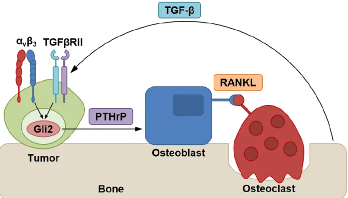
This vicious cycle of tumor-induced bone disease (TIBD) results in severe comorbidities, including extreme bone pain, spinal cord compression, hypercalcemia, and pathologic fractures that significantly decrease patient quality of life and increase mortality. This chapter will discuss integrin αvβ3 in the context of bone metastatic cancers, particularly how αvβ3 modulates tumor cell response to the bone microenvironment as well as downstream signaling pathways that promote tumor-induced bone destruction.
The Biology of Integrin αvβ3
Interestingly, while integrin αvβ is also expressed in primary bone cancers such as osteosarcoma and chondrosarcoma, high expression of αvβ3 has been shown to primarily promote lung metastasis of these tumors [ 50 , 51 ]. Integrin αvβ3 is a heterodimeric transmembrane glycoprotein that mediates cell adhesion to the extracellular matrix (ECM) by recognizing conserved arginine-glycine-aspartic acid (RGD) motifs in various ligands, including osteopontin, vitronectin, and fibronectin [53].
Integrin αvβ3 is Upregulated in Cancers that Metastasize to Bone
Results showed that integrin αvβ3 was overexpressed in B02 cells compared to the parental MDA-MB-231 cells while the cell surface expression of other integrins did not differ significantly between the two cell lines. In a more recent report, de novo expression of integrin αvβ3 in tumor cells that typically metastasize to the lungs was sufficient to promote homing to bone [ 72 ].
Expression of Tumor-Specific αvβ3 Promotes Bone Destruction
Taken together, these data demonstrate that integrin αvβ3 contributes to the osteotropism of metastatic cancer cells. In summary, these studies illustrate that increased αvβ3 expression in metastatic cancer cells contributes to the pathophysiology of tumor-induced bone destruction.
Integrin αvβ3 Modulates Tumor Response to the Rigid Bone Matrix
Fluorescence resonance energy transfer and coimmunoprecipitation assays showed that the colocalization of integrin αvβ3 and TGFβRII was significantly increased in tumor cells cultured on stiff substrates. Inhibition of integrin αvβ3 in MDA-MB-231 cells using an shRNA or the monoclonal antibody LM609 significantly reduced both Gli2 and PTHrP expression.
Targeting Integrin αvβ3-Expressing Tumors in Bone
The cyclic RGD-mimetic peptide cilengitide was first developed for the treatment of glioblastoma multiforme [82, 83] but has been investigated for use in patients with advanced solid tumors including prostate cancer, non-small cell lung cancer and carcinoma of squamous cells. Recently, the small molecule GLPG0187 was evaluated for its effects in patients with progressive gliomas and other advanced solid malignancies [ 85 ].
Concluding Remarks
Despite success in early clinical trials, many of these therapies have not yielded clinically meaningful results compared with standard chemoradiotherapy; however, few studies have specifically targeted cancer patients with bone metastases. To evaluate the efficacy of new or existing αvβ3 antagonists against bone metastases, future trials will need to include more patients with TIBD.
Abstract
Importantly, GANT58-NPs reduced tumor cell proliferation but did not alter mesenchymal stem cell proliferation or osteoblast mineralization in vitro, nor was there evidence of cytotoxicity after repeated in vivo treatment. Thus, inhibition of Gli2 using GANT58-NPs is a potential therapy to reduce bone resorption that should be considered for further testing and development toward clinical translation.
Introduction
Cells were then induced with MSC osteogenic differentiation medium (PromoCell) treated with vehicle (DMSO), free GANT58, Empty-NP, or GANT58-NP. For the biodistribution of polymer NPs, Cy7-grafted GANT58-NPs were injected by tail vein injection (8 mg/kg GANT58).
Results
Immunofluorescent analyzes revealed significantly reduced colocalization of Gli2 with DAPI-stained nuclei in cells treated with GANT58 or GANT58-NPs compared to the Empty-NP control (Fig. 3.4A). In addition, MDA-MB-231 bone cells treated with increasing concentrations of free GANT58 or GANT58-NPs showed decreased nuclear Gli2 protein levels as shown by immunoblotting (Fig. 3.4B). BV/TV was also significantly higher in the mice treated with GANT58-NPs than those treated with Empty-NPs (Fig. S3.4B).
3D µCT views of representative tibiae from each group again demonstrate the tumor-induced bone destruction in the mice treated with Empty-NPs compared to those treated with GANT58-NPs (Fig. S3.4F).
Discussion
There was no significant increase in aspartate aminotransferase (AST), alanine aminotransferase (ALT), or blood urea nitrogen (BUN) levels above two standard deviations from the mean levels reported by the animal supplier (Envigo) (Fig. S3.5A). GANT58 treatment of mouse bone marrow-derived cells co-cultured with MDA-MB-231 bone tumor cells significantly inhibited osteoclastogenesis (Figure 3.5D-F). Matrix rigidity in the bone microenvironment is a key mediator of Gli2 expression in bone-metastatic tumor cells.
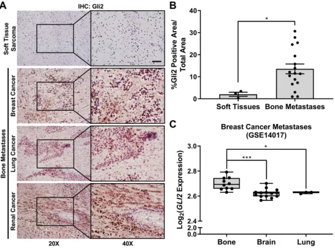
Similarly, we show that Gli2 is also overexpressed in bone metastatic tumors from various primary sites (Fig. 3.1A-B).
Conclusions
In patients with metastatic cancer, PTHrP is expressed in > 90% of bone-resident tumors compared with < 20%. The occurrence of Gli2 and PTHrP expression in bone suggests that GANT58 treatment is likely to be effective in a broad spectrum of patients suffering from bone metastases, which is supported by our observations of bone protection by GANT58-NPs across of both breast and lung cancer models. Thus, combined delivery of GANT58, which blocks the transition to the bone-destructive phenotype, with targeted therapies or conventional chemotherapeutics that block tumor growth in the bone marrow could potentially slow the progression of TIBD and improve the patient's quality of life.
Western blots of Gli2 cytoplasmic protein after 72 hours of treatment with (A) free GANT58 and (B) GANT58 NPs.

Abstract
GLI ANTAGONIST blocks bone invasion by oral squamous cell carcinomas regardless of resistance to EGFR INHIBITORS. Similarly, micro-CT analyzes revealed that both CAL27 and C27R tumor-bearing mice treated with GANT58-MPs had significantly higher trabecular bone volume (p < 0.001) compared to control-treated mice. Taken together, these data suggest that Gli inhibition via GANT58-MPs is a potential strategy for the treatment of OSCC patients with bone invasive tumors, regardless of their resistance to existing EGFR inhibitors.
Introduction
Genetic inhibition of Gli2 attenuates PTHrP expression and blocks osteoclast-mediated bone destruction induced by OSCC cells in vivo, suggesting that Gli2 is an ideal therapeutic target for advanced oral cancer [172]. Treatment with the EGFR TKI erlotinib significantly reduced Gli2 and PTHrP expression in multiple OSCC lines, but not in erlotinib-resistant cells. However, treatment with GANT58 inhibited Gli2 and PTHrP expression in cells sensitive or resistant to erlotinib.
We found that GANT58-MPs reduced bone destruction caused by OSCC tumors, independent of their response to EGFR inhibition.
Results
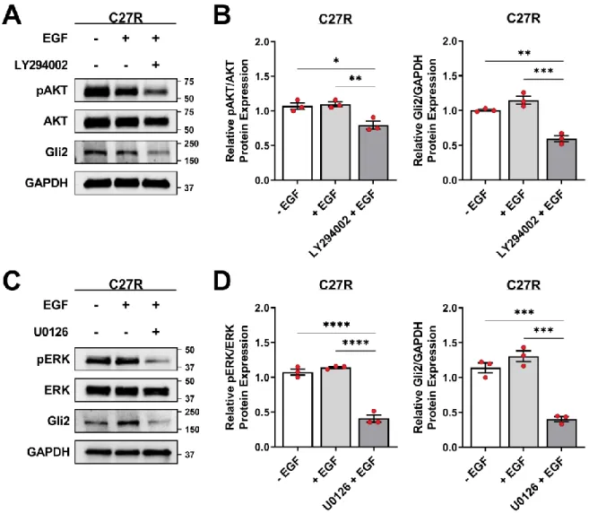
Inhibition of pAKT or pERK was sufficient to reduce Gli2 protein expression in C27R cells (Fig. S4.3A–D), suggesting that erlotinib-resistant cells reactivate these downstream kinases to maintain Gli2 and PTHrP expression. In contrast, the mesenchymal marker VIM and EMT-promoting transcription factor ZEB2 were upregulated in C27R cells (Fig. 4.4C). As expected, treatment with 10 μM erlotinib significantly reduced CAL27 cell proliferation, while no significant change was observed in C27R cells (Fig. 4.5E).
Immunohistological analyzes also showed a significant decrease in intratumoral PTHrP protein levels with GANT58-MP treatment (Fig. 4.6H-I).

Discussion
Taken together, these results demonstrate that inhibition of Gli2 via GANT58-MPs treatment blocks osteolytic signaling and subsequent bone invasion of tumors regardless of their resistance to erlotinib. We show that inhibition of Gli2 using the small molecule inhibitor GANT58 not only reduces EGF-stimulated PTHrP expression (Fig. 4.5) but also attenuates bone destruction by tumors in vivo (Fig. 4.6). We saw a significant decrease in PTHrP expression with GANT58 treatment as measured by qRT-PCR and immunohistochemistry in both CAL27 and C27R cells (Fig. 4.5-6).
Finally, GANT58 treatment inhibited tumor proliferation of CAL27 and C27R cells in vitro but did not reduce tumor burden in vivo.
Supplementary Data
DESIGNING 3D MODELS OF TUMORS AND BONE TO UNDERSTAND TUMOR-INDUCED BONE DISEASES AND IMPROVE TREATMENTS. Engineering 3D models of tumors and bones to understand tumor-induced bone diseases and improve treatments.
Abstract
Introduction
Co-cultivation of MG-63 human osteoblast-like cells and MDA-MB-231 human breast cancer cells on the silk scaffolds resulted in a decrease in the MG-63 population compared to MDA-MB-231 despite being seeded at a 1:1 ratio. This suggests that the breast cancer cells inhibited the growth of the osteoblasts, a finding that is supported by previous studies [262]. To take these results a step further, the effect of the breast cancer cells on matrix mineralization was investigated using the same co-culture.
This formulation was then introduced into the 3D co-culture system to test the efficacy of the drug in vitro as well as its targeting ability.
Future of 3D Models


