The deduced amino acid sequences of the F protein cleavage site revealed a unique virulent cleavage site of 112RRQKR↓F117. Sequence of the cleavage site of rNDV (virulent) as well as the altered cleavage site of rNDV-Fmut (avirulent).
General Introduction and review of literature 1
Etiology.…
Newcastle disease virus
- Nucleocapsid (N) protein
- Phosphoprotein (P)
- Matrix (M) protein
- Fusion (F) protein
- Hemagglutinin-neuraminidase protein (HN)
- Large polymerase protein (L)
P protein is also known to play a role in virulence depending on the strain (Huang et al., 2003). The HN protein is the primary determinant of tropism and virulence of NDV (Huang et al., 2004b).

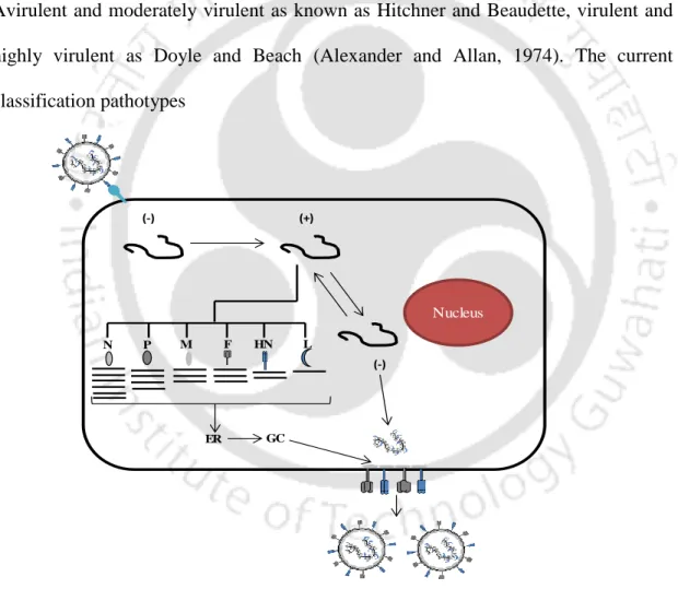
NDV infection and replication
Pathotypes of NDV …
Host range
NDV infections are accidental with mild conjunctivitis-like symptoms (Hunter et al., 1951; Quinn et al., 1952).
Clinical signs and symptoms
Diagnosis
- Viral isolation
- Serology …
- Molecular diagnosis
In some studies, the authors have mainly targeted the cleavage site of the F gene and followed by restriction fragment length polymorphism using BglI, Based on the pattern the isolates are classified into lentogenic, mesogenic and velogenic strains (Nathakumar et al., 2000). Molecular-based pathotyping of NDV isolates by RT-PCR of the F cleavage site and sequencing is preferred.
Vaccination
- Inactivated vaccine
- Live attenuated vaccine
- Recombinant vaccine
DNA vaccine expressing the F gene of NDV was developed in chicken and a high titer of the antibody was observed (Sakaguchi et al., 1996). Recombinant chickenpox expressing NDV F protein has shown protection, but preexisting chickenpox immunity in chickens interferes with vaccine efficacy (Taylor et al., 1990).
The rationale of the study
However, much remains to be explored about the oncolytic potential of the strains isolated from India.
Specific objectives
Review of literature
- Global scenario
- Indian scenario
- Reverse genetics
- NDV as an Oncolytic virus
For example, a role for the F protein cleavage site in NDV pathogenicity has been determined (Panda et al., 2004). NDV is known to induce apoptosis in various cancer cells of different origins (Elankumaran et al., 2006).
Molecular characterization of NDV strains from India 22
- Introduction
- Material and methods
- Samples
- Virus isolation and determination of virulence
- NDV F gene sequencing and phylogenetic analysis
- Results and Discussion
- Pathogenicity of NDV isolates
- F gene sequencing and features of NDV isolates
- Phylogenetic analysis based F gene sequence
- Conclusion
The deduced amino acid sequence of the F protein cleavage site and phylogenetic analysis of all five isolates indicated circulating genotype XIIIb NDV viruses. The close genetic similarity of all isolates suggested the circulation of virulent NDV strains of the same ancestor in and around central India. Phylogenetic analysis of the F-marked gene sequence was done by maximum likelihood with 500 bootstrap replicates in Molecular Evolutionary Genetic Analysis (MEGA) version 6 (Tamura et al., 2013).
Based on the pathogenicity index tests, the five isolates from this study were found to be highly virulent and velogenic in nature. Common necropsy findings included proventricular and cecal tonsil hemorrhage (Table 2.1). The velogenic nature of NDV isolates from Nagpur suggested the endemicity of genotype XIII viruses in India (Shabbir et al., 2013). The widespread nature of genotype XIII NDV strains in India has the potential of a new panzootic (Miller et al., 2015).
The long period of the outbreak and the isolates of the same ancestral origin suggest the circulation of the virus throughout the period.

Complete genome sequence of virulent NDV strain from India 30
- Introduction
- Material and methods
- Virus isolation and pathogenicity tests
- Viral RNA isolation and complete genome sequencing
- Phylogenetic analysis
- Results and discussion
- Pathogenicity of NDV/Chicken/Bareilly/01/10
- Genomic, protein features and nucleotide identity of NDV/Chicken/
- Conclusion
The MDT and ICPI were performed to determine the pathogenicity of the NDV strain NDV/Chicken-/Bareilly/01/10. The 3' leader and 5' trailer sequences of NDV/Chicken/Bareilly/01/10 were determined using the rapid amplification of cDNA ends (RACE) technique. Phylogenetic analysis of the complete genome sequence of strain NDV/Chicken/Bareilly/01/10 with 76 full-length genomes of other NDV strains of both classes I and II (genotype I to XVI) was performed by Molecular Evolutionary Genetics Analysis software ( MEGA 6 ).
The NDV/Chicken/Bareilly/01/10 primer is 114 nt long, the same length as other NDV isolates. Phylogenetically, NDV/Chicken/Bareilly/01/10 was closely related to the Stera/Ast/2755/2001 strain than to other NDV strains. The nt identity and phylogenetic relatedness of NDV/Chicken/Bareilly/01/10 to the mesogenic strain R2B suggest that.
NDV/Chicken/Bareilly/01/10 from India will be useful in further understanding pathogenicity and vaccination failure.

Construction of reverse genetics of genotype XIII NDV strain
Introduction
Cleavage of the F protein in a wide range of tissues is responsible for the systemic spread of NDV (Collins et al., 1993; Garten et al., 1980). The F proteins of virulent NDV poly-basic in their cleavage site (RRQKR↓F) were as non-virulent strains of NDV are monobasic (GRQGR↓L) (Nagai et al., 1976; Panda et al., 2004). The use of F cleavage site confirmed reverse genetics as a major determinant for virulence (de Leeuw et al., 2003; Peeters et al., 1999).
Recent studies have shown that highly virulent strains of genotype XIII are responsible for these outbreaks in India (Khorajiya et al., 2015; Morla et al., 2016; Nath et al., 2016). We have published the complete genome sequence of a genotype XIII strain from northern India in commercially vaccinated chickens, causing up to 35%. Divergence between circulating genotype XIII strains and the vaccine strain may account for incomplete protection and the outbreak in India.
In the present study, we constructed a reverse genetics system for the genotype XIII strain from India to generate an F splice site mutant, a likely genotype-matched vaccine candidate.
Material and methods
- Cells and Virus
- Construction of full length and support plasmids of genotype XIII
- Mutation of F cleavage site of full-length plasmids of genotype XIII
- Genetic stability and pathogenic assessment of the recombinant virus
A synthetic construct was synthesized (GenScript, USA) containing a unique linker of the set restriction enzymes present in the entire genome of the NDV strain Bareilly in the pUC19 vector (Figure 4.1). A pNDV restriction map was generated using SnapGene screening software (SnapGene, USA), and individual multi-site restriction enzymes were used to verify clone integrity. Full-length ORFs of N, P and L genes were amplified from NDV Bareilly strain using Phusion high fidelity polymerase (NEB, USA) and cloned into pcDNA3.1 (Invitrogen, USA).
The full-length antigenomic cDNA of the NDV strain Bareilly was cloned into pUC19, cloned between the T7 promoter and HDR. The fragment containing the virulent F splice site was mutated into a virulent F splice site by overlapping PCR. The mutated F fragment was designed with AgeI and PsiI restriction enzymes and used to replace the corresponding fragment in the full-length NDV (Figure 4.2).
The modified fragment is replaced in pNDV and named pNDV-Fmut using restriction enzymes AgeI and PsiI.

Results and discussion
- Generation of full-length clones and support plasmids of genotype XIII
Recovered viral RNA was collected and genetic marker and cleavage site F sequencing analysis was performed and found to be stable. In vitro growth kinetics were performed for both rNDV and rNDV-Fmut viruses, and there were no differences in replication between the two viruses (Figure 4.5A). The pathogenicity of rNDV and rNDV-Fmut was evaluated by the MDT test, and a standard pathogenicity test was performed.
In vitro growth kinetics of rNDV and rNDV-Fmut in DF-1 cell line (A), Western blot analysis of the rNDVs using polyclonal NDV antiserum and β-actin antibodies (B). We recently classified the virulent NDV strains circulating in India as belonging to genotype ; Nath et al., 2016). In some previous studies using reverse genetics, the virulent F splice site was mutated to avirulent due to viral attenuation (Kim et al., 2017; Manoharan et al., 2018).
The F cleavage site mutant virus was stable in cell culture and grows at high titers in embryonated eggs, which is a requirement for vaccine production.

Abstract
Introduction
Tumor cells generally have a defective antiviral response that supports NDV replication (Fiola et al., 2006). The development of NDV as a vector for the expression of foreign genes has been investigated to enhance its oncolytic activity (Krishnamurthy et al., 2000). Matrix metalloproteinases (MMPs) are Zn2+-dependent proteases that play a key role in the degradation of the extracellular matrix and induce the migration of cancer cells (Liotta, 1986; Liotta et al., 1980).
The mutation of APC is involved in β-catenin-mediated carcinogenesis and enhanced cancer cell migration ( Lowy et al., 2003 ; Zhai et al., 2002 ). APC and β-catenin are considered potential diagnostic markers for malignant transformation in oral squamous cell carcinoma (Chaw et al., 2012). MMP-7, a downstream target of β-catenin, has been implicated in metastasis by immunohistochemistry of pancreatic and oral cancer (Jones et al., 2004).
Previous studies have shown a role for β-catenin/MMP-7 in cancer invasion and progression (Shao et al., 2017; Zucker and Vacirca, 2004).
Material and methods
- Virus and cells
- Apoptosis study in cancer cells
- Wound healing assay
- Real time-PCR
- Transfection studies
- Immunoblotting
- Luciferase reporter assay
- Nuclear and cytoplasmic fractionation
- Statistical analysis
The SAS cells were infected with NDV at an MOI of 0.1 and the cells were collected at regular intervals. Cells were infected with NDV at an MOI of 0.1 and stained with JC-1 dye at 48 h postinfection. The wound was made using a 200 µl tip and the cells were washed with PBS to remove detached cells.
SAS cells were seeded in 6-well plates and treated with NDV at an MOI of 0.1 for 48 h. SAS cells (104/well) were seeded in 24-well plate and transfected with 400 ng of TOPFlash or FOPFlash plasmids with 40 ng of pRenillaTK using Lipofectamine 2000 (Invitrogen, USA). Cells were infected with 0.1 MOI of NDV or mock infected six hours after transfection.
Cells were stimulated with 20 mM LiCl or vehicle for 12 h, and cell lysates were collected 24 h postinfection.

Results
- Effect on cell viability and apoptosis upon infection with NDV
- NDV inhibits migration of oral cancer cells
- Investigation of MMP-7 mediated migration inhibition in oral cancer
DNA laddering in SAS cells infected with NDV started at 48 hours and was evident at 72 hours post-infection (Figure 5.3B). By Hoechst 33342 staining, chromatin condensation was readily observed in SAS cells 48 hours after infection with NDV (Figure 5.3C). Similarly, cleavage of PARP and caspase 3 was observed after infection of NDV in SAS cells (Figure 5.3E).
Open wound area was calculated at regular intervals post-NDV infection in SAS cells. Western blot was performed at different time points to decipher the role of NDV infection on β-catenin levels in SAS cells. NDV infection decreased the levels of p-Akt, p-GSK-3β and MMP-7 in a time-dependent manner in SAS cells (Figure 5.6D).
Cytoplasmic and nuclear fractions were collected from SAS cells 48 h after infection with NDV (0.1 MOI) and analyzed for β-catenin by Western blot.

Discussion
Mutational susceptibility changes of glycoprotein F protease of Newcastle disease virus: effects on pathogenicity. Evaluation of Newcastle disease virus fusion protein cleavage site sequences in genotype-matched vaccines. Detection and molecular characterization of Newcastle disease virus in peacocks (Pavo cristatus) in Haryana state, India.
Molecular characterization of an apoptotic strain of Newcastle disease virus isolated from an outbreak in India. Proteolytic cleavage of viral glycoproteins and its significance for Newcastle disease virus virulence. Newcastle disease virus (NDV) is the causative agent of Newcastle disease (ND) in many avian species.

Conclusion