저작자표시-비영리-변경금지 2.0 대한민국 이용자는 아래의 조건을 따르는 경우에 한하여 자유롭게
l 이 저작물을 복제, 배포, 전송, 전시, 공연 및 방송할 수 있습니다. 다음과 같은 조건을 따라야 합니다:
l 귀하는, 이 저작물의 재이용이나 배포의 경우, 이 저작물에 적용된 이용허락조건 을 명확하게 나타내어야 합니다.
l 저작권자로부터 별도의 허가를 받으면 이러한 조건들은 적용되지 않습니다.
저작권법에 따른 이용자의 권리는 위의 내용에 의하여 영향을 받지 않습니다. 이것은 이용허락규약(Legal Code)을 이해하기 쉽게 요약한 것입니다.
Disclaimer
저작자표시. 귀하는 원저작자를 표시하여야 합니다.
비영리. 귀하는 이 저작물을 영리 목적으로 이용할 수 없습니다.
변경금지. 귀하는 이 저작물을 개작, 변형 또는 가공할 수 없습니다.
의학박사 학위논문
Establishment of
a Parkinson’s disease model and evaluation of therapeutic effects of
dopaminergic precursor cells in a MPTP-treated common marmoset
MPTP 투여 마모셋 파킨슨병 모델의 확립과 도파민성 신경전구세포의 치료 효과 평가
2020년 8월
서울대학교 대학원 의학과 중개의학전공
안 재 범
의학박사 학위논문
Establishment of
a Parkinson’s disease model and evaluation of therapeutic effects of
dopaminergic precursor cells in a MPTP-treated common marmoset
MPTP 투여 마모셋 파킨슨병 모델의 확립과 도파민성 신경전구세포의 치료 효과 평가
2020년 8월
서울대학교 대학원 의학과 중개의학전공
안 재 범
MPTP 투여 마모셋 파킨슨병 모델의 확립과 도파민성 신경전구세포의 치료 효과 평가
Establishment of a Parkinson’s disease model and evaluation of therapeutic effects of
dopaminergic precursor cells in a MPTP-treated common marmoset
지도교수 강 병 철
이 논문을 의학박사 학위논문으로 제출함
2020년 5월
서울대학교 대학원
의학과 중개의학전공
안 재 범
안재범의 박사 학위논문을 인준함 2020년 7월
위 원 장 (인)
부 위 원 장 (인)
위 원 (인)
위 원 (인)
ABSTRACT
Establishment of
a Parkinson’s disease model and evaluation of therapeutic effects of
dopaminergic precursor cells in a MPTP-treated common marmoset
Jae-Bum Ahn, D.V.M.
Department of Medicine Translational Medicine Major The Graduate School Seoul National University
Parkinson's disease (PD) is one of the most important neuro- degenerative diseases. Studies investigating cell transplantation as an alternative to L-3,4-dihydroxyphenylalanine administration or deep brain stimulation surgery are being actively conducted.
Many PD animal models are used for PD treatment or prevention. However, most of them are rodent models, and the most representative is the model established with 1-meth-
yl-4-phenyl-1,2,3,6-tetrahydropyridine (MPTP). Compared to other models, nonhuman primate (NHP) MPTP-treated models show clinical symptoms similar to human patients and facilitate behav- ioral evaluation, suggesting the use of various MPTP injection models according to experimental needs. Most NHP MPTP-treat- ed models are optimized for short-term studies within three months and are not suitable for long-term studies such as cell transplantation. Since fetal mesenchymal cell transplantation in early studies, studies using mesenchymal stem cells or embryonic stem cells (ESCs) have been conducted. Studies have also been conducted using induced pluripotent stem cells, which can resolve ethical concerns and immune rejection. Despite advances in effi- cacy evaluation and safety of cell transplantation, studies on dif- ferentiation and discovery of homogeneous classification marker have yet to be investigated systematically since the degree of differentiation and homogeneity of cells after differentiation are directly related to clinical recovery and reduction of side effects.
Accordingly, a Parkinson's disease model was established by subcutaneous administering "2-2-1-1-1" mg/kg of MPTP to common marmosets (Callithrix jacchus) to induce a long-term and stable clinical manifestations. Daily observation showed sta- ble and persistent clinical symptoms. The results of tower test also reduced the motor function compared with pre-treatment with MPTP. In striatal positron emission tomography (PET) im- ages, radioactivity was significantly reduced compared with prior MPTP administration. Immunohistrochemical analysis showed loss
of tyrosine hydroxylase (TH)-positive cells and fibers in sub- stantia nigra. In addition, 2.0 × 10⁶ cells were implanted intra- cranially into the stratum of marmoset PD model to evaluate the therapeutic effect of dopaminergic (DAergic) precursor cells from human ESCs differentiating into DAergic neurons associated with PD symptoms using trophoblast glycoprotein, a new differ- entiation marker. The results of daily observation showed that the clinical symptoms recovered significantly from the third week after the cell transplant compared with the group exposed to MPTP. The tower test result confirmed that significant increase in the number of levels the marmosets climbed from the seventh week after the cell transplant. In the striatal PET image, the specific uptake ratio value was significantly increased from the fourteenth week after the cell transplant compared to the MPTP treatment group. The histopathological analysis revealed no ex- cessive inflammatory reactions or tumor-like neoplasms, and TH-positive cells developed from implanted DAergic precursor cells in the cell transplant site. Based on the above results, it is purposed that the marmoset model produced by the new MPTP treatment method is suitable for long-term studies such as cell transplantation, and it is suggested that DAergic precursor cells represent potential as PD treatments for human patients.
Keywords: Parkinson’s disease; nonhuman primates; common marmoset; cell therapeutics; animal model; stem cell; cell trans- plantation
Student Number: 2012-31146
CONTENTS
ABSTRACT ··· ⅰ CONTENTS ··· ⅳ LIST OF TABLES ··· ⅷ LIST OF FIGURES ··· ⅹ LIST OF ABBREVIATIONS ··· ⅹⅱ
LITERATURE REVIEW ··· 1
CHAPTER Ⅰ
Establishment of a novel Parkinson’s disease model in common marmoset for cell therapy evaluation
ABSTRACT ··· 52
INTRODUCTION ··· 55
MATERIALS AND METHODS ··· 61
Animals ···61
MPTP-induced PD model ···61
Behavioral assessment ···64
PET imaging analysis ···67
Microscopic assessment ···69
Statistical analysis ···69
RESULTS ··· 71
Stable parkinsonian symptoms without death for 32 weeks after MPTP treatment with novel method ···71
Motor dysfunctions without recovery for 32 weeks after MPTP treatment with novel method ···73
Amelioration of clinical symptoms temporarily after administration of L-DOPA following MPTP treatment ···77
Lower radioactivity in the striatum based on 18F-FP-CIT PET images without recovery for 32 weeks after MPTP treatment with novel method ···79
Loss of tyrosine hydroxylase-positive cells and fibers i n t h e s ubstan ti a n igra a nd stri atum a fte r M PT P treatment with novel method··· 81
DISCUSSION···83
CHAPTER Ⅱ
Evaluation of therapeutic effects of human embryonic stem cell-derived dopaminergic precursor cells transplanted into
a marmoset model of Parkinson’s disease
ABSTRACT···92
INTRODUCTION···94
MATERIALS AND METHODS···99
Animals ···99
MPTP-induced PD model ···100
Cell collection ···102
Cell transplantation ···102
Behavioral assessment ···105
PET-CT imaging and analysis ···107
Histopathologic examination ···108
Statistical analysis ···110
RESULTS···112
N o si gn if i ca nt d i ff e re nc e i n bod y we i gh t be t we e n MPTP-treated and cell-transplanted marmosets due to intensive care··· 112
Progressive recovery of motor symptoms in MPTP pre-treated cell-transplanted marmosets compared to MPTP-treated marmosets··· 115
Significant, but not full recovery of motor function in c e l l - t r a n s p l a n t e d P D m a r m o s e t s c o m p a r e d t o MPTP-treated marmosets··· 117
Weak recovery pattern in striatal PET images and SUR
in cell-transplanted marmosets··· 119
No tumor-like lesions, but increase d TH-positive neurons and fibers at transplant sites at 28 weeks after cell transplantation in MPTP-treated marmosets··· 122
Identification of cells expressing DAergic markers in transplanted cells at 28 weeks after cell transplantation ··· 126
DISCUSSION···129
REFERENCES···138
ABSTRACT IN KOREAN (국문초록) ···203
LIST OF TABLES
Table 1. Characteristics of animal models used in PD ··· 7 Table 2. NHP PD model characteristics according to various
MPTP treatment methods ··· 17 Table 3. Human α-syn overexpression NHP models ··· 22 Table 4. Clinical symptoms similar to human PD patients
observed in MPTP-treated NHP models ··· 25 Table 5. Gene therapy studies for PD ··· 31 Table 6. Tissues and cells used in cell transplantation studies
for PD therapy ··· 33 Table 7. Therapeutic effect of cell transplantation using NHP
PD models ··· 36 Table 8. Biomarkers related oxidative stress in human ··· 46 Table 9. Biomarkers related to abnormal protein accumulation
and aggregation in human ··· 50 Table 10. Various MPTP treatment methods used in common
marmoset PD models ··· 59 Table 11. Evaluation items and scores of MPTP-treated common
marmosets ··· 66 Table 12. Cell transplant studies using NHP models of PD ··· 90 Table 13. Cell therapy studies employing NSCs, DAergic progenitor
or precursor cells ··· 98 Table 14. Cell transplant site coordinates using a stereotaxic
device ··· 104
Table 15. Scoring scale for daily monitoring of MPTP-treated common marmoset models ··· 106
LIST OF FIGURES
Figure 1. Species distribution in animal models for PD ··· 6 Figure 2. Methods used in animal models for PD from 1974
to 2019 ··· 8 Figure 3. Methods used to create NHP model of PD from
1974 to 2019 ··· 16 Figure 4. The scheme of MPTP treatment regimen ··· 63 Figure 5. The experiment protocol ··· 68 Figure 6. Behavioral assessment score of MPTP-treated
common marmosets ··· 72 Figure 7. Changes in the body weight of MPTP-treated
common marmosets. ··· 74 Figure 8. Tow er test results of M PTP-treated common
marmosets ··· 76 Figure 9. Behavioral assessment and tower test result of
L-DOPA administration to MPTP-treated common marmosets ··· 78 Figure 10. The striatal 18F-FP-CIT PET images (A) and
radioactivity changes (B) after MPTP treatment in common marmosets ··· 80 Figure 11. Immunohistochemistry of TH in the substantia nigra of
MPTP-treated common marmosets ··· 82 Figure 12. The experiment protocol ··· 101
Figure 13. Monitoring body weight of M PTP-treated and cell-transplanted marmosets ··· 114 Figure 14. B ehavioral assessm ent in M PT P-treated and
cell-transplanted marmosets ··· 116 Figure 15. C o m p a r is o n o f ju m p in g a b ilit y b e t w e e n
M PTP-treated and cell-transplanted com mon marmosets ··· 118 Figure 16. Striatal 18F-FP-CIT-PET-CT images (A) and
SUR changes (B) of MPTP-treated and cell-transplanted marmosets ··· 121 Figure 17. Comparison of H&E stain results in the brain tissue of
MPTP-treated and cell-transplanted marmosets ··· 123 Figure 18. C om parison of anti- TH IH C results in brain
tissue of M PTP-treated and cell-transplanted marmosets ··· 125 Figure 19. IF staining of the caudate in cell-transplanted
marmosets to identify the origin (A), survival, function (B),and lineage (C) of the transplanted cells ··· 128
LIST OF ABBREVIATIONS
PD Parkinson’s disease
DAergic Dopaminergic
SN Substantia nigra
L-DOPA L-3,4-dihydroxyphenylalanine; Levodopa
DBS Deep brain stimulation
6-OHDA 6-hydroxydopamine
MPTP 1-methyl-4-phenyl-1,2,3,6-tetrahydropyridine
ESC Embryonic stem cell
NHP Nonhuman primate
PET Positron emission tomography
TH Tyrosine hydroxylase
TPBG Trophoblast glycoprotein
LITERATURE REVIEW
Parkinson’s Disease (PD)
Parkinson’s disease (PD) is one of the most important neuro- degenerative disorder triggered by dopaminergic (DAergic) cell death in the substantia nigra (SN) and dopamine depletion sub- stantially, leading to manifestations such as tremor, rigidity and slow movement. PD was described by Dr. James Parkinson in 1817 in the essay, “Shaking Pasly”, and was named in 1862 by Jean-Martin Charcot in honor of Dr. Parkinson (1). PD is one of the top three geriatric diseases, along with Alzheimer's disease (AD) and stroke (2), and is the second-largest degenerative brain disease with a prevalence of 200 per 100,000 people worldwide (3, 4). The number of patients in their 60s and over increases rap- idly (5).
Classification and Etiology of PD
According to Jean-Martin Charcot, PD is characterized by tremor and rigid/akinesia depending on the clinical motor symptoms;
however, PD patients do not necessarily need to manifest tremor (1). PD is classified into different types depending specific fea- tures; idiopathic, inherited, and other atypical PD (6). Idiopathic type of PD is the most common although underlying etiology is unknown, unfortunately. Some studies have reported that PD is caused by factors such as smoking, coffee and tea consumption,
and exposure to pesticides, traumatic brain damages, organic sol- vents, uric acid, daily products, nonsteroidal anti inflammatory drugs, statin and calcium channel blockers (7). Other studies have reported that diabetes and vitamin D deficiencies are also associated with PD (8). However, it is attributed to a combina- tion of genetic and environmental conditions, and not to a specif- ic etiological factor alone.
Five to 10% of PD patients present with the inherited type, and specific genes are known to be associated with PD such as Parkin, DJ-1, PINK1 and others (3, 9, 10). PARK genes 1 to 18 are currently linked to familial PD, since the publication of the first mutant genetic map associated with possible PD in 1996 (11). PARK8 and PARK17 were associated with general PD oc- curring in older age groups, with corresponding mutant genes LRRK2 and VPS35, respectively. Early-onset PD is associated with PARK2 (Parkin), PARK6 (PINK1), PARK7 (DJ-1), PARK9 (ATP13A2), PARK14 (PLA2G6), and PARK15 (FBXO7) excluding PARK1, whose gene name is SNCA. Among them, SNCA, LRRK2, Parkin, PINK1, DJ-1, and ATP13A2, are linked to PD, and the identity of the rest is still being investigated for possible linkage to PD (12).
Motor and non-motor symptoms of PD
Several neuropathological and neurochemical studies have shown that major clinical symptoms of PD, including motor symptoms are associated with dopamine. Subsequent studies have reported that motor symptoms do not appear until dopamine levels in the striatum are reduced significantly along with extensive loss of DAergic neuron in the SN (13). In the “subclinical state” charac- terized by symptoms of motor abnormalities, the dopamine level in the striatum decreases to 80%, and nearly 60% of DAergic cells in the SN appear to be lost, with eventual dopamine reduc- tion and loss of DAergic neurons. However, it is known that motor symptoms do not occur due to various compensatory mechanisms until the disease is at an advanced stage (14). Thus, major motor symptoms such as tremors appear at late stage of disease, and it is very difficult to treat or prevent progressive disease because of the loss of a large number of DAergic neurons. Therefore, treatment initiation before the onset of
“subclinical state” with motor symptoms can most effectively prevent disease progression (15, 16). However, it is very difficult to identify specific early symptoms, especially non-motor symp- toms such as sleep disorder, anxiety and depression, because those symptoms vary among patients and it is not easy to real- ize specific non-motor symptoms (17, 18). Nonmotor symptoms can be largely classified into neuropsychological abnormalities, sleep disorders, autonomic neurological abnormalities, sensory ab-
normalities, and pain, and according to Witjas et al., anxiety, se- vere sweating, delayed information processing, fatigue, hyper- sensitivity, and hallucinations appear to be approximately 50% of those in PD patient (19). Non-motor symptoms are important not only as an indicator for early diagnosis in PD patients, but also in terms of quality of life in both patients and caregivers as well as motor symptoms (20).
Animal models of PD
In vitro and in vivo models are used to study PD mechanisms and treatments. In vitro models facilitate rapid pathological in- vestigations inexpensively, and without the ethical concerns asso- ciated with animal models. In addition, genetic manipulation is easier and the reproducibility is high as large-scale experiments can be performed in a short time. However, since actual PD oc- curs via interaction with various neurons and other cells or tis- sues in addition to dopamine neurons, it can be studied only in living animal models. For this reason, results obtained from the in vitro model for this reason should be verified in studies using animal models (21). A search of SCOPUS (Elsevier) with the keywords “Parkinson ’s disease” and “Animal models” returned about 13,000 articles. In particular, in the last 5 years, the num- ber of studies using animal models has increased, with about 1,000 articles in each year (Figure 1). The rat model for the PD study was first developed in 1970 by Ungerstedt and Arbuthnott
by administering a neurotoxic substance called 6-hydroxydop- amine (6-OHDA) into the brain (22). In 1971, Hockman estab- lished a cat model by including thermal damage in the brain tis- sue surgically (23). Subsequently, studies using dog and rat models have been conducted, and primate models have been re- ported in Japan in 1979 (24). The PD animal models include acute models developed via neurotoxin administration and chronic model involving genetically modified animals. The animal model used for most PD studies is established via a neurotoxin admin- istration (25) (Table 1, Figure 2).
Figure 1. Species distribution in animal models for PD. As a re- sult of literature search in SCOPUS with “Parkinson’s disease”
and “animal model”, rodent models such as mice and rats were mostly used in PD studies, and primate models were used within 10%. (A) Total period (1974-2019). (B) Last 5 years (2015-2019).
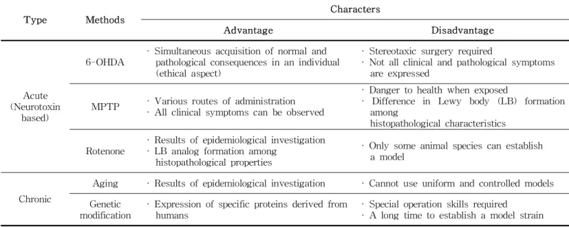
Table 1. Characteristics of animal models used in PD
Type Methods
Characters
Advantage Disadvantage
Acute (Neurotoxin
based)
6-OHDA
· Simultaneous acquisition of normal and pathological consequences in an individual (ethical aspect)
· Stereotaxic surgery required
· Not all clinical and pathological symptoms are expressed
MPTP · Various routes of administration
· All clinical symptoms can be observed
· Danger to health when exposed
· Difference in Lewy body (LB) formation among
histopathological characteristics Rotenone
· Results of epidemiological investigation
· LB analog formation among histopathological properties
· Only some animal species can establish a model
Chronic
Aging · Results of epidemiological investigation · Cannot use uniform and controlled models Genetic
modification
· Expression of specific proteins derived from humans
· Special operation skills required
· A long time to establish a model strain
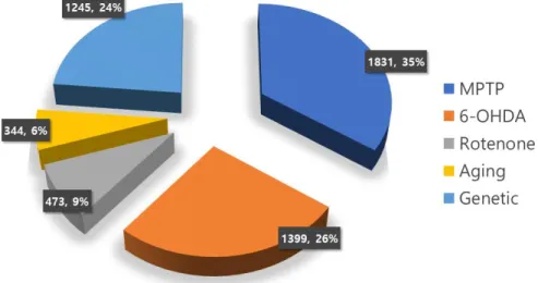
Figure 2. Methods used in animal models for PD from 1974 to 2019. As a result of literature search in SCOPUS with
“Parkinson’s disease” and “animal model”, most studies were conducted using the MPTP treatment (35%), followed by 6-OHDA treatment (26%), and the genetic manipulation (24%).
6-OHDA models
The 6-OHDA model is established via topically application of a chemical neurotoxin, and various models including mice, cats, dogs, and monkeys as well as rats have been developed (26).
Although studies using 6-OHDA models were conducted until 1985, administering 6-OHDA directly to brain tissue was techni- cally difficult because 6-OHDA cannot cross the blood-brain barrier. Particularly, a bilateral lesion induced via intraventricular or intracranial administration results in death, due to motor symptoms and the inability to feed or drink water (27).
Therefore, the 6-OHDA model is most commonly used to admin- ister 6-OHDA directly to the substantia nigra, the nigrostriatal tract, or the striatum. In addition to the model retention rate due to reduced mortality, a single animal can be used in the ex- perimental and animal groups simultaneously (28).
MPTP models
The MPTP model was constructed in 1982 by Langston et al., who found symptoms similar to PD in drug addicts who injected MPTP-contaminated heroin (29). In particular, most of the histo- pathological findings along with the characteristic clinical symp- toms in PD patients were observed in the nonhuman primate (NHP) model, and in the mouse model, DAergic neurons degen- eration was observed, although no representative clinical symp- toms were observed. However, the rat model was inadequate be-
cause it was resistant to MPTP due to the species characteristics (30). In addition, in the case of the mouse model, no inclusion body similar to the Lewy body (LB), one of hallmarks of PD, was found, although the model was established with acute, sub- chronic, and chronic MPTP treatment via injection or using an osmotic pump (31, 32). However, acute or subacute MPTP treat- ment leads to necrosis of DAergic neurons in the SN, but is limited by a spontaneous recovery due to the reversible response.
Other neurotoxin models
As studies investigated the role of environmental factors in hu- man PD, a model was developed by chemical treatment the as- sumption that exposure to herbicides or pesticides may be a cause. Among the herbicides, paraquat (N, N'-dimethyl-4-4'-bi- piridinium) was structurally similar to MPP+ (1-methyl-4-phenyl- pyridinium), a metabolite of MPTP, and therefore used in mouse studies. Decreased DAergic nerve cell fibers in the striatum and neurons of the SN were reported in patients with decreased mo- tor abilities (33). In contrast, only a small number of necrotic ni- grostriatal DAergic neurons were observed in other studies (34) without behavioral abnormalities or neural circuit destruction (35).
However, based on several models developed using the method reported by Betarbet et al., (36) only about half of the rats treat- ed with rotenone showed necrosis of nigrostriatal neural cells, and the model was not established in mice or monkeys except
rats (37).
In recent years, inflammation has been reported as an important factor contributing to PD. A rat model in which the endotoxin lipopolysaccharide (LPS) was directly injected to the nigrostriatal pathway has been developed based on inflammation in the ni- grostriatal pathway triggered by neurotoxins administered (38).
LPS administered topically to the SN or striatum is not directly toxic to DAergic neurons, but cytotoxins released by microglia activation disrupted the dopamine neural circuit. Hunter et al. (39) confirmed a decrease in dopamine concentration and accumulation of α-synuclein (α-syn) in the striatum along with SN cell ne- crosis in a mouse model administered LPS in the striatum.
Genetic models
After studies using mouse and rat transgenic animal models ex- posed to oxidative stress, which believed to cause PD, cell trans- plantation therapy was developed using genetically modified ani- mals (40, 41). In the late 1990s, various genetically modified mouse models were used to investigate pathological mechanisms:
MAO-B transformation or knockout (KO) mouse model (42, 43), neuronal nitric oxide synthase or inducible NOS KO mouse model (44, 45), and dopamine transfer factor (DAT) or dopamine re- ceptor KO mice (46, 47). However, this strategy is difficult to ac- cept as practical transgenic PD models. In the early 2000s, mice with knockout of α-syn (48, 49), DJ-1 (50), Parkin (51), PINK1
(52), and other genes were investigated. However, a non-mam- malian transgenic animal model was developed as an alternative to compensate for the poor nigrostriatal neuronal necrosis in transgenic mouse models. The representative non-mammalian transgenic animal models, fruit flies (Drosophila) (53, 54), Caenorhabditis elegans (55), and zebrafish (56) models are eco- nomical in terms of model development and maintenance com- pared with rodent or NHP models, and a large number of such models can be tested simultaneously (57). As a result, an optimal model for evaluating the effect of α-syn deletion or neuro- protection was developed while maintaining the nigrostriatal pathway, which increases the concentration of dopamine and dop- amine metabolites in the striatum and increases the concentration of α-syn in the striatum (58).
Limitation of rodent models of PD
In general, rodents have been utilized as animal models compared with NHP based on accessibility, easy handling and manipulation, ease of husbandry, and economics of management. As an animal model for PD studies, the 6-OHDA-treated rat model presents behavioral disorders such as limb movement abnormalities, ataxia, and sensory-motor disorders; however, the standard 6-OHDA model carries a hemisphere lesion, which is different from the patient's symptoms (59). The MPTP-treated mouse model ex- hibits motor symptoms such as tremor and gait abnormalities,
but unlike the primate model, it does not show dyskinesia, be- cause the nerve pathways associated with MPTP damages are presumed to differ from those of the mouse and NHP, and sev- eral therapies may explain the difference in interpretation of re- sults in mouse models and human patients (60). The transgenic mouse model is easy to develop and maintain compared to a neurotoxin-treated model, except that genetic engineering techni- ques are essential. In addition, in a mouse model, the over- expression or deletion a gene underlying hereditary PD represents an optimal condition to identify the causative role of a specific gene. However, the common intrinsic genetic factor underlying dopamine neuronal necrosis is not observed in the characteristic mouse species (61).
NHP models of PD
The ideal animal model of PD should show motor or non-motor symptoms in PD patients and responses to therapies used clin- ically, and similar histopathologic lesions in the brain (62-64).
However, no animal models meeting the above conditions com- pletely are available. Therefore, NHP models have an advantage over other species models because NHP models are the closest genetically with human, exhibit similar anatomy of central nerv- ous system (CNS) including the brain and other organs, and op- erate arms or legs like human (29, 65, 66).
Neurotoxin models
–
6-OHDA, MPTPNeurotoxin-treated primate models were mainly developed by MPTP and 6-OHDA, especially 6-OHDA models of baboons (Papio papio) (67) and rhesus monkeys (Macaca mulata) (68), but mainly common marmosets (Callithrix jacchus). The charac- teristics of 6-OHDA NHP model vary according to the location and frequency of administration of 6-OHDA (69-76). In general, the striatum, SN, and medial forebrain bundle, which is the ni- grostriatal pathway, are the treatment sites. New models have been developed to control dose frequency due to the nature of recovery after about 10 weeks after treatment with 6-OHDA similar to MPTP (77). In order to develop the 6-OHDA model, although a very small needle is used in the brain tissue, it is asscociated with the risk of physical damage following multiple injections, and appropriate stereotaxic surgery is required to in- ject it in the correct position. In addition, administering 6-OHDA to induce symptoms in only one hemisphere, the various clinical symptoms seen in human PD patients do not appear.
However, the MPTP model was created using old world monkeys such as velvet monkey (Chlorocebus aethiops), rhesus monkey, and cynomolgus monkey (Macaca fasicularis), in addition to new world monkeys such as squirrel monkey (Saimiri sciureus) and common marmoset, and is the most used model (77) (Figure 3).
In addition, a model development method in which MPTP admin- istration route, dosage, and frequency or duration of admin-
istration is very diverse was introduced depending on the clinical symptom progression and maintenance period of the model (Table 2). Unlike other models, in the MPTP model, it affects only the DAergic neurons in the brain (78), and leads to the accumulation of α-syn in the dopaminergic neurons associated with LBs (79).
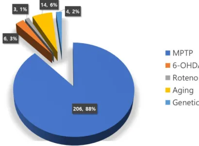
Figure 3. Methods used to create NHP model of PD from 1974 to 2019. As a result of literature search in SCOPUS with
“Parkinson’s disease”, “animal model” and “primate” or “monkey”, most studies were conducted using the MPTP treatment model (88%), followed the Aging model (6%), and the 6-OHDA treat- ment model (3%).
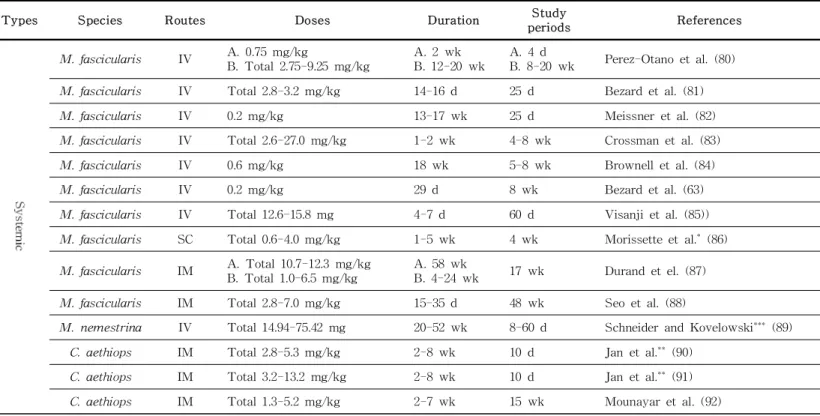
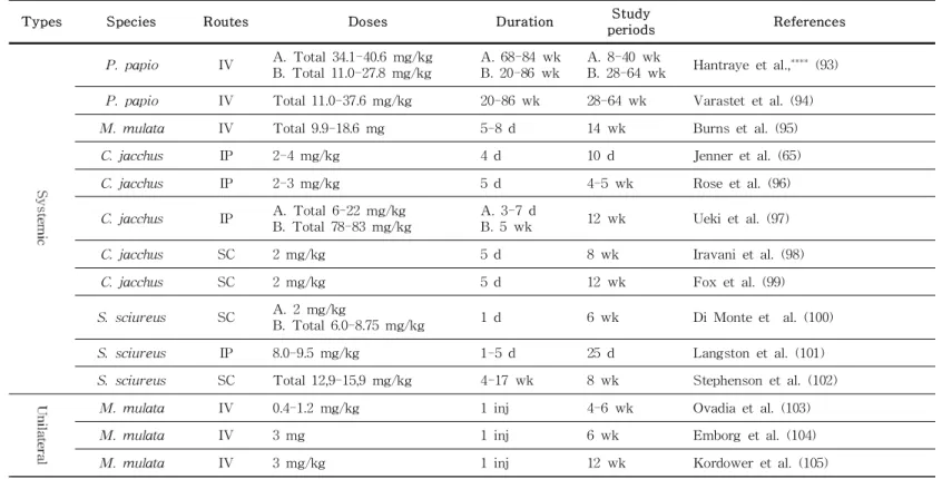
Table 2. NHP PD model characteristics according to various MPTP treatment methods
Types Species Routes Doses Duration Study
periods References
M. fascicularis IV A. 0.75 mg/kg
B. Total 2.75-9.25 mg/kg
A. 2 wk B. 12-20 wk
A. 4 d
B. 8-20 wk Perez-Otano et al. (80)
M. fascicularis IV Total 2.8-3.2 mg/kg 14-16 d 25 d Bezard et al. (81)
M. fascicularis IV 0.2 mg/kg 13-17 wk 25 d Meissner et al. (82)
M. fascicularis IV Total 2.6-27.0 mg/kg 1-2 wk 4-8 wk Crossman et al. (83)
M. fascicularis IV 0.6 mg/kg 18 wk 5-8 wk Brownell et al. (84)
M. fascicularis IV 0.2 mg/kg 29 d 8 wk Bezard et al. (63)
M. fascicularis IV Total 12.6-15.8 mg 4-7 d 60 d Visanji et al. (85))
M. fascicularis SC Total 0.6-4.0 mg/kg 1-5 wk 4 wk Morissette et al.* (86)
M. fascicularis IM A. Total 10.7-12.3 mg/kg B. Total 1.0-6.5 mg/kg
A. 58 wk
B. 4-24 wk 17 wk Durand et el. (87)
M. fascicularis IM Total 2.8-7.0 mg/kg 15-35 d 48 wk Seo et al. (88)
M. nemestrina IV Total 14.94-75.42 mg 20-52 wk 8-60 d Schneider and Kovelowski*** (89)
C. aethiops IM Total 2.8-5.3 mg/kg 2-8 wk 10 d Jan et al.** (90)
C. aethiops IM Total 3.2-13.2 mg/kg 2-8 wk 10 d Jan et al.** (91)
C. aethiops IM Total 1.3-5.2 mg/kg 2-7 wk 15 wk Mounayar et al. (92)
(Continues)
Table 2. (continued)
Types Species Routes Doses Duration Study
periods References
P. papio IV A. Total 34.1-40.6 mg/kg B. Total 11.0-27.8 mg/kg
A. 68-84 wk B. 20-86 wk
A. 8-40 wk
B. 28-64 wk Hantraye et al.,**** (93) P. papio IV Total 11.0-37.6 mg/kg 20-86 wk 28-64 wk Varastet et al. (94)
M. mulata IV Total 9.9-18.6 mg 5-8 d 14 wk Burns et al. (95)
C. jacchus IP 2-4 mg/kg 4 d 10 d Jenner et al. (65)
C. jacchus IP 2-3 mg/kg 5 d 4-5 wk Rose et al. (96)
C. jacchus IP A. Total 6-22 mg/kg B. Total 78-83 mg/kg
A. 3-7 d
B. 5 wk 12 wk Ueki et al. (97)
C. jacchus SC 2 mg/kg 5 d 8 wk Iravani et al. (98)
C. jacchus SC 2 mg/kg 5 d 12 wk Fox et al. (99)
S. sciureus SC A. 2 mg/kg
B. Total 6.0-8.75 mg/kg 1 d 6 wk Di Monte et al. (100)
S. sciureus IP 8.0-9.5 mg/kg 1-5 d 25 d Langston et al. (101)
S. sciureus SC Total 12,9-15,9 mg/kg 4-17 wk 8 wk Stephenson et al. (102)
M. mulata IV 0.4-1.2 mg/kg 1 inj 4-6 wk Ovadia et al. (103)
M. mulata IV 3 mg 1 inj 6 wk Emborg et al. (104)
M. mulata IV 3 mg/kg 1 inj 12 wk Kordower et al. (105)
(Continues)
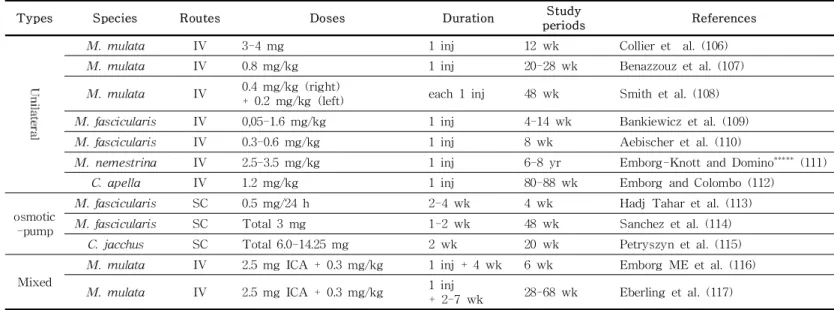
Table 2. (continued)
Types Species Routes Doses Duration Study
periods References
M. mulata IV 3-4 mg 1 inj 12 wk Collier et al. (106)
M. mulata IV 0.8 mg/kg 1 inj 20-28 wk Benazzouz et al. (107)
M. mulata IV 0.4 mg/kg (right)
+ 0.2 mg/kg (left) each 1 inj 48 wk Smith et al. (108)
M. fascicularis IV 0,05-1.6 mg/kg 1 inj 4-14 wk Bankiewicz et al. (109)
M. fascicularis IV 0.3-0.6 mg/kg 1 inj 8 wk Aebischer et al. (110)
M. nemestrina IV 2.5-3.5 mg/kg 1 inj 6-8 yr Emborg-Knott and Domino***** (111)
C. apella IV 1.2 mg/kg 1 inj 80-88 wk Emborg and Colombo (112)
osmotic -pump
M. fascicularis SC 0.5 mg/24 h 2-4 wk 4 wk Hadj Tahar et al. (113)
M. fascicularis SC Total 3 mg 1-2 wk 48 wk Sanchez et al. (114)
C. jacchus SC Total 6.0-14.25 mg 2 wk 20 wk Petryszyn et al. (115)
Mixed
M. mulata IV 2.5 mg ICA + 0.3 mg/kg 1 inj + 4 wk 6 wk Emborg ME et al. (116)
M. mulata IV 2.5 mg ICA + 0.3 mg/kg 1 inj
+ 2-7 wk 28-68 wk Eberling et al. (117) inj: injection; d: day; wk: week; yr: year
* 2-3 mg injections at weekly intervals
** 0.4 mg/kg 4 daily injections in each of the first 2 weeks followed by 1 or 2 injections per week
*** 0.010-0.175 mg/kg injections up to 3 times per week
**** 0.4 mg/kg 5 daily injections followed 5-6 months later by weekly injections of 0.2-0.5 mg/kg for 8-18 months
***** Freezing was not observed in all MPTP-infused monkeys.
Aging models
The NHP PD model can be largely classified into three types: an aging model, a neurotoxin administration model, and a genetic modification model. The aging model was developed based on the fact that PD in most human patients occurs at an older age and aging is presumed to be the cause of PD. The model was devel- oped by several investigators using rhesus monkeys (118-126) and squirrel monkeys (127, 128). In the aging model, postural and gait abnormalities and mild tremors were detected in human PD cases, whereas histopathological degenerative changes involving nigrostriatum such as dopamine reduction in the striatum, dop- amine neuronal necrosis in the SN, and deposition of lipofuscin were observed. However, aging is not a disease, but a natural change, and among NHP, it is difficult to develop a specific model because the aging period vary with each strain and individual. It is difficult to associate motor symptoms with ab- normalities involving the DAergic neuropathy, as it is possible to develop not only PD symptoms, but also other aging diseases of the musculoskeletal system or metabolism, in addition to the high cost required for model management.
Genetic models
In humans, α-syn is a major component associated with the SNCA gene mutation in patients with hereditary familial PD and the LBs found in patients with idiopathic PD. Unfortunately, in
NHPs, no human-like spontaneous mutation is known. Therefore, a model was established by directly injecting the human α-syn gene into the brain, and similar to the 6-OHDA model, intra- cranial administration was included (129, 130). Thus, most models use marmoset monkeys instead of other strains. In the model overexpressing normal α-syn and mutant α-syn, degenerated dopamine neurons were found in the striatum, whereas DAergic neuronal necrosis in the ventral midbrain region was higher in the model over-expressing the mutant α-syn than in the model over-expressing the normal α-syn (129). In order to introduce the α-syn gene, the degree of overexpression and peaking time vary depending on the type of carrier used in the viral vector (131, 132) (Table 3). Overexpression of the introduced gene and symp- tom manifestation occur after a few days to weeks. In addition, it is necessary to introduce a gene directly into the brain tissue similar to the 6-OHDA model, which may lead to physical dam- age in the absence of adequate surgical expertise. However, the symptoms observed in human familial or idiopathic PD patients cannot be detected as the gene introduced only in one hemisphere is expressed, which is a model limitation.
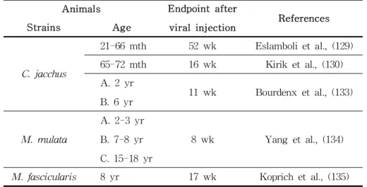
Table 3. Human α-syn overexpression in NHP models
Animals Endpoint after
viral injection References
Strains Age
C. jacchus
21-66 mth 52 wk Eslamboli et al., (129) 65-72 mth 16 wk Kirik et al., (130) A. 2 yr
B. 6 yr 11 wk Bourdenx et al., (133)
M. mulata
A. 2-3 yr B. 7-8 yr C. 15-18 yr
8 wk Yang et al., (134)
M. fascicularis 8 yr 17 wk Koprich et al., (135) wk: week; mth: month; yr: year
Advantages of MPTP-treated NHP models
NHP PD models involving NHPs compared with other animal species are mostly developed via MPTP administration. Various NHP models such as cynomolgus monkey model were developed (136), since Langston et al. (101) developed the squirrel monkey model and Jenner et al., (65) developed the common marmoset model in 1984 (137). Cynomolgus and rhesus monkeys, which are most frequently used as old-world monkey MPTP models, exhibit individual differences in sensitivity to MPTP. Therefore, several investigators have introduced models to showcase different routes and dosages (111, 138-140). However, most of the marmoset monkeys used as the new world monkey MPTP models were developed via subcutaneous administration for 5 consecutive days, and some researchers modified the MPTP treatment method for the desired condition (97, 141, 142) (Table 2).
The NHP MPTP model can be used to evaluate in a variety of combinations such as bradykinesia, postural abnormalities, facial expression changes, tremors, and rigidity in human PD patients.
It also facilitates the evaluation of cognitive abilities, fine move- ments, tremors, and excessive blinking using tools similar those adopted for human PD patients, as well as the Wisconsin General Apparatus Test (89, 116, 143-145).
However, the NHP MPTP model develops dyskinesia caused by L-DOPA administration similar to that of human PD patients.
The varying severity of dyskinesia in the NHP model, caused by
L-DOPA after MPTP administration, is clinically relevant as mentioned in chapter 4 of the Unified Parkinson’s Disease Rating Scale (UPDRS), used to evaluate human PD patients (146, 147).
In particular, 70% or more of drugs evaluated using a NHP model, compared with rodents or other animal models, have sci- entific and ethical advantages in predicting efficacy in phase 2 clinical trials of dyskinesia treatment (148, 149). The NHP MPTP model is useful not only for the study of motor symptoms but also for non-motor symptoms. In particular, in the case of the marmoset model, unlike other species, psychosis-like behavior was numerically evaluated (150, 151). Since non-motor symptoms such as hallucinations in human PD patients are important in- dicators of disease severity (152), they are invaluable in new drug development or research studies evaluating excessive body care or hallucinogenic symptoms with varying severity. In addi- tion, rapid eye movements (REM) can be observed in the NHP MPTP model in sleep (153) and cognitive disorders (154), which are very common in human PD patients (155, 156). Further, evi- dence of lost cognitive ability related to the frontal lobe (157) suggests its advantage as a diagnostic tool (Table 4).
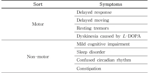
Table 4. Similar clinical symptoms of human PD and MPTP-treated NHP models
Sort Symptoms
Motor
Delayed response Delayed moving Resting tremors
Dyskinesia caused by L-DOPA
Non-motor
Mild cognitive impairment Sleep disorder
Confused circadian rhythm Constipation
Current PD therapy
Currently, PD therapy is based on drugs such as Levodopa (L-3,4-dihydroxyphenylalanine; L-DOPA) for the management of symptoms underlying motor disorders and deep brain stimulation (DBS), which is a surgical method of electrostimulation to specif- ic parts of the brain that control motor functions. Individual medications are used to relieve symptoms associated with non-motor abnormalities.
L-DOPA and supportive medication for PD therapy
L-DOPA has been used as a combination of L-DOPA and DOPA-decarboxylase inhibitor (DDCI) to increase the duration of drug efficacy for nearly 20 years since the approval by the United States Food and Drug Administration (FDA) as a PD therapy in 1970 after Gerge Cotzias confirmed its clinical useful- ness in 1967 (158). Subsequently, as a treatment of advanced PD, ergoline dopamine agonists (DAs) such as pergolide or cabergo- line, which directly affect the dopamine receptor alter the intrinsic neurotransmitter (159), and monoamine oxidase B (MAO-B) in- hibitors such as selegiline or rasagiline, which showed efficacy in MPTP-treated animals were used with L-DOPA (160). In the 1990s, catecholmethyltransferase inhibitors such as entacapone or trocapone were used to prevent degradation of L-DOPA in blood or across blood-brain barrier (161), in addition to non-ergoline DAs such as ropinirole and pramipexole (159). L-DOPA is still
used as a golden standard for PD therapy. Evidence supports the use of exogenous dopamine against endogenous dopamine deple- tion to ameliorate symptomatic parkinsonism but not to prevent progression of disorder (162-164).
Surgical approach for PD therapy
In addition, surgical treatment is used instead of drug admin- istration, and the first reported surgical treatment involved pallid- otomy (165, 166) and thalamotomy (167), which were used in ear- ly 1950s to excised the underlying basal nucleus. Since 1980, Brice and McLellan developed DBS, a reversible, controllable, and safer surgical method has been developed to suppress the disease progression via electrical stimulation to the midbrain and basal ganglia, operating in the most common mode (168). In the early stages of DBS, electrodes were also inserted into the ventral in- termediate nucleus of thalamus, which is effective both tremor and dyskinesia, without affecting other parkinsonisms (169).
Thus, electrical stimulation is commonly performed in the sub- thalamic nucleus and globus pallidus pars interna (170) during DBS. Additional insertion of electrodes into the pedunculopontine nucleus, assists with walk and balance (171).
Limitation of current therapy and alternative trials for PD
It is reported that side effects such as “Wearing-off” and
L-DOPA-induced dyskinesia may occur when PD patients are treated with L-DOPA long term (172, 173). In general, after an average of 5 years of L-DOPA treatment, at least 40% of pa- tients develop severe dyskinesia and motor fluctuations (174), as well as significant increases in non-motor symptoms during that period, according to several studies (19, 175). Therefore, novel and variable formulations to minimize the unexpected adverse effects associated with long-term L-DOPA treatment have been inves- tigated in field conditions; however, the formulation that can be used to exclude all adverse effects is not still exist (176, 177).
However, DBS may alleviate or control motor symptoms, but it can not resolve the underlying cause of PD and the mechanism of motor symptom relief is not fully understood (171). In addition, there is a limitation that not all PD patients undergo DBS sur- gery, but only patients who meet the criteria such as clinically defined age, duration and progression of PD, and especially, re- sponse to L-DOPA (178). Further, in most patients undergoing DBS surgery, the therapeutic effects on motor symptoms are recognizable; however, non-motor symptoms such as hallucina- tions and memory loss persist (179).
Due to limitations of current pharmacological and surgical thera- pies, multiple complementary or alternative therapies has been of- fered additionally to enhance the quality of patient’s life (180).
Most of the currently available pharmacological or alternative therapies may be used to managed major motor symptoms, and
molecular approaches such as neurotrophic factor therapy (181, 182), gene therapy (183), or cell replacement therapy (184-186) are emphasized. Neuromodulator therapy is based on the role of neurotrophins, peptides secreted from neuroglia, support cells, such as astrocytes or dendritic cells, in regulating growth and differentiation of nervous system (187, 188). Although neurtrophins associated with PD have been reduced or neuronal necrosis is not reported in human patients, neurotrophins may be of ther- apeutic value by promoting neuronal growth and function and disrupting neurotoxic processes. Neutrophins that are expected to have therapeutic effects include nerve growth factor, as well as brain-derived neurotrophic factor (BDNF), neurotrophin 3 and 4/5, glial cell-derived neurotrophic factor (GDNF), cerebral dopamine neurotrophic factor (CDNF), and recently discovered mesen- cephalic astrocyte-derived neurotrophic factor (MANF) (189).
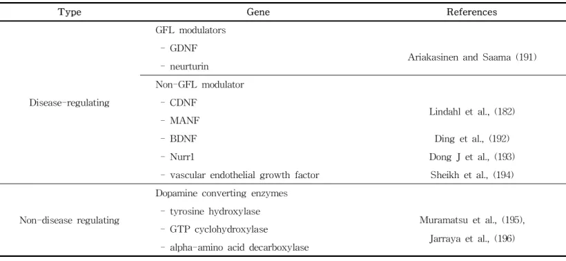
Gene therapy involves a combination of methods for disease reg- ulation and non-regulation. Most studies involves in disease con- trol aim to control cell necrosis associated with PD and re- generate necrotic cells by overexpressing neuronal regulatory substances with neuroprotective effects in the SN region.
However, non-disease regulation entails normalization of abnormal nerve signals in the basal nucleus by expressing DAergic or γ-aminobutryic acidergic enzymes, and control of motor dysfunc- tion rather than regulating the underlying cause of PD (190) (Table 5). Although those therapies are tested pre-clinically and
clinically, no remedy is available for complete patient recovery.
Table 5. Gene therapy studies for PD
Type Gene References
Disease-regulating
GFL modulators - GDNF
Ariakasinen and Saama (191) - neurturin
Non-GFL modulator - CDNF
Lindahl et al., (182) - MANF
- BDNF Ding et al., (192)
- Nurr1 Dong J et al., (193)
- vascular endothelial growth factor Sheikh et al., (194)
Non-disease regulating
Dopamine converting enzymes - tyrosine hydroxylase
Muramatsu et al., (195), Jarraya et al., (196) - GTP cyclohydroxylase
- alpha-amino acid decarboxylase
Development of stem cell therapy for PD
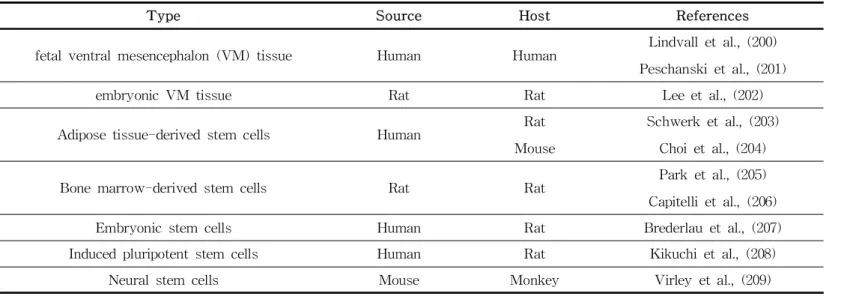
Among the alternative therapies introduced above, cell therapy facilitates cellular transplantation to replace necrotic or apoptotic DAergic cells affected by unknown causes and restore its func- tion (Table 6). Since a study published in 1979 demonstrating, improved dyskinesia and graft survival and differentiation after transplanting nerve cells containing DAergic cells derived from rat fetus in a rat model (197), fetal nigral cells transplantation has been reported to be effective not only in supplying dopamine but also to induce the formation of a neural network between the transplanted cells and existing cells. However, using aborted fe- tuses to obtain embryo-derived cells triggered ethical and im- munological concerns (198). To address these challenges, a meth- od of transplanting autologous cells was introduced, and clinical studies using various tissues to synthesize dopamine were also conducted (199). Two main approaches to cell transplantation therapy are availabe. First, neurorestoration, in which the trans- planted cells or tissues might play a direct role in existing cells or tissues, alleviating or regulating motor dysfunction caused by necrotic cells via secretion and release of neurotransmitters, syn- apse formation, and neural circuit formation. Second, the trans- planted cells and tissues modify host environment indirectly re- sulting in neuroprotection or neurorescue via anti-inflammatory, angiogenic or neurogenic, and immune regulatory effects (199).
Table 6. Tissues and cells used in cell transplantation studies for PD therapy
Type Source Host References
fetal ventral mesencephalon (VM) tissue Human Human Lindvall et al., (200) Peschanski et al., (201)
embryonic VM tissue Rat Rat Lee et al., (202)
Adipose tissue-derived stem cells Human Rat
Mouse
Schwerk et al., (203) Choi et al., (204)
Bone marrow-derived stem cells Rat Rat Park et al., (205)
Capitelli et al., (206)
Embryonic stem cells Human Rat Brederlau et al., (207)
Induced pluripotent stem cells Human Rat Kikuchi et al., (208)
Neural stem cells Mouse Monkey Virley et al., (209)
Implications of stem cell therapy
Stem cells can be divided into multipotent adult stem cells, pluri- potent embryonic stem cells (ESCs), and induced pluripotent stem cells (iPSCs) according to differentiation ability. Most of the cell transplantation therapies are expected to replace necrotic cells via differentiation or play a supplemental role in case of lost function.
However, the biggest risk is that of neoplasm due to excessive differentiation or hyperproliferation (210). Cell transplantation therapy must ensure the safety of the transplanted cells. General chemical-based therapy can identify adverse reactions within a short time or days after administration, in contrast to cell transplantation. The absolute physical time for proliferation and differentiation to an appropriate number ranges from a few weeks and months to a long time until the cell is settled at an appro- priate position in the recipient with functional expression.
Therefore, it cannot be evaluated via a safety evaluation method based on general chemical compounds (211).
Unlike other organs or tissues, the brain, in particular, has unique anatomical and histological characteristics. The blood-brain barrier prevents the direct entry of bacteria or cells, which are polymers greater than a nm in size. Therefore, a cell transplantation method for the treatment of brain diseases includ- ing PD requires, effectively delivery of cells other than direct transplantation into a specific brain region, and has yet to developed. Several studies have reported the improvement of mo-
tor function and the presence of DAergic neurons in the NHP model used in the experiment. Other studies have reported that only some of the animals used in the experiment were less ef- fective or not at all. In particular, the effects on survival, differ- entiation and proliferation, and transplanted individuals after transplantation differed depending on the origin, lineage, and de- gree of differentiation of the cells used in transplantation (Tab1e 7).
Table 7. Therapeutic effects of Cell transplantation using NHP PD models
Models Transplanted cells Results References
MPTP Monkey neural progenitor cells ․motor symptoms recovery was observed
․no recovery pattern was observed in PET images Takagi et al. (212) MPTP Human iPSC-derived neural
progenitor cells
․no recovery pattern was observed in PET images
․survival and differentiation of transplanted tissues were observed at 6 months after transplantation
Kikuchi et al. (213)
MPTP Human ESC-derived neural progenitor cells
․new tissues of D14-graft cells were observed
․dopamine-PET images of D42-graft cells were observed
․no motor symptoms recovery was observed
Doi et al. (214)
MPTP Monkey iPSC-derived DAergic precursor cells
․hyperplasia or new tissues was not observed
․no recovery pattern in PET images or motor symptoms were observed
Emborg et al (215)
MPTP Monkey bone marrow-derived mesenchymal stem cells
․motor symptoms recovery was gradually observed
․no new tissues were observed in biopsy and PET images
Hayashi et al. (216)
MPTP Monkey iPSCs
․motor symptoms recovery was observed in only 1 out of 3 animals
․no recovery pattern was observed in PET images
Hallett et al. (217)
6-OHDA Monkey embryonic nigra tissue
․motor symptoms recovery was observed at 6 months after transplantation
․Turning movements related to caudate nucleus and limb movements related to putamen were confirmed
Annett et al. (218)
The immune response of transplanted recipients is the most im- portant factor in the therapeutic effect of cell transplantation. In studies using rat models to date, the survival rate of DAergic neurons was only 3-20%, suggesting it is not possible to expect the effect because it cannot produce enough dopamine to improve motor symptoms. Most of the transplanted cells die within a week after transplantation. Within 24 hours, the cells are elimi- nated by apoptosis due to hypoxia or lack of nutrients, or phag- ocytosis by neutrophils. The immune response by microglia or astorcytes leads to loss of cells from day 3 after transplantation (219, 220). Therefore, as a strategy to maximize the effect on cell transplantation, various studies have been conducted to ensure that the transplanted cells survive as much as possible, and the cell survival rate is increased or rather decreased by simulta- neous treatment with GDNF, the known neurotrophic factor, or drugs that control the inflammatory response (221, 222). However, immunosuppressants administered to control the recipient's im- mune response can lead to infections or tumors induced by bac- teria or viruses, and an increased risk of teratoma formation by transplanted cells. Therefore, it is necessary to study the optimal immunosuppression strategy that can maximize the survival of transplanted cells while reducing the risk of the recipient (223, 224). The immune response is also an important factor in the se- lection of the animal model used in cell therapy studies. NHP models that can confirm the survival and function of transplanted
cells without immunosuppression when transplanting hu- man-derived cells as well as allogeneic cells, is the best choice for studying the effects of cell transplantation on motor symp- toms recovery or treatment (225).
Importance of early PD diagnosis and available biomarkers
Most people with PD manifest weak clinical symptoms are diag- nosed with PD after an average of 10 years since the actual on- set of PD. By then, approximately 70% of DAergic neurons are already lost, resulting in poor treatment nad prognostic outcomes based on a diagnosis of clinical symptoms (226). PD is not a le- thal disease, but because patients can lives for at least 20 years after the intervention, it is important to ensure that the quality of life is not deteriorated by various complications caused by motor and non-motor symptoms, and the increased familial, social and economic burdens of the patient (227). Therefore, early diagnosis of PD guarantees the patient's quality of life to the maximum extent possible, as it increases the range of options available for appropriate treatment to alleviate both movement and non-motor symptoms and delay the progress of the disease. In addition, there is a great advantage in reducing the costs of patient care (15).
The difficulty in diagnosis of PD based on motor symptoms
Current studies about treatment or prevention may be focused on finding specific early symptoms (228, 229) or biomarkers (230-232) for diagnosis and potential inhibition of disease progression. However, to date, there is no biomarker for early di- agnosis, and unfortunately, motor symptoms such as resting tremor, rigidity, posture impairment in PD are found in other CNS degenerative diseases such as multiple system atrophy (MSA), progressive supranuclear palsy (PSP), corticobasal degen- eration (CBD), dementia with LBs, essential tremor, and drug-in- duced Parkinsonism. Therefore, it has been reported that 6-8% of misdiagnosis is attributed to neurology specialists and 47% of misdiagnosis due to general practitioners diagnosing PD with on- ly movement symptoms (233-238).
Current imaging diagnostics for early stage PD
Currently, techniques for early diagnosis used clinically include imaging and biomarker detection using biologically derived samples. Diagnostic methods using imaging equipment include single photon emission computed tomography (SPECT), and posi- tron emission tomography (PET) using radioactive isotopes, and magnetic resonance imaging (MRI), magnetic resonance spectro- scopy (MRS), and transcranial sonography (TSC) without radio- active isotopes (239).
SPECT and PET
For SPECT and PET using radioactive isotopes, changes in dop- aminergic nerve pathways such as the SN and striatum are identified and used in diagnosis. SPECT is generally less ex- pensive than PET, facilitating its application in clinical trials.
Although the dopamine activity is reduced in patients at early stages of PD (240), it cannot differential diagnosis of, such as MSA, PSP, and CBD (241). However, PET has recently been ap- proved as a method for the diagnosis of PD by the USA FDA [18F]F-DOPA has been used to measure the activity of L-DOPA decarboxylase. Various radioisotope markers such as iodine ([123I]FP-CIT) and technetium ([99mTc]TRODAT) as well as fluo- rine ([18F]FP-CIT) related to dopamine transporter in dopamine neurons were used clinically (242-244). In particular, most studies using dopamine transporters reported a sensitivity higher than 95% and a specificity of 90-100%, and suggested that changes in intake of dopamine transporters were related to the severity of motor symptoms (245). However, about 15% of patients with early PD show normal findings in PET images of dopamine car- riers (246, 247).
MRI and MRS
MRI that does not use radioactive isotopes is used for diagnosis by identifying structural changes in PD-related region such as striatum or SN in the brain. It is particularly useful for differ-
ential diagnosis of atypical PD patients, and is used as an alter- native diagnostic to SPECT or PET. Recently, functional MRI with blood oxygen level dependent signals has been used as an indirect diagnostic method because the signal changes and patho- logical mechanisms are not fully understood (248). MRS is an- other diagnostic method using nuclear magnetic resonance for identifying metabolic changes in the brain. MRS is useful not only for diagnosis but also identification of effective biomarkers (249, 250). In addition, several studies have reported that it is particularly useful in differentiating PD and other neuro- degenerative diseases that exhibit similar movement symptoms (251).
TCS
TCS, another diagnostic method that does not use radioactive isotopes, focuses on the discovery of hyperechoic images in the SN of PD patients. van de Loo et al. (252) reported showed a diagnostic sensitivity of 79% and a specificity of 81%. Differential diagnosis is possible with diseases showed similar motor symp- toms of PD based on the change in the echo of the basal nu- cleus or lens nucleus except the SN (253, 254).
Biomarkers in biological fluids for early diagnosis of PD
Research is ongoing to explore the specific biomarkers for early
diagnosis of PD. However, unfortunately, biomarkers for accurate prediction of PD outbreaks in preclinical stages applicable in clin- ical trials have yet to be identified. In general, biomarkers are used to evaluate pathophysiology, and pharmacological responses to treatment attempts. Many studies have been conducted using cerebrospinal fluid (CSF) and blood to detect biomarkers for dif- ferential and early diagnosis of PD or other diseases with Parkinsonian symptoms, however, the results were inconsistent due to the large heterogeneity of the sample population and the method of obtaining non-standardized samples (255). Blood, sali- va, and biopsy tissues are sources of realistic biomarkers, be- cause cerebrospinal fluid is limited by the risks and costs asso- ciated with obtaining a specimen.
Biomarkers related oxidative stress in CSF and serum
Biomarkers for early diagnosis of PD to date can be divided into oxidative stress-related biomarkers, which are related to patho- genesis and progression of PD, and biomarkers related to abnor- mal protein accumulation and aggregation. Representative bio- markers related to oxidative stress include DJ-1 protein, uric acid, homocysteine (Hcy), and 8-hydroxydeoxyguanosine (8-OHdG) (Table 8).
DJ-1
DJ-1 protein has a neuroprotective effect against oxidative stress,
and DJ-1 dysfunction is known to induce various oxidative stress-related diseases including PD (256). In addition, several studies have been conducted in relation to the DJ-1 concentration and PD symptoms in the blood (257) or CSF (258), as mutations in the DJ-1 gene are associated with hereditary PD (259). In an early study, it was found that the concentration of DJ-1 in CSF or plasma in PD patients was higher than in the normal control group and was proportional to the Hoehn-Yahr (H & Y) score, another clinical diagnostic measure. However, studies using recent techniques have shown that the concentration of DJ-1 in plasma (260) and CSF (261) of PD patients is lower than that of normal controls, possibly due to errors in sample acquisition and purifi- cation in that most of the DJ-1 occurs in hemoglobin and plate- lets of red blood cells. Although the relationship between the progression of PD disease and DJ-1 is not clearly known (262), it is considered as an important potential biomarker for the diag- nosis of PD based on the results showing that the DJ-1 concen- tration in PD patients is lower than the CSF of patients with AD or MSA, other neurodegenerative disorders (263).
Uric acid
It is known that uric acid has an antioxidant effect as the free radicals (264), such as reactive oxygen species, produced during an inflammatory reaction presumed to be the cause of PD, gen- erate oxidative stress and necrosis of dopamine neurons in the
SN (265). In vitro experiments revealed that uric acid not only prevents DAergic neuronal necr