저작자표시-비영리-변경금지 2.0 대한민국 이용자는 아래의 조건을 따르는 경우에 한하여 자유롭게
l 이 저작물을 복제, 배포, 전송, 전시, 공연 및 방송할 수 있습니다. 다음과 같은 조건을 따라야 합니다:
l 귀하는, 이 저작물의 재이용이나 배포의 경우, 이 저작물에 적용된 이용허락조건 을 명확하게 나타내어야 합니다.
l 저작권자로부터 별도의 허가를 받으면 이러한 조건들은 적용되지 않습니다.
저작권법에 따른 이용자의 권리는 위의 내용에 의하여 영향을 받지 않습니다. 이것은 이용허락규약(Legal Code)을 이해하기 쉽게 요약한 것입니다.
Disclaimer
저작자표시. 귀하는 원저작자를 표시하여야 합니다.
비영리. 귀하는 이 저작물을 영리 목적으로 이용할 수 없습니다.
변경금지. 귀하는 이 저작물을 개작, 변형 또는 가공할 수 없습니다.
의학박사 학위논문
Analysis of various blood group genotypes and unexpected antibodies in Korean population for the establishment
of the rare blood program
국내 희귀혈액프로그램 구축을 위한 다양한 혈액형 유전형 및
비예기항체 분석
2016 년 8 월
서울대학교 대학원 의학과 검사의학 전공
홍 윤 지
i
Abstract
It is often difficult to supply adequate amounts of blood for patients with rare phenotypes in Korea. Moreover, the definition of rare blood is ambiguous, and much remains to be clarified. In this study, the prevalences of various human erythrocyte antigens (HEA) were determined from the donor viewpoint, and the demands for specific antigen-negative blood were estimated from the patient viewpoint. The data will aid the establishment of a Rare Blood Program in Korea (KRBP).
HEA genotyping of 419 blood donors (306 D-positives and 113 D-negatives) was performed using a Lifecodes RBC/RBC-R typing kit (Immucor, Norcross, GA, USA).
A national recipient registry website has been established where each hospital-based blood bank voluntarily enters data on detected and identified antibodies, as well as the outcomes of specific antigen testing. The availabilities of specific antigen-negative blood components were calculated based on these registry data, and the prevalences of the various HEAs were predicted by genotyping.
The prevalences of various red blood cell (RBC) antigens in the D-negative population were determined for the first time, and the Cartwright, Scianna, Dombrock, Colton, Landsteiner-Wiener, Cromer, and Knops blood group systems were identified.
The availabilities of specific antigen-negative units differed when the calculations were based on serotyping or genotyping, especially in the D-negative group. “Rare blood”
was defined as “blood units without antigens that are prevalent in less than 1% of the population”. According to the definition, Fya− blood, Dib− blood, and D− blood were defined as rare blood. In multiple antigen-negative blood, D+C−E− blood, D+c−e−
ii
blood, and S−s− blood were defined. In consideration of the multicenter registry, M−
AND E− AND (Jka− and/or Jkb−) AND (Lea− and/or Leb−) blood were defined as rare blood.
Data on the prevalences of various blood antigens are essential to estimate the availabilities of blood components that are appropriate for use by patients expressing relevant antibodies. Blood banks would then be able to efficiently supply safe blood products.
Keywords: Korean Rare Blood Program, KRBP, rare blood, unexpected antibody, recipient registry
Student number: 2013-21715
iii
Contents
Abstract
∙∙∙∙∙∙∙∙∙∙∙∙∙∙∙∙∙∙∙∙∙∙∙∙∙∙∙∙∙∙∙∙∙∙∙∙∙∙∙∙∙∙∙∙∙∙∙∙∙∙∙∙∙∙∙∙∙∙∙∙∙∙∙∙∙∙∙∙∙∙∙∙∙∙∙∙∙∙∙∙∙∙∙ iContents
∙∙∙∙∙∙∙∙∙∙∙∙∙∙∙∙∙∙∙∙∙∙∙∙∙∙∙∙∙∙∙∙∙∙∙∙∙∙∙∙∙∙∙∙∙∙∙∙∙∙∙∙∙∙∙∙∙∙∙∙∙∙∙∙∙∙∙∙∙∙∙∙∙∙∙∙∙∙∙∙∙∙ iiiList of tables
∙∙∙∙∙∙∙∙∙∙∙∙∙∙∙∙∙∙∙∙∙∙∙∙∙∙∙∙∙∙∙∙∙∙∙∙∙∙∙∙∙∙∙∙∙∙∙∙∙∙∙∙∙∙∙∙∙∙∙∙∙∙∙∙∙∙∙∙∙∙∙∙∙∙∙∙ vList of figures
∙∙∙∙∙∙∙∙∙∙∙∙∙∙∙∙∙∙∙∙∙∙∙∙∙∙∙∙∙∙∙∙∙∙∙∙∙∙∙∙∙∙∙∙∙∙∙∙∙∙∙∙∙∙∙∙∙∙∙∙∙∙∙∙∙∙∙∙∙∙∙∙∙∙ viIntroduction
∙∙∙∙∙∙∙∙∙∙∙∙∙∙∙∙∙∙∙∙∙∙∙∙∙∙∙∙∙∙∙∙∙∙∙∙∙∙∙∙∙∙∙∙∙∙∙∙∙∙∙∙∙∙∙∙∙∙∙∙∙∙∙∙∙∙∙∙∙∙∙∙∙ 1Materials and Methods
∙∙∙∙∙∙∙∙∙∙∙∙∙∙∙∙∙∙∙∙∙∙∙∙∙∙∙∙∙∙∙∙∙∙∙∙∙∙∙∙∙∙∙∙∙∙∙∙∙∙∙∙∙∙ 41.1 Study population for human erythrocyte antigen (HEA) typing ∙∙∙∙∙∙∙∙∙∙∙∙∙∙∙∙∙∙∙∙∙∙∙ 4 1.2 DNA extraction and molecular analysis for HEA typing ∙∙∙∙∙∙∙∙∙∙∙∙∙∙∙∙∙∙∙∙∙∙∙∙∙∙∙∙∙∙∙∙∙∙∙ 6 1.3 Statistics ∙∙∙∙∙∙∙∙∙∙∙∙∙∙∙∙∙∙∙∙∙∙∙∙∙∙∙∙∙∙∙∙∙∙∙∙∙∙∙∙∙∙∙∙∙∙∙∙∙∙∙∙∙∙∙∙∙∙∙∙∙∙∙∙∙∙∙∙∙∙∙∙∙∙∙∙∙∙∙∙∙∙∙∙∙∙∙∙∙∙∙∙∙∙∙∙∙∙∙∙∙∙∙∙∙∙∙∙∙∙∙∙∙∙∙∙∙∙ 6 2.1 Study population for DEL typing ∙∙∙∙∙∙∙∙∙∙∙∙∙∙∙∙∙∙∙∙∙∙∙∙∙∙∙∙∙∙∙∙∙∙∙∙∙∙∙∙∙∙∙∙∙∙∙∙∙∙∙∙∙∙∙∙∙∙∙∙∙∙∙∙∙∙∙∙∙∙∙∙∙∙∙ 6 2.2 Development of real-time PCR and melting curve analysis for DEL detection ∙ 8 2.3 Real-time PCR for RHD complete deletion and RHD-CE-D hybrid variants ∙∙ 10 2.4 Multiplex single-base primer extension reaction for DEL confirmation ∙∙∙∙∙∙∙∙∙∙∙∙ 10 3. Establishment of a multicenter registration system ∙∙∙∙∙∙∙∙∙∙∙∙∙∙∙∙∙∙∙∙∙∙∙∙∙∙∙∙∙∙∙∙∙∙∙∙∙∙∙∙∙∙∙∙∙∙∙∙∙ 11
Results
∙∙∙∙∙∙∙∙∙∙∙∙∙∙∙∙∙∙∙∙∙∙∙∙∙∙∙∙∙∙∙∙∙∙∙∙∙∙∙∙∙∙∙∙∙∙∙∙∙∙∙∙∙∙∙∙∙∙∙∙∙∙∙∙∙∙∙∙∙∙∙∙∙∙∙∙∙∙∙∙∙∙ 131.1 Genotypes of 22 blood group antigens ∙∙∙∙∙∙∙∙∙∙∙∙∙∙∙∙∙∙∙∙∙∙∙∙∙∙∙∙∙∙∙∙∙∙∙∙∙∙∙∙∙∙∙∙∙∙∙∙∙∙∙∙∙∙∙∙∙∙∙∙∙∙∙∙∙∙∙∙ 13 1.2 Expected phenotypes of blood groups ∙∙∙∙∙∙∙∙∙∙∙∙∙∙∙∙∙∙∙∙∙∙∙∙∙∙∙∙∙∙∙∙∙∙∙∙∙∙∙∙∙∙∙∙∙∙∙∙∙∙∙∙∙∙∙∙∙∙∙∙∙∙∙∙∙∙∙∙ 16 2.1 RhD genotypic profiles in serologically D-negative Koreans ∙∙∙∙∙∙∙∙∙∙∙∙∙∙∙∙∙∙∙∙∙∙∙∙∙∙∙∙∙∙ 24
iv
2.2 DEL confirmation using multiplex single-base primer extension reaction ∙∙∙∙∙∙∙∙ 27 2.3 RhCEce profiles ∙∙∙∙∙∙∙∙∙∙∙∙∙∙∙∙∙∙∙∙∙∙∙∙∙∙∙∙∙∙∙∙∙∙∙∙∙∙∙∙∙∙∙∙∙∙∙∙∙∙∙∙∙∙∙∙∙∙∙∙∙∙∙∙∙∙∙∙∙∙∙∙∙∙∙∙∙∙∙∙∙∙∙∙∙∙∙∙∙∙∙∙∙∙∙∙∙∙∙∙∙∙∙∙∙∙∙ 27 3. Likelihoods of obtaining specific antigen-negative bloods ∙∙∙∙∙∙∙∙∙∙∙∙∙∙∙∙∙∙∙∙∙∙∙∙∙∙∙∙∙∙∙∙∙∙∙∙∙∙∙ 30
Discussion
∙∙∙∙∙∙∙∙∙∙∙∙∙∙∙∙∙∙∙∙∙∙∙∙∙∙∙∙∙∙∙∙∙∙∙∙∙∙∙∙∙∙∙∙∙∙∙∙∙∙∙∙∙∙∙∙∙∙∙∙∙∙∙∙∙∙∙∙∙∙∙∙∙∙∙∙∙ 32References
∙∙∙∙∙∙∙∙∙∙∙∙∙∙∙∙∙∙∙∙∙∙∙∙∙∙∙∙∙∙∙∙∙∙∙∙∙∙∙∙∙∙∙∙∙∙∙∙∙∙∙∙∙∙∙∙∙∙∙∙∙∙∙∙∙∙∙∙∙∙∙∙∙∙∙∙∙ 40Abstract in Korean
∙∙∙∙∙∙∙∙∙∙∙∙∙∙∙∙∙∙∙∙∙∙∙∙∙∙∙∙∙∙∙∙∙∙∙∙∙∙∙∙∙∙∙∙∙∙∙∙∙∙∙∙∙∙∙∙∙∙∙∙∙∙ 46v
List of Tables
Table 1. Anti-D antisera used in this study ∙∙∙∙∙∙∙∙∙∙∙∙∙∙∙∙∙∙∙∙∙∙∙∙∙∙∙∙∙∙∙∙∙∙∙∙∙∙∙∙∙∙∙∙∙∙∙∙∙∙∙∙∙∙∙ 5 Table 2. PCR primers and probes used for RHD genotyping ∙∙∙∙∙∙∙∙∙∙∙∙∙∙∙∙∙∙∙∙∙∙∙∙ 9 Table 3. HEA genotypes in the Korean population ∙∙∙∙∙∙∙∙∙∙∙∙∙∙∙∙∙∙∙∙∙∙∙∙∙∙∙∙∙∙∙∙∙∙∙∙∙∙∙∙ 15 Table 4. Prevalence of the Rh phenotype in Koreans ∙∙∙∙∙∙∙∙∙∙∙∙∙∙∙∙∙∙∙∙∙∙∙∙∙∙∙∙∙∙∙∙∙∙∙∙∙∙ 18 Table 5. Prevalences of expected MNS phenotypes in Koreans ∙∙∙∙∙∙∙∙∙∙∙∙∙∙∙∙∙∙∙∙ 19 Table 6. Prevalences of expected phenotypes of various HEA in Koreans ∙ 20 Table 7. Expected HEA phenotypes for each antigen in D-positives and D-
negatives∙∙∙∙∙∙∙∙∙∙∙∙∙∙∙∙∙∙∙∙∙∙∙∙∙∙∙∙∙∙∙∙∙∙∙∙∙∙∙∙∙∙∙∙∙∙∙∙∙∙∙∙∙∙∙∙∙∙∙∙∙∙∙∙∙∙∙∙∙∙∙∙∙∙∙∙∙∙∙∙∙∙∙∙∙∙∙∙∙∙∙∙∙∙∙∙∙∙∙∙∙ 22 Table 8. Molecular characteristics of serologically D-negative samples in
Koreans∙∙∙∙∙∙∙∙∙∙∙∙∙∙∙∙∙∙∙∙∙∙∙∙∙∙∙∙∙∙∙∙∙∙∙∙∙∙∙∙∙∙∙∙∙∙∙∙∙∙∙∙∙∙∙∙∙∙∙∙∙∙∙∙∙∙∙∙∙∙∙∙∙∙∙∙∙∙∙∙∙∙∙∙∙∙∙∙∙∙∙∙∙∙∙∙∙∙∙∙∙∙ 26 Table 9. Available proportions of specific antigen-negative units in blood banks (%), calculated by serological antigen testing and via genotyping ∙∙∙∙∙ 31 Table 10. Proposal of rare blood units based on various blood group genotypes and unexpected antibodies in Korean population ∙∙∙∙∙∙∙∙∙∙∙∙∙∙∙∙∙∙∙∙∙∙∙∙∙∙∙∙∙∙∙∙39
vi
List of Figure
Figure 1. The homepage of KRBP website of recipient registry ∙∙∙∙∙∙∙∙∙∙∙∙∙∙∙∙∙∙∙∙∙∙∙∙∙∙∙∙ 12 Figure 2. Differentiation of RHD (c.1222T>C) DEL, RHD (c.1227G>A) DEL,
weak D and RHD absence by melting curve analysis ∙∙∙∙∙∙∙∙∙∙∙∙∙∙∙∙∙∙∙∙∙∙∙∙∙∙∙∙∙∙∙∙∙∙ 25 Figure 3. Multiplex single-base primer extension reaction of RHD ∙∙∙∙∙∙∙∙∙∙∙∙∙∙∙∙∙∙∙∙∙∙ 28 Figure 4. RhCEce phenotype of 199 serologically RhD-negative individuals ∙∙∙∙∙ 29
1
Introduction
The main role of blood bank is to provide compatible blood components for patients.
Today, the focus is on “immune compatibility” with the recipient as well as prevention of transfusion-transmitted disease.
As acute hemolytic reactions are occasionally life-threatening, the ABO blood type of a transfused component is always matched to that of a patient. Whenever possible, D-positive patients should be transfused with D-positive blood because D-negative blood should be reserved for D-negatives. D-negatives, especially females in their childbearing years, must receive only D-negative blood, to prevent alloimmunization and production of anti-D antibodies (Roback, Mark K. Fung et al. 2014). For blood groups other than ABO and Rh, the transfusion policy varies among institutions, ranging from crossmatching of compatible blood supplies unless clinically significant alloantibodies are detectable to specific antigen-matching in certain patients suffering from sickle-cell disease or hematological malignancies (Ribeiro, Guarnieri et al. 2009, Li, Jiang et al. 2010, Chou 2013).
If alloantibodies are evident, a blood bank will seek to locate blood that lacks the antigen recognized by that antibody. However, it is often difficult to source suitable blood components if such antigens are highly prevalent in populations, or when multiple antigens should be excluded. If all such attempts fail, blood banks inevitably release units that are at least somewhat incompatible (Li, Jiang et al. 2010).
In the time since Mourant emphasized the need to establish an international panel of rare donors at the general assembly of the International Society of Blood Transfusion
2
(ISBT) in the 1960s, the importance of such a panel has been recognized worldwide (Mourant 1965). An ISBT Working Party on Rare Donors has been established and regular formal meetings are held. Moreover, many countries have organized local programs for rare blood donors when scarcities associated with ethnicity are in play (Nance 2009).
In Korea, it is undeniably true that the definition of “rare blood” is ambiguous, and much remains to be learned prior to the establishment of a rare blood program. Most research on the frequencies of various blood groups in Koreans was performed decades ago using serological methods and targeting only a few antigens (Lee 1965, Lee 1975, Kim, Suh et al. 2003). Moreover, the actual means by which frontline transfusion services select blood components remains unclear; it is uncertain how often blood banks successfully identify immunologically compatible blood, and what procedures are followed if problems are encountered.
In the Korean population, the ABO blood type is evenly distributed and ABO blood type components are readily available. For the Rh blood group, however, the circumstances are different. In East Asian populations the frequency of D-negative individuals is very low (<1%). Among Koreans it is even rarer, less than 0.5% of the total population (Delaney, Harris et al. 2015). In routine practice, it is often difficult to supply adequate amounts of components for D-negative patients, and the situation is rendered worse when blood banks receive requests for D-negative components for patients with multiple alloantibodies.
Moreover, among the people typed as D-negative by the indirect antiglobulin test (IAT) in East Asia, 15–30% carry an D variant known as DEL (Chen, Lin et al. 2004, Kim, Kim et al. 2005). DEL is associated with extremely low levels of D antigen
3
expression and thus the presence of this antigen is confirmed indirectly using adsorption-elution techniques. Owing to these difficulties, anti-D alloimmunization has been reported in D-negative recipients after the transfusion of mistyped DEL blood components (Wagner, Kormoczi et al. 2005, Kim, Kim et al. 2009, Yang, Lee et al.
2015).
It is critical to identify DEL individuals among serologically D-negative blood donors. However, DEL typing using adsorption-elution techniques is prone to technical errors (Nuchnoi, Thongbus et al. 2014). Therefore, there is a growing interest in molecular assays to detect DEL.
In the present study, the author surveyed the demand for specific blood components from the patient viewpoint, and explored the prevalence of various red blood cell antigens from the donor viewpoint, to define both “rare blood” and “rare donor” in Korea in the future. Especially, the author gave weight to D-negatives, and the prevalence of various red blood cell antigens was calculated based on two separate groups, D-positives and D-negatives. In addition, the author investigated RHD variants, focused on DEL, in Korean population by developing a convenient molecular technique to detect DEL. Such information is essential for the establishment of a rare blood program in Korea.
4
Materials and Methods
1.1 Study population for human erythrocyte antigen (HEA) typing
Peripheral venous blood was collected from 419 healthy blood donors into EDTA- containing tubes; the sample contained 306 D-positives and 113 D-negatives. All donors were of Korean origin; other races were excluded via questionnaire. Using routine tube methods, D antigens were screened using a mixture of IgM + IgG monoclonal anti-D (Merck Millipore, Darmstadt, Germany). When the D-antigen status was negative, further tests using seven different antisera were performed to rule out weak D or partial D (Table 1). The study was approved by the Institutional Review Board of the Seoul National University Bundang Hospital (B-1103-123-005) and the participants were able to provide written informed consent.
5 Table 1. Anti-D antisera used in this study.
Product name Type of
Immunogl obulin
Clone Manufacturer Reagent
type
BIOSCOT Anti-D IgM/IgG blend IgM, IgG TH-28
MS-26
Merck Millipore LPMB
immuClone Anti-D rapid IgM IgM RUM-1 Immucor LPM
NOVACLONE Anti-D IgM + IgG Monoclonal Blend IgM, IgG D175-2 D415 1E4
Immucor LPMB
Anti-D Bioclone IgM, IgG MAD-2 blend with polyclonal Ortho Clinical Diagnostics LPMPB
ANTI-D TOTEM IgM, IgG P3X61
P3X21223B10 P3X290
P3X35
Diagast LPMB
Anti-D (RH1) IgG IgG HM16 Diagast LPM
IgM anti-D IgM MS-201 Grifols* LPM
Combi anti-D Mono-Type IgM, IgG LDM3
ESD1
Grifols* LPMB
LPM, low protein monoclonal; LPMB, low protein monoclonal blend; LPMPB, low protein monoclonal/polyclonal blend
*Formerly known as Medion Diagnostics
6
1.2 DNA extraction and molecular analysis for HEA typing
Genomic DNA was extracted from 200 μL whole blood using a DNA purification kit (QIAmp Blood Mini Kit, Qiagen, Hilden, Germany) according to the manufacturer’s instructions. PCR was performed on final volumes of 25 µL containing genomic DNA (100 ng), master mix (10 µL), and Taq polymerase (0.25 µL), with the aid of a PTC- 200 Thermal Cycler (MJ Research, Watertown, MA) under the following PCR conditions: 95°C for 4 min; 45 cycles of 95°C for 30 s, 51°C for 45 s, and 65°C for 30 s; and 65°C for 5 min. DNA amplicon (5 µL) and probe mix (15 µL) were incubated at 56°C for 22 min prior to the addition of 170 µL Dilution Solution/PE-Streptavidin mixture provided by Lifecodes RBC/RBC-R Typing Kit (Immucor). Fluorescence emission by amplified products was analyzed using MATCH IT!-RBC software (Immucor) on a Luminex platform (Luminex, Austin, TX).
1.3 Statistics
The chi-squared test and Fisher’s exact test were run using SPSS version 21. A P value
≤ 0.05 was considered to reflect statistical significance.
2.1 Study population for DEL typing
A total of 325 RhD-negative individuals were involved in this study. Specimens were collected in two separate periods. Period 1 included 199 individuals: RhD negativity was confirmed using 8 different reagents (Table 1), and RhCEce typing was further evaluated with antisera (Diagast, Loos, France). In period 2, RhD typing was performed on 126 individuals using two of the following three antisera: BIOSCOT
7
Anti-D IgM/IgG blend (Merck Millipore), Anti-D Bioclone (Ortho Clinical Diagnostics, Raritan, NJ, USA), and NOVACLONE Anti-D IgM + IgG (Immucor, Norcross, GA, USA). All phenotyping tests were performed in accordance with the manufacturer’s instruction.
8
2.2 Development of real-time PCR and melting curve analysis for DEL detection
DEL variants with RHD (c.1222T>C) or RHD (c.1227G>A) were detected using real- time PCR and melting curve analysis. The real-time PCR procedure for DEL was as follows: 50 ng DNA template mixed with 2 μL LC FastStart DNA Master HybProbe (Roche, Penzberg, Germany), 0.3 μM each primer, 0.3 μL each probe (Table 2), and 4.0 mM MgCl2 in a final volume of 20 μL. The following PCR program was used: 10 minutes at 95°C and 35 cycles of 5 seconds at 95°C, 10 seconds at 60°C, and 10 seconds at 72°C. The program for analytical melting was 30 se9conds at 95°C and 2 minutes at 40°C and an increase to 80°C at a ramp rate of 0.4°C per second. PCR amplification was checked and the melting temperature was analyzed. In the presence of RHD variants such as weak D and DEL, fragments were amplified and could be distinguished based on melting temperature. In the absence of RHD, no amplification occurred.
9 Table 2. PCR primers and probes used for RHD genotyping.
Name of primer Nucleotide sequence Position
RHK409-F 5'-AAAATATGGAAAGCACCTCATGA-3' 1,171-1,193
RHK409-R 5'-ATGGATTTGTTTCTCCTCTAGTT-3' 1,359 - 1,381
RHK409-D LC Red 640-ATCTTACCTTCCAGAAAACTTGGTCATCAA-Phosphate 1,205 - 1,234
RHK409-A CATGCACTCAAAATCTATCACGTTAATAGGTGAA-Fluorescein 1,237 - 1,270
RHZ10-F 5'-CCTCTCACTGTTGCCTGCATT-3' 3’-UTR
RHZ10-R 5'-AGTGCCTGCGCGAACATT-3' 3’-UTR
RHZ10-P 5'-FAM-TACGTGAGAAACGCTCATGACAGCAAAGTCT-TAMRA-3' 3’-UTR
RHD1222Ty-F 5’-AAATATTTGATGACCAAGTTTTC-3’ 1,198 - 1,221
RHD1227Ty-R 5’-T10-ACGTTAATAGGTGAAAAATCTTAC-3’ 1,228 - 1,251
10
2.3 Real-time PCR for RHD complete deletion and RHD-CE- D hybrid variants
Real-time PCR targeted to the 3’ untranslated region (3’-UTR) of exon 10 was performed to confirm the presence of complete deletion of RHD and RHD-CE-D hybrid variants (Kim, Kim et al. 2009). A real-time PCR assay for exon 10 of RHD genes was designed to screen for RhD deletion. The primers and probes are listed in Table 2. PCR was completed according to the following conditions: 100 ng DNA template was mixed with 4 μL LC TaqMan Master (Roche, Penzberg, Germany), 0.4 μM each primer, and 0.4 μL probe in a final volume of 20 μL. The PCR program was as follows: 10 minutes at 94°C and 40 cycles of 10 seconds at 94°C, 15 seconds at 55°C, and 10 seconds at 72°C. PCR products were amplified in the presence of RHD- CE-D hybrid variants and not amplified in the presence of complete RHD deletion.
2.4 Multiplex single-base primer extension reaction for DEL confirmation
RHD genotyping was performed for two sites, RHD (c.1222T>C) and RHD (c.1227G>A). The DEL specimens were included in the RHD single nucleotide extension assay. Amplicons produced using real-time PCR were used after ExoSAP purification (ExoSap-it, Affymetrix, Cleveland, OH, USA). Multiplex single-base primer extension reactions were performed using an ABI PRISM SNaPshot Multiplex kit (Applied Biosystems, Foster City, CA, USA) and the primers shown in Table 2.
Reactions were performed in a final volume of 10 µL, containing 1 µL each template,
11
5 µL reaction mix, and 0.4 µL primers for RHD (c.1222T>C) or RHD (c.1227G>A).
Cycling conditions were as follows: 25 cycles at 96°C for 10 seconds, 50°C for 5 seconds, and 60°C for 30 seconds. The products were treated with shrimp alkaline phosphatase (SAP) and separated using 0.15 µL GeneScan-120 LIZ size standard with an ABI PRISM 3100 Genetic Analyzer, and data were analyzed using GeneMapper Analysis Software version 2.0 (Applied Biosystems).
3. Establishment of a multicenter registration system
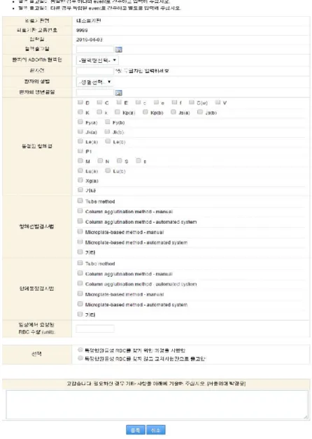
A national recipient registry website is part of a nationwide transfusion initiative that commenced in July 2013. Each hospital blood bank was asked to voluntarily register data on antibody detection and identification, and specific antigen testing, on the website (http://safeblood.or.kr). The frequencies at which tests for specific antigens were run to locate antigen-negative units were also entered on the website (figure 1).
Information entered from July 2013 to February 2014 was analyzed, to define the characteristics of blood components sought and available (Chung, Hong et al. 2014).
12
Figure 1. The homepage of KRBP website of recipient registry to investigate current state of alloantibodies detected in the recipient viewpoint. It is designed to input alloantibodies detected, the number of units requested from clinicians, and so on.
13
Results
1.1 Genotypes of 22 blood group antigens
The prevalences of the HEA genotypes of 22 blood group antigens in 419 individuals are described below. In terms of the S and s alleles of the MNS blood group system, one specimen was indeterminate in terms of call, and three presented with a S− s−
phenotype due to GYPB deletion. The numbers of GYPA*M/GYPA*M homozygotes, GYPA*M/GYPA*N heterozygotes, and GYPA*N/GYPA*N homozygotes in the GYPA
alleles were 68 (22.2%), 162 (53.0%), and 76 (24.8%) among D positives, and 19 (16.8%), 65 (57.5%), and 29 (25.7%) among D negatives, respectively. The distribution of S/s genotypes were 2 (0.7%), 28 (9.3%), and 272 (90.0%) among D positives, and 0 (0.0%), 4 (3.5%), and 109 (96.5%) among D negatives.
In terms of the Rh blood group system, RHCE*C/RHCE*C homozygotes, RHCE*C/RHCE*c heterozygotes, and RHCE*c/RHCE*c homozygotes of the C and
c alleles numbered 127 (41.5%), 140 (45.8%), and 39 (12.7%) among D positives, and 3 (2.7%), 26 (23.0%), and 84 (74.3%) among D negatives. Those with the combination of the E and e alleles numbered 35 (11.4%), 113 (36.9%), and 158 (51.7%) among D positives, and 1 (0.9%), 15 (13.3%), and 97 (85.8%) among D negatives.
In terms of the Duffy blood group system, FY*A/FY*A homozygotes, FY*A/FY*B heterozygotes, and FY*B/FY*B homozygotes numbered 249 (81.4%), 56 (18.3%), and 1 (0.3%) among D positives, and 96 (85.0%), 17 (15.0%), and 0 (0.0%) among D-
14
negatives. In particular, one D positive donor had a c.265C>T single nucleotide polymorphism. In the Kidd and Lutheran blood group systems, JK*A/JK*A homozygotes, JK*A/JK*B heterozygotes, and JK*B/JK*B homozygotes, LU*A/LU*A homozygotes, LU*A/LU*B heterozygotes, LU*B/LU*B homozygotes numbered 57(18.6%), 172 (56.2%), 77(25.2%), 0 (0.0%), 5 (1.7%), and 293 (98.3%) among D positives, and 25 (22.1%), 56 (49.6%), 32 (28.3%), 0 (0.0%), 0 (0.0%), and 113 (100.0%) among D negatives.
In terms of the Dombrock, Cartwright, and Diego blood groups system, AA homozygotes, AB heterozygotes, and BB homozygotes in the DO*A/DO*B, YT*A/YT*B, and DI*A/DI*B alleles numbered 6 (2.0%), 51 (16.7%), 249 (81.3%),
304 (99.7%), 1 (0.3%), 0 (0.0%), 1 (0.3%), 44 (14.4%), and 261 (85.3%) among D positives, and 0 (0.0%), 22 (19.5%), 91 (80.5%), 113 (100.0%), 0 (0.0%), 0 (0.0%), 2 (1.8%), 8 (7.1%), and 103 (91.1%) among D negatives. All subjects were BB homozygotes for the three K/k, Kpa/Kpb/Kpc, Jsa/Jsb polymorphisms at the KEL locus, and AA homozygous for all polymorphisms tested in the LW, SC, CROM, KN blood group systems. In addition, one specimen did not possess any allele relevant to the Cartwright blood group system. The Lutheran blood group system, for which six subjects were indeterminate and for which two test failures were encountered, exhibited the highest number of specimens for which the genotype could not be determined (Table 3).
15 Table 3. HEA genotypes in the Korean population.
Blood group system
Antigen D positive D negative
Allele AA AB BB AA AB BB
MNS M/N 68 162 76 19 65 29
S/s* 2 28 272 0 4 109
GYPB 230C/230T 306 0 0 113 0 0
GYPB +5G/+5T 306 0 0 113 0 0
Rh c/C 39 140 127 84 26 3
e/E 158 113 35 97 15 1
Lutheran LUA/LUB† 0 5 293 0 0 113
Kell K/k 0 0 306 0 0 113
KPA/KPB/KPC 0 0 306 0 0 113
JSA/JSB 0 0 306 0 0 113
Duffy FYA/FYB 249 56 1 96 17 0
FY 265C/265T(FYX) 305 1 0 113 0 0
FY -33T/-33C(GATA) 306 0 0 113 0 0
Kidd JKA/JKB 57 172 77 25 56 32
JK 306 0 0 113 0 0
Diego DIA/DIB 1 44 261 2 8 103
WRA/WRB 0 0 306 0 0 113
Cartwright YTA/YTB‡ 304 1 0 113 0 0
Scianna SC1/SC2 306 0 0 113 0 0
Dombrock DOA/DOB 6 51 249 0 22 91
DO 323G/323T 306 0 0 113 0 0
DO 350C/350T 306 0 0 113 0 0
Colton COA/COB 306 0 0 113 0 0
Landsteiner LWA/LWB 306 0 0 113 0 0
Cromer CR 679G/679C 306 0 0 113 0 0
Knops KNA/KNB 306 0 0 113 0 0
McCA/McCB 306 0 0 113 0 0
SL1/SL2 306 0 0 113 0 0
306 113
* One specimen showed ID, and 3 were failed to analyze.
† Six specimens showed ID, and 2 were failed to analyze.
‡ One specimen was failed to analysis.
16
1.2 Expected phenotypes of blood groups
The expected phenotypes of blood antigens are shown in Table 4 – Table 6. In the Rh blood group systems, the DCe/DCe (R1/R1) phenotype was the most common (41.3%) followed by DCe/DcE(R1/R2) (35.4%), DcE/DcE (R2/R2) (11.5%), and DCe/dce (R1/r) (10.2%). The D+C−E+c+e+, D+C+E+c−e+, and D+C−E−c+e+ phenotypes were rare. On the other hand, of all D negatives, 64.9% were D−C−E−c+e+ (the most common phenotype). The second most common phenotype was D−C+E−c+e+(19.8%), followed by D−C−E+c+e+ (9.0%), D−C+E+c+e+ (2.7%), D−C+E−c−e+ (1.8%), D−C−E+c+e− (0.9%), and D−C+E+c−e+ (0.9%). Neither D+C+E+c+e− nor D+C+E+c−e−, corresponding to D−C+E+c+e− and D−C+E+c−e−
in D-negatives, was detected (Table 4).
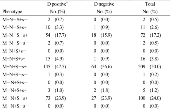
Of the 419 individuals, 25.1% and 20.8% were M- and N-negative, respectively, and in terms of the S and s antigens, which could not be detected in one specimen, 91.9% and 1.2% were negative for S and s, respectively. In terms of combinations of M and N antigens, the M+N+ phenotype was the most prevalent (54.2%). The frequencies of the phenotypes S+s−, S+s+, S−s+, and S−s− were 0.5%, 7.7%, 91.1%, and 0.7%, respectively. The most common phenotypic combination in the MNS blood group was M+N+S−s+ (50%) (Table 5).
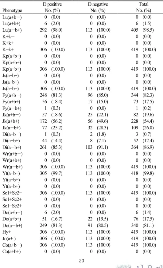
The expected phenotypes in terms of other blood groups ([a+b−], [a+b+], and [a−b+]) were as follows: Lutheran, 0.0%, 1.5%, and 98.5%; Duffy, 82.3%, 17.5%, and 0.2%;
Kidd, 19.6%, 54.4%, and 26.0%; Diego, 0.7%, 12.4%, 86.9%; and Dombrock 1.4%, 17.5%, and 81.1%. One individual, with an FYA 265 mutation evident upon genotyping, failed to yield phenotypic data. Wr(a−b+), one of the Diego blood group
17
antigens, was expressed in all individuals, as were Hy+ and Jo(a+) of the Dombrock blood group system. All individuals were identical in terms of Kell, Cartwright, Scianna, Colton, Landsteiner-Wiener, Cromer, and Knops blood grouping (Table 6 &
Table 7).
18 Table 4. Prevalence of the Rh phenotype in Koreans.
Phenotype No. of
specimens (%)
Most probable
genotype Other possible genotype
D positive
D+C+E−c−e+ 126 (41.3) R1R1 R1 r′
D+C+E+c+e+ 107 (35.1) R1R2 Rzr, R2r′,R1r″, RzR0, R0ry
D+C−E+c+e− 35 (11.5) R2R2 R2r′
D+C+E−c+e+ 32 (10.5) R1r R1R0, R0r′
D+C−E+c+e+ 3 (1.0) R2r R2R0, R0r′
D+C+E+c−e+ 1 (0.3) R1Rz Rzr′, R2ry
D+C+E+c+e− 0 (0.0) R2Rz Rzr′, R2ry
D+C−E−c+e+ 1 (0.3) R0r R0R0
D+C+E+c−e− 0 (0.0) RzRz Rzry
D negative
D−C+E−c−e+ 2 (1.8) r′r′
D−C+E+c+e+ 3 (2.7) r′r″, rry
D−C−E+c+e− 1 (0.9) r″r″
D−C+E−c+e+ 22 (19.8) rr′
D−C−E+c+e+ 10 (9.0) rr″
D−C+E+c−e+ 1 (0.9) r′ry
D−C+E+c+e− 0 (0.0) r″ry
D−C−E−c+e+ 72 (64.9) rr
D−C+E+c−e− 0 (0.0) ryry
19
Table 5. Prevalences of expected MNS phenotypes in Koreans.
D positive* D negative Total
Phenotype No. (%) No. (%) No. (%)
M+N−S+s− 2 (0.7) 0 (0.0) 2 (0.5)
M+N−S+s+ 10 (3.3) 1 (0.9) 11 (2.6)
M+N−S−s+ 54 (17.7) 18 (15.9) 72 (17.2)
M+N−S−s− 2 (0.7) 0 (0.0) 2 (0.5)
M+N+S+s− 0 (0.0) 0 (0.0) 0 (0.0)
M+N+S+s+ 15 (4.9) 1 (0.9) 16 (3.8)
M+N+S−s+ 145 (47.5) 64 (56.6) 209 (50.0)
M+N+S−s− 1 (0.3) 0 (0.0) 1 (0.2)
M−N+S+s− 0 (0.0) 0 (0.0) 0 (0.0)
M−N+S+s+ 3 (1.0) 2 (1.8) 5 (1.2)
M−N+S−s+ 73 (23.9) 27 (23.9) 100 (24.0)
M−N+S−s− 0 (0.0) 0 (0.0) 0 (0.0)
*One specimen from D+ individual was indeterminate.
20
Table 6. Prevalences of expected phenotypes of various HEA in Koreans.
D positive D negative Total
Phenotype No. (%) No. (%) No. (%)
Lu(a+b−) 0 (0.0) 0 (0.0) 0 (0.0)
Lu(a+b+) 6 (2.0) 0 (0.0) 6 (1.5)
Lu(a−b+) 292 (98.0) 113 (100.0) 405 (98.5)
K+k− 0 (0.0) 0 (0.0) 0 (0.0)
K+k+ 0 (0.0) 0 (0.0) 0 (0.0)
K−k+ 306 (100.0) 113 (100.0) 419 (100.0)
Kp(a+b−) 0 (0.0) 0 (0.0) 0 (0.0)
Kp(a+b+) 0 (0.0) 0 (0.0) 0 (0.0)
Kp(a−b+) 306 (100.0) 113 (100.0) 419 (100.0)
Js(a+b−) 0 (0.0) 0 (0.0) 0 (0.0)
Js(a+b+) 0 (0.0) 0 (0.0) 0 (0.0)
Js(a−b+) 306 (100.0) 113 (100.0) 419 (100.0)
Fy(a+b−) 248 (81.3) 96 (85.0) 344 (82.3)
Fy(a+b+) 56 (18.4) 17 (15.0) 73 (17.5)
Fy(a−b+) 1 (0.3) 0 (0.0) 1 (0.2)
Jk(a+b−) 57 (18.6) 25 (22.1) 82 (19.6)
Jk(a+b+) 172 (56.2) 56 (49.6) 228 (54.4)
Jk(a−b+) 77 (25.2) 32 (28.3) 109 (26.0)
Di(a+b−) 1 (0.3) 2 (1.8) 3 (0.7)
Di(a+b+) 44 (14.4) 8 (7.1) 52 (12.4)
Di(a−b+) 261 (85.3) 103 (91.1) 364 (86.9)
Wr(a+b−) 0 (0.0) 0 (0.0) 0 (0.0)
Wr(a+b+) 0 (0.0) 0 (0.0) 0 (0.0)
Wr(a−b+) 306 (100.0) 113 (100.0) 419 (100.0)
Yt(a+b−) 305 (99.7) 113 (100.0) 418 (99.8)
Yt(a+b+) 0 (0.0) 0 (0.0) 0 (0.0)
Yt(a−b+) 0 (0.0) 0 (0.0) 0 (0.0)
Sc1+Sc2− 306 (100.0) 113 (100.0) 419 (100.0)
Sc1+Sc2+ 0 (0.0) 0 (0.0) 0 (0.0)
Sc1−Sc2+ 0 (0.0) 0 (0.0) 0 (0.0)
Do(a+b−) 6 (2.0) 0 (0.0) 6 (1.4)
Do(a+b+) 51 (16.7) 22 (19.5) 76 (17.5)
Do(a−b+) 249 (81.3) 91 (80.5) 340 (81.1)
Hy+ 306 (100.0) 113 (100.0) 419 (100.0)
Jo(a+) 306 (100.0) 113 (100.0) 419 (100.0)
Co(a+b−) 306 (100.0) 113 (100.0) 419 (100.0)
Co(a+b+) 0 (0.0) 0 (0.0) 0 (0.0)
21
Co(a−b+) 0 (0.0) 0 (0.0) 0 (0.0)
LW(a+b−) 306 (100.0) 113 (100.0) 419 (100.0)
LW(a+b+) 0 (0.0) 0 (0.0) 0 (0.0)
LW(a−b+) 0 (0.0) 0 (0.0) 0 (0.0)
Cr(a+) 306 (100.0) 113 (100.0) 419 (100.0)
Kn(a+b−) 306 (100.0) 113 (100.0) 419 (100.0)
Kn(a+b+) 0 (0.0) 0 (0.0) 0 (0.0)
Kn(a−b+) 0 (0.0) 0 (0.0) 0 (0.0)
McC(a+b−) 306 (100.0) 113 (100.0) 419 (100.0)
McC(a+b+) 0 (0.0) 0 (0.0) 0 (0.0)
McC(a−b+) 0 (0.0) 0 (0.0) 0 (0.0)
Sl1+Sl2− 306 (100.0) 113 (100.0) 419 (100.0)
Sl1+Sl2+ 0 (0.0) 0 (0.0) 0 (0.0)
Sl1−Sl2+ 0 (0.0) 0 (0.0) 0 (0.0)
22
Table 7. Expected HEA phenotypes for each antigen in D-positives and D-negatives Blood
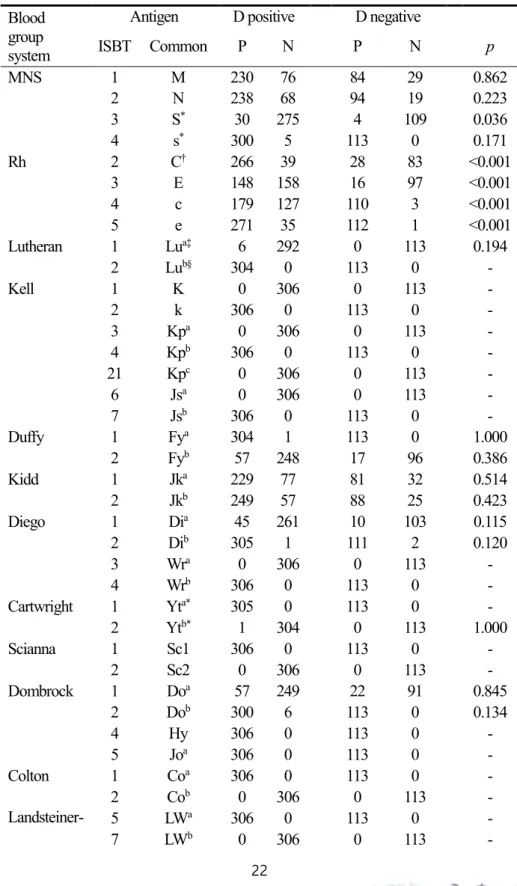
group system
Antigen D positive D negative
ISBT Common P N P N p
MNS 1 M 230 76 84 29 0.862
2 N 238 68 94 19 0.223
3 S* 30 275 4 109 0.036
4 s* 300 5 113 0 0.171
Rh 2 C† 266 39 28 83 <0.001
3 E 148 158 16 97 <0.001
4 c 179 127 110 3 <0.001
5 e 271 35 112 1 <0.001
Lutheran 1 Lua‡ 6 292 0 113 0.194
2 Lub§ 304 0 113 0 -
Kell 1 K 0 306 0 113 -
2 k 306 0 113 0 -
3 Kpa 0 306 0 113 -
4 Kpb 306 0 113 0 -
21 Kpc 0 306 0 113 -
6 Jsa 0 306 0 113 -
7 Jsb 306 0 113 0 -
Duffy 1 Fya 304 1 113 0 1.000
2 Fyb 57 248 17 96 0.386
Kidd 1 Jka 229 77 81 32 0.514
2 Jkb 249 57 88 25 0.423
Diego 1 Dia 45 261 10 103 0.115
2 Dib 305 1 111 2 0.120
3 Wra 0 306 0 113 -
4 Wrb 306 0 113 0 -
Cartwright 1 Yta* 305 0 113 0 -
2 Ytb* 1 304 0 113 1.000
Scianna 1 Sc1 306 0 113 0 -
2 Sc2 0 306 0 113 -
Dombrock 1 Doa 57 249 22 91 0.845
2 Dob 300 6 113 0 0.134
4 Hy 306 0 113 0 -
5 Joa 306 0 113 0 -
Colton 1 Coa 306 0 113 0 -
2 Cob 0 306 0 113 -
Landsteiner- Wiener
5 LWa 306 0 113 0 -
7 LWb 0 306 0 113 -
23
Cromer 1 Cra 306 0 113 0 -
Knops 1 Kna 306 0 113 0 -
2 Knb 0 306 0 113 -
3 McCa 306 0 113 0 -
6 McCb 0 306 0 113 -
4 Sl1 306 0 113 0 -
7 Sl2 0 306 0 113 -
P, antigen-positive; N, antigen-negative
* One specimen from D+ individual was indeterminate.
† One specimen from D+ individual was indeterminate, and 2 specimens from D- individuals were indeterminate
‡ Eight specimens from D+ individual were indeterminate.
§Two specimens from D+ individual were indeterminate.
24
2.1 RhD genotypic profiles in serologically D-negative Koreans
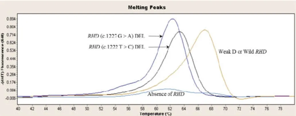
Among 325 serologically D-negative individuals, 260 (80.0%) individuals with absence of RHD were identified by rapid DEL genotyping. Of the remaining 65 individuals, 53 (16.3%) individuals with RHD (c.1227G>A) DEL were identified using a characteristic melting curve, which has a plateau at 54–56°C and a peak melting temperature of 61.95°C. Three (0.9%) individuals with RHD (c.1222T>C) DEL exhibited peak melting temperatures of 62.62°C (figure 2). Nine individuals (2.8%) with weak D were amplified by DEL rapid genotyping and had a melting temperature of 66.88°C, which is typical in the presence of RHD. Of 268 individuals that did not yield amplification products by rapid DEL genotyping, 16 (4.9%) were amplified by real-time PCR targeted to 3’-UTR and revealed to be RHD-CE-D hybrid variants (Table 8).
25
Figure 2. Differentiation of RHD (c.1222T>C) DEL, RHD (c.1227G>A) DEL, weak D and RHD absence by melting curve analysis. The RHD (c.1227G>A) DEL variant has a melting temperature of around 61.95°C, RHD (c.1227G>A) DEL of around 62.62°C, and weak D of around 66.88°C. RHD-absent samples are not amplified by rapid DEL genotyping targeted to exon 9 of RHD gene
26 Table 8. Molecular characteristics of serologically D-negative samples in Koreans.
RHD Exon 9
amplification*
Exon 9 Tm (°C)
Exon 10 genotyping**
Period 1 (no.)
Period 2 (no.)
Total (no.)
Percentage (%) Absence
Complete deletion NA NA 145 99 244 75.1
Hybrid NA Amplified 16 0 16 4.9
Presence
DEL 1222T>C Amplified 62.62 Amplified 1 2 3 0.9
DEL 1227G>A Amplified 61.95 Amplified 36 17 53 16.3
Weak D Amplified 66.88 Amplified 1 8 9 2.8
199 126 325 100.0
27
2.2 DEL confirmation using multiplex single-base primer extension reaction
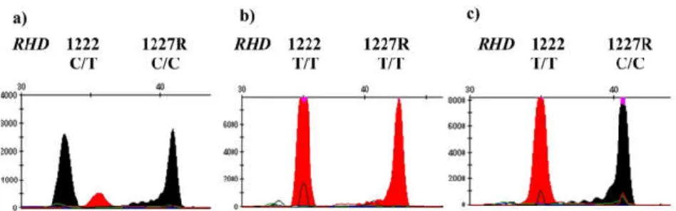
Fifty-three specimens, which had melting curve plateaus at 54–56°C and peaks at 61.95°C, were confirmed as RHD (c.1227G>A) by multiplex single-base primer extension reaction. Three specimens with melting temperatures of 62.62°C were confirmed as RHD (c.1222T>C). The newly developed rapid DEL genotyping method correctly distinguished RHD (c.1222T>C) DEL and RHD (c.1227G>A) DEL through comparison with multiplex single-base primer extension reactions (Figure 3).
2.3 RhCEce profiles
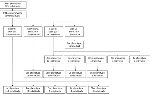
During study period 1, 199 of 325 individuals were tested by extended Rh phenotyping. In the RHD-absent individuals, their Rh phenotypes (other than D) were ce, Ece, Cce, Ec, and CEce, in order of prevalence. Overall, 37 individuals with DEL variants had Cce, CEce, Ce, and CEe phenotypes, in order of prevalence. Further Rh information is presented in Figure 4.
28
Figure 3. Multiplex single-base primer extension reaction of RHD. a) RHD (c.1222T>C) DEL have both C and T peaks on the site of 1222. b) RHD (c.1227G>A) DEL have T/T homozygotes on the site of 1227. As 1227 primers are reverse primer, it can be interpreted to A/A homozygotes. c) Wild RHD that have 1222T and 1227G.
29
Figure 4. RhCEce phenotype of 199 serologically RhD-negative individuals.
30
3. Likelihoods of obtaining specific antigen-negative bloods
A total of 599 events in 266 patients from eight institutions were registered on the website. In one case of anti-K alloimmunization, it was easy to obtain compatible blood because all blood units tested were K negative. E-, P1-, and Lea -negative blood could be sourced about half of the time. The number of units that had to be typed before identifying compatible blood increased in the order: anti-Fyb, anti-Jka, anti-Jkb, anti-Leb, anti-C, anti-M, and anti-S.
In terms of multiple alloantibodies, the highest demand was for blood that was both E- and c-negative. Such units were sourced 38% of the time. The maximum number of alloantibodies reported to the registry was four, and the likelihood of sourcing specific antigen-negative units was 4.7% when anti-E, anti-Jka, anti-Lea, and anti-M coexisted (Table 4).
31
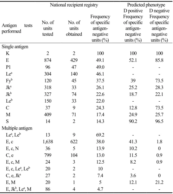
Table 9. Available proportions of specific antigen-negative units in blood banks (%), calculated by serological antigen testing and via genotyping.
National recipient registry Predicted phenotype D positive D negative Antigen tests
performed
No. of units tested
No. of units obtained
Frequency of specific antigen- negative units (%)
Frequency of specific antigen- negative units (%)
Frequency of specific antigen- negative units (%) Single antigen
K 2 2 100 100 100
E 874 429 49.1 52.1 85.8
P1 96 47 49.0 - -
Lea 304 140 46.1 - -
Fyb 120 45 37.5 39 73.5
Jka 318 33 26.1 25.2 28.3
Jkb 327 74 22.6 18.7 22.1
Leb 150 33 22.0 - -
C 37 9 24.3 12.8 73.5
M 409 71 17.4 24.9 25.7
S 14 2 14.3 90.2 96.5
Multiple antigen
Lea, Leb 13 9 69.2 - -
E, c 1,638 622 38.0 41.3 1.8
E, c, N 36 5 13.9 10.2 0
C, e 799 104 13.0 11.5 0.9
E, c, M 24 3 12.5 8.2 0.9
E, c, Lea, Leb 20 2 10 - -
C, e, Jka 27 2 7.4 3.6 0
E, M 20 1 5 12.1 21.2
E, Jkb, Lea, M 86 4 4.7 - -
32
Discussion
Transfusion medicine is concerned with the safety of both donors and patients.
Although from the donor viewpoint distribution of blood components is the first action of a transfusion service, a request from a patient is a prerequisite for distribution. In the present study, the author explored both the supply and demand of blood components to establish the KRBP.
From the donor viewpoint, the author examined the prevalence of HEA in various blood groups in Korea by analyzing targeted alleles. Several other studies have explored the prevalence of HEA, but without reference to D antigen status, and the phenotypes of D negatives have been overlooked because such subjects are very rare.
In the present study, the author included a number of D negatives to clarify the prevalences of various HEA.
Using the modified Wiener nomenclature, the author found that the predicted haplotype combinations in the Rh blood group system were R1R1, R1R2, and R2R2 in order of prevalence, followed by R1R0 (or R1r) and R2R0 (or R2r). The first and second most common haplotype combinations are in agreement with data of a previous study (Lee 1975). In addition, the incidence of the rr haplotype, the most common in D negatives worldwide, was 64.7% (Roback, Mark K. Fung et al. 2014). Notably, one D negative individual exhibited a very rare CE phenotype, the predicted haplotype of which was r′ry.
M+N+S−s+ is the most common phenotype of the MNS blood group system in Koreans, Caucasians, and Africans (Reid 2009). The rate of s-antigen negativity in Korea was 1.2% in the present study, similar to previous results (Lee 1965). It is
33
remarkable that three of five s-negative individuals had GYPB deletions yielding the S−s− phenotype. In addition, no subject was negative for both the s and D antigen.
Only one Fya-negative (0.3%) subject was encountered; blood components for that person would be extremely difficult to source. This finding is in agreement with other work performed in Korea and other Asian populations. Further analysis of one subject is required; that subject had a 265C>T substitution in an allele of the FY gene. This substitution, which is very rare in Asians, is a dominant feature of the FY*X genotype, which has a weak Fyb phenotype. The expected phenotype of the observed polymorphism is Fy(a+b+) (Hashmi, Shariff et al. 2007). It is surprising that the antigen prevalences of the Dombrock, Cartwright, Scianna, Colton, Landsteiner- Wiener, Cromer, and Knops blood groups system have only now been explored in the Korean population.
The author developed a rapid DEL genotyping method targeting both RHD (c.1222T>C) and RHD (c.1227G>A) simultaneously, based on the fact that the two variants are located within 5 bp on exon 9. This newly developed assay uses real-time PCR and melting curve analysis. The melting curve of RHD (c.1227G>A) had a peak temperature of 61.95°C, while the melting temperature of RHD (c.1222T>C) had a peak temperature of 62.62°C. Although there has been studies about DEL detection using melting curve analysis (Sun, Liu et al. 2008), this is the first study which evaluate simultaneous detection of both RHD (c.1222T>C) and RHD (c.1227G>A). The difference in melting temperatures enables the discrimination of DEL variants in serologically D-negative individuals from complete RHD deletions and RHD-CE-D hybrid variants. It rapid compared to labor-intensive sequencing-based analysis.
34
Because it requires neither purification nor sequencing steps after amplification, researchers can quickly and easily carry out DEL genotyping.
Until now, up to twenty DEL alleles had been registered (Gardener, Legler et al.
2012). RHD (c.1222T>C) and RHD (c.1227G>A) were the only DEL alleles found in Korean populations (Luettringhaus, Cho et al. 2006, Kim, Kim et al. 2009, Yang, Lee et al. 2015), accounting for 15.9% and 0.4% of serologically D-negative Koreans based on IAT.
Detection of DEL is important regarding transfusions, because DEL red cells have the ability to alloimmunize D-negative individuals (Wagner, Kormoczi et al. 2005, Kim, Kim et al. 2009, Yang, Lee et al. 2015). To do this, the implementation of DEL genotyping assay is inevitable because it is impossible to detect DEL using routine serological methods. Alternative methods for the detection of DEL, adsorption-elution techniques, could be considered, but it is too cumbersome to screen large population such as blood donor group.
RHD (c.1227G>A) DEL exhibits the Ce haplotype (Flegel, von Zabern et al. 2009,
Srijinda, Suwanasophon et al. 2012). Some laboratories have utilized the characteristics of the RhCEce phenotype to detect DEL variants when molecular typing of DEL is not available. Therefore, the author performed RhCEce typing of DEL variants to determine whether this typing strategy is suitable for the Korean population. Although all DEL variants had the C antigen on their RBCs, their RhCEce phenotypes were inconsistent. Overall, 7 (4.8%) of 145 RhCEce-typed individuals with complete RHD deletion, and 13 (81.3%) of 16 individuals with the RHD-CE-D hybrid variant, expressed the C antigen. DEL variant detection using RhCEce phenotyping therefore causes unnecessary exclusion of complete RHD deletions and
35
RHD-CE-D hybrid variants. Because RHD absence is very rare in East Asian populations, DEL variant detection with RhCEce phenotyping should be reconsidered.
If rapid DEL genotyping by melting curve analysis is applied to blood donor tests, it will help to identify DEL blood samples among D negatives in blood donor centers or hospital blood banks and facilitate appropriate usage of serologically D-negative blood units
The author also designed primers for the exon 10 3’-UTR. The amplification of exon 10 3’-UTR can differentiate RHD-CE-D hybrid variants from RHD complete deletions in samples (N = 260) that were not amplified by DEL rapid genotyping.
Among these samples, 16 (6.2%) were amplified by exon 10 3’-UTR real-time PCR, which implies the presence of RHD-CE-D hybrid variants.
In routine blood bank practice, specific antigen-negative blood components are selected when an unexpected alloantibody is detected. It is often a challenge to source suitable blood. The author calculated the likelihoods of sourcing specific antigen- negative blood units by antigen testing. The distributions of unusual antibodies were similar to those noted in a previous study (Ok, Kim et al. 2013). As in that work, anti-E antibody was the most frequently reported. In addition, the greater the number of alloantibodies, the lower the likelihood of sourcing appropriate blood components. For this reason, although it depends on the circumstances, the widely accepted definition of rare blood components usually has two categories: high prevalence antigen-negative components and multiple antigen-negative components.
In Korea, the prevalence of D negatives is approximately 0.1%, thus differing notably from the levels in Caucasians and Africans. The author believe that it is appropriate to define blood components that are concurrently negative for D and other
36
highly prevalent antigens as rare blood units. For example, blood banks would be challenged when asked to source units for D negatives with alloantibodies to the c or e antigens; the author found only three and one individual(s) who were c- and e-negative, respectively, among the 113 D-negative subjects.
In the time since the ISBT Working Party on Rare Donors was established, efforts have been made to establish classification systems and to explore the antigenic profiles of donors. Today, many researchers use molecular methods rather than immunohematological techniques to study the biology of red cell antigens (Kormoczi, Wagner et al. 2007). Nevertheless, many authors have focused on one to two blood groups of major interest, and on common well-known antigens (Nance 2009). It is noteworthy that the author used commercially available molecular tests to initially establish the rare donor program. In the D-positive group, the results were similar when actual antigen typing by serological methods reported previously (Choi, Kim et al. 1984) and virtual typing by molecular methods, were used to evaluate most blood groups, except the S antigen.
In addition, the registry showed that the likelihood of sourcing S-negative blood units was 14.3%; however, the prevalence of S antigen negativity in D positives in Korea is 90.2%. To verify the genotyping results of GYPB, and usefulness of S antigen prediction, the author compared the results of S antigen typed by antiserum and by prediction based on genotype. Among 51 individuals, all genotype results were perfectly matched with that performed by serotyping. It is thought that the S-antigen negativity data in the registry were the only one-time event, and the selection bias cannot be excluded.
37
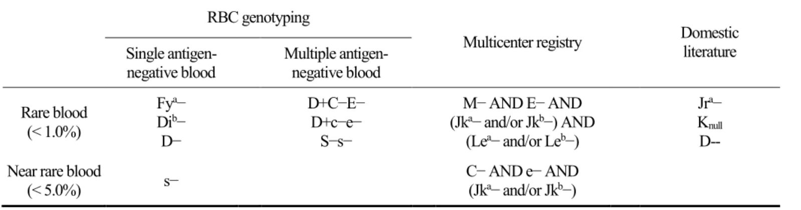
Because the program was at an early stage, it was hard to defined the term “rare blood” adequately and clearly. Most countries consider blood as rare when its prevalence is less than 1 in 1,000 (Reesink, Engelfriet et al. 2008). In reality, it is true that obtaining Fya− or Dib− blood component is tough. According to the study, Fya− or Dib− donor were estimated 0.2% and 0.7% of D-positive group. No individual had Fya− or Dib− red cells in D-negative group. In Korean blood donation and transfusion system, it would be suitable to define “rare blood” as “blood units without antigens that are prevalent less than 1% of the population.” Not only about the single antigens, multiple antigen-negative units such as S−s− units, D+C−E − units, and D+c−e− units were also considered as “rare blood.” The prevalence of s− only units were calculated 1.7%. Although these units are prevalent in more than 1.0% of Korean population, those unites are not sufficiently supplied to the recipients. Thus, the units without antigens that are prevalent less than 5% of the population were classified to “Near rare blood.”
In consideration of multicenter registry, 4 units of M−E−Jkb−Lea− blood were obtained by testing 86 units. In analysis of genotyping data, 3 D-positive individuals (0.9%) and 4 D-negative individuals (3.5%) were M−E−Jka− simultaneously. If Lewis blood group was considered, the prevalence of multiple antigen-negative units would be lesser than that of simulated. The 2 units of C−e−Jka− could be obtained among 27 units, and the prevalence of 7.4% is predicted. However, based on genotyping data, 11 individuals (3.4%) among the 416 individuals had C−e−Jka− blood. This type of units also should be considered as worthy (Table 10).
The limitation of the study is that other minor blood group systems, including the Gerbich, Jr, and Vel systems, which are known to cause hemolytic transfusion
38
reactions or hemolytic disease in newborns, have not yet been targeted (van Gammeren, Overbeeke et al. 2008, Kim, Park et al. 2010, Baughn, Whitacre et al.
2011). In addition, blood group antigens with carbohydrate epitopes could not be genotyped. To achieve the purpose of the KRBP, as more work is performed, the author will get closer to the goal.
Only data on antibody detection and identification, and the numbers of antigen tests performed to source specific antigen-negative units, are registered in the centralized database. Personal information (including patient identity) is protected. However, several studies on centralized databases with information on patients with decreased alloantibody titers have appeared; the databases seek to prevent anamnestic hemolytic transfusion reactions (Harm, Yazer et al. 2014). Expansion of the nationwide database and ensuring database stability and accuracy will reduce the adverse effects of transfusions.
There is no absolute definition of what constitutes rare blood. The term has different meanings in different places, depending on the accessibility of blood donors or blood units. Herein, the author describes the efforts to establish the KRBP, a national rare blood program, from both the patient and donor perspectives. The author believe that the experience may be of interest to both blood banks and patients; the efficient supply of safe blood products is our goal.
39
Table 10. Proposal of rare blood units based on various blood group genotypes and unexpected antibodies in Korean population RBC genotyping
Multicenter registry Domestic literature Single antigen-
negative blood
Multiple antigen- negative blood Rare blood
(< 1.0%)
Fya− Dib− D−
D+C−E−
D+c−e−
S−s−
M− AND E− AND (Jka− and/or Jkb−) AND
(Lea− and/or Leb−)
Jra− Knull
D-- Near rare blood
(< 5.0%) s− C− AND e− AND
(Jka− and/or Jkb−)
40
References
Baughn, M. R., P. Whitacre, G. S. Lo, S. Pandey and T. A. Lane (2011). "A mild acute hemolytic transfusion reaction in a patient with alloanti-Ge3: a case report and review of the literature." Transfusion 51(9): 1966-1971.
Chen, J.-C., T.-M. Lin, Y. L. Chen, Y.-H. Wang, Y.-T. Jin and C.-T. Yue (2004). "RHD 1227A Is an Important Genetic Marker for RhDel Individuals." Am J Clin Pathol 122(2): 193-198.
Choi, S. Y., S. I. Kim and H. I. Cho (1984). "Study on Gene Frequencies of Blood Groups in Koreans." Korean J Hematol 19(1): 63-75.
Chou, S. T. (2013). "Transfusion therapy for sickle cell disease: a balancing act."
Hematology Am Soc Hematol Educ Program 2013: 439-446.
Chung, Y., Y. J. Hong, S. M. Hwang, T. S. Kim, K. U. Park, J. Song and K. S. Han (2014). "Probability of Obtaining Specific Antigen-Negative Blood Units through Antigen Testing in Korean Medical Institutes." Korean J Blood Transfus 25(1): 34-40.
Delaney, M., S. Harris, A. Haile, J. Johnsen, G. Teramura and K. Nelson (2015). "Red blood cell antigen genotype analysis for 9087 Asian, Asian American, and Native American blood donors." Transfusion 55(10): 2369-2375.
41
Flegel, W. A., I. von Zabern and F. F. Wagner (2009). "Six years' experience performing RHD genotyping to confirm D- red blood cell units in Germany for preventing anti-D immunizations." Transfusion 49(3): 465-471.
Gardener, G. J., T. J. Legler, J. A. Hyett, Y. W. Liew, R. L. Flower and C. A. Hyland (2012). "Anti-D in pregnant women with the RHD(IVS3+1G>A)-associated DEL phenotype." Transfusion 52(9): 2016-2019.
Harm, S. K., M. H. Yazer, G. F. Monis, D. J. Triulzi, J. P. Aubuchon and M. Delaney (2014). "A centralized recipient database enhances the serologic safety of RBC transfusions for patients with sickle cell disease." Am J Clin Pathol 141(2): 256-261.
Hashmi, G., T. Shariff, Y. Zhang, J. Cristobal, C. Chau, M. Seul, P. Vissavajjhala, C.
Baldwin, K. Hue-Roye, D. Charles-Pierre, C. Lomas-Francis and M. E. Reid (2007).
"Determination of 24 minor red blood cell antigens for more than 2000 blood donors by high-throughput DNA analysis." Transfusion 47(4): 736-747.
Kim, H., M.-J. Park, T.-J. Sung, J. S. Choi, J. Hyun, K. U. Park and K.-S. Han (2010).
"Hemolytic Disease of the Newborn Associated with Anti-JraAlloimmunization in a Twin Pregnancy: The First Case Report in Korea." The Korean Journal of Laboratory Medicine 30(5): 511.
Kim, I. T., I. B. Suh, K. R. Ma, C. S. Lim, Y. K. Kim and K. N. Lee (2003). "The genotyping of Kell, Duffy, and Kidd System in Korean." Korean J Blood Transfus 14(1): 9-19.
42
Kim, J. Y., S. Y. Kim, C. A. Kim, G. S. Yon and S. S. Park (2005). "Molecular characterization of D- Korean persons: development of a diagnostic strategy."
Transfusion 45(3): 345-352.
Kim, K. H., K. E. Kim, K. S. Woo, J. Y. Han, J. M. Kim and K. U. Park (2009).
"Primary anti-D immunization by DEL red blood cells." Korean J Lab Med 29(4):
361-365.
Kormoczi, G. F., T. Wagner, C. Jungbauer, M. Vadon, N. Ahrens, W. Moll, A.
Muhlbacher, S. Ozgul-Gulce, T. Kleinrath, S. Kilga-Nogler, D. Schonitzer and C.
Gassner (2007). "Genetic diversity of KELnull and KELel: a nationwide Austrian survey." Transfusion 47(4): 703-714.
Lee, S. Y. (1965). "Further Analysis of Korean Blood Types." Yonsei Med J 6(1): 16- 25.
Lee, S. Y. (1975). "Possible problems on the blood-typological studies of Korean caused by inter-racial marriages with Caucasians." Korean J Hematol 10(1): 33-39.
Li, B. J., Y. J. Jiang, F. Yuan and H. X. Ye (2010). "Exchange transfusion of least incompatible blood for severe hemolytic disease of the newborn due to anti-Rh17."
Transfus Med 20(1): 66-69.
Luettringhaus, T. A., D. Cho, D. W. Ryang and W. A. Flegel (2006). "An easy RHD genotyping strategy for D- East Asian persons applied to Korean blood donors."
Transfusion 46(12): 2128-2137.
43
Mourant, A. E. (1965). "The establishment of na international panel of blood donos of rare types." Vox Sang 10: 129-132.
Nance, S. T. (2009). "How to find, recruit and maintain rare blood donors." Curr Opin Hematol 16(6): 503-508.
Nuchnoi, P., J. Thongbus, A. Srisarin, U. Kerdpin and V. Prachayasittikul (2014).
"Clinical and laboratory update on the DEL variant." Lab Med 45(4): 285-290.
Ok, S. J., S. Y. Kim, I. S. Kim, E. Y. Lee and H. H. Kim (2013). "Eleven Years' Experience with Unexpected Antibody Screening Tests Including a Di(a) Cell in Transfusion Candidates." Korean J Blood Transfus 24(1): 64-70.
Reesink, H. W., C. P. Engelfriet, H. Schennach, C. Gassner, S. Wendel, R. Fontão- Wendel, M. A. de Brito, P. Sistonen, J. Matilainen, T. Peyrard, B. N. Pham, P. Rouger, P.
Y. Le Pennec, W. A. Flegel, I. von Zabern, C. K. Lin, W. C. Tsoi, I. Hoffer, K.
Barotine-Toth, S. R. Joshi, K. Vasantha, V. Yahalom, O. Asher, C. Levene, M. A. Villa, N. Revelli, N. Greppi, M. Marconi, Y. Tani, C. C. Folman, M. de Haas, M. M. W.
Koopman, E. Beckers, D. S. Gounder, P. Flanagan, L. Wall, E. Aranburu Urtasun, H.
Hustinx, C. Niederhauser, C. Flickinger, S. J. Nance and G. M. Meny (2008). "Donors with a rare pheno (geno) type." Vox Sanguinis 95(3): 236-253.
Reid, M. E. (2009). "MNS blood group system: a review." Immunohematology 25(3):
95-101.