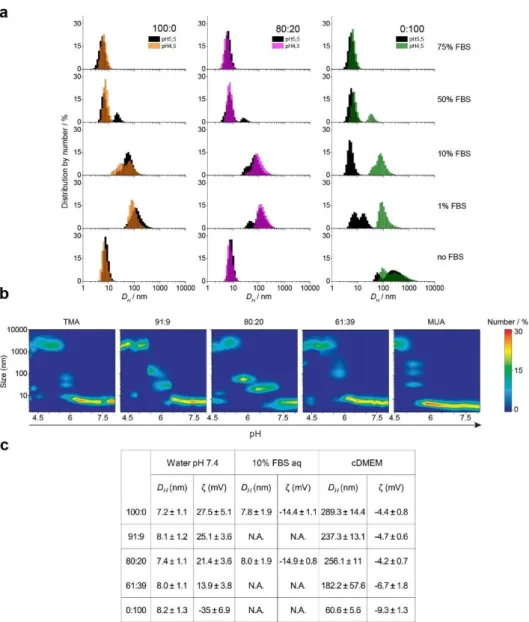
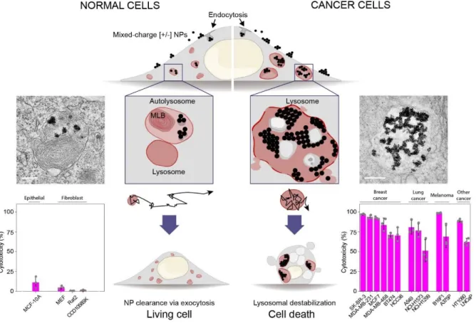
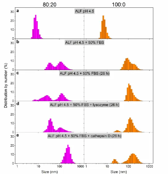
Aggregation properties of mixed-charge nanoparticles depend on concentration of proteins, pH or in cell culture media. Cellular uptake and differential intracellular aggregation of mixed charge NPs in cancer versus normal cells.
Introduction
- Overview of cancer treatment
- Nanomaterials for biomedical applications
- Mitochondria targeting
- Choice of fluorescent dye for mitochondria labeling
- Thesis structure
- Research objectives
- References
Cellular uptake and cytotoxicity of positively charged chitosan gold nanoparticles in human lung adenocarcinoma cells. Engineering gram selectivity of mixed-charge gold nanoparticles by tuning the balance of surface charges.
Targeted crystallization of mixed-charge nanoparticles in lysosomes induces selective death
Abstract
Introduction
In contrast, in normal cells, [+/-] NPs reside in lysosomes only transiently and rapidly move to autolysosomes, causing little damage to these cells (Figure 2.1, left). Overview of how crystallization of mixed-charge NPs in cancer lysosomes leads to selective killing of cancer cells.
Results
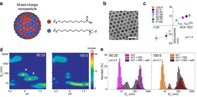
- Physiochemical characteristics of mixed-charge nanoparticles
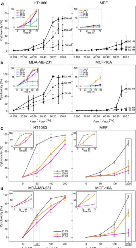
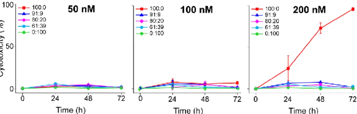
- Selective killing of cancer cells by mixed-charge nanoparticles
- The impact of the aggregation of mixed-charge nanoparticles on lysosomal
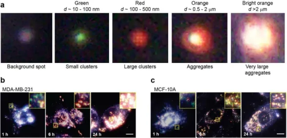
- Intracellular aggregation of mixed-charge NPs and lysosome “swelling”
- Impact of mixed-charge NPs on lysosome movements
- Selective destabilization of lysosomal membranes by mixed-charge NPs
- Accumulation and clearance of mixed-charge NPs
The results summarized in Figure 2.11e,f show that the uptake of different types of [+/-] NPs further sensitized lysosomes of both HT1080 and MDA-MB-231 cells to damage by photooxidation (indicated by the increase in green fluorescence ). We further investigated the function of lysosomes in the cancer-specific toxicity of [+/-] NPs by treating cancer cells with [+/-] NPs and lysosomal inhibitors bafilomycin A (Baf) or chloroquine (Cq) (Figure 2.11g, f).
Discussion
- Biochemical effects
Petersen et al.47,48 proposed that lysosome stability is determined by the lipid composition of these internal lysosomal membranes, which include high concentrations of anionic phospholipid bis(monoacylphosphoglycerol) phosphate (BMP), with no reported differences in BMP levels at cancer vs normal. cells). In response to 80:20 NPs, acid ceramidase levels increase in normal cells (which can prevent ceramide build-up), but not in cancer cells (Figure 2.20). Furthermore, we identified significant changes in lysosomal proteins in cancer versus normal cells in response to [+/-]NP crystallization (Figure 2.20b).
Analysis of differences in lysosomal proteins upon mixed-charge nanoparticle treatment of normal and cancer cells. a) Two lysosomal fractions were extracted and analyzed from NP-treated cells -. In contrast, the amounts of RagC and mTORα were increased in the lysosomes of normal cells (MCF10A: Lyso and NP).
Conclusion
Appendix
- Appendix Note A1
- Mixed-charge nanoparticle aggregation in solutions mimicking lysosomal
- Appendix Note A2
- The role of protein corona in the internalization of mixed-charge
- Appendix Note A3
- pH differences in cancer vs normal cells’ lysosomes
- Discussion on the choice of nanoparticle size
- Justification of the choice of cell lines
Proteins in coronas were examined using SDS-PAGE electrophoresis to define and compare protein coronas adsorbed on pure-TMA/100:0 vs. 13 cancer cell lines were used in this study, six of which were breast cancer cell lines representing different subtypes of breast cancer: MDA-MB-231, MDAMB-468 and HCC38 are triple negative (ER-; PR-; Her receptor, HerR-) basal subtype adenocarcinomas , while MCF7 is Luminal A subtype (ER+, PR+) adenocarcinoma, BT -474 is Luminal B subtype invasive ductal. Mixed nanoparticles were also found to be effective against lung cancer cell lines A549, NCI-H1299 and NCI-H1573.
Because MEF fibroblasts are “more similar” to HT1080 in terms of cell size and amount of lysosomes per cell than normal human fibroblasts, we selected them as one of the main “normal” cell lines. Three cancer cell lines sensitive to 80:20 NPs have KRAS mutations that make cancer resistant to traditional treatments.
Materials and Methods
An IncuCyte ZOOM system with a 20 objective was used to automatically acquire images of live (green) and dead (red) cells (IncuCyte ZOOM Live Cell Analysis System, Essen BioScience). The Cell Counter plugin for ImageJ software (NIH) was used to count the number of live/dead cells, and cytotoxicity was expressed as the percentage of dead cells. Red + Green + Blue) 3 was used to calculate the RGB values for each bright point and the surrounding background points.
The BCA Protein Assay Kit was used to determine the protein concentrations in the samples obtained. X-Y midplane confocal images were used to measure the mean diameter of LysoTracker red-labeled vesicles.
Efficient Gene Delivery Vectors by Tuning the Surface Charge Density of Amino Acid Functionalized Gold Nanoparticles. Nanoparticle size and surface properties define the protein corona with potential implications for biological effects. Size-dependent protein-nanoparticle interactions on citrate-stabilized gold nanoparticles: Protein corona display.
The enzymatic conversion of sphingomyelin to ceramide increases the viscosity of the shear membrane at the air-water interface. Protein Corona affects cellular uptake of gold nanoparticles by phagocytic and non-phagocytic cells in a size-dependent manner.
Mixed-Charge Nanocarriers Allow for Selective Targeting of Mitochondria by Otherwise
Abstract
Introduction
Even though recent advances have been made using these existing probes to detect short-term intra-mitochondrial dynamics at high spatiotemporal resolution12,17,18 thanks to super-resolution fluorescence microscopy, the low photostability, high background fluorescence and high photoinduced toxicity12,15 ,17 associated with mitochondrial fragmentation and swelling remain the main problems limiting its use for long-term tracking of mitochondria. Furthermore, because mitochondrial rearrangements occur on such a wide time scale (ranging from minutes to days, ~102 to 105 s)19, no single mitochondria tracer dye can meet all the requirements. Only a few of these probes (MitoTracker dyes and MitoPB Yellow based on C-Naphox) are retained in the mitochondria by covalent binding to mitochondrial proteins12,15, and even fewer remain for long periods in the mitochondria20, while the majority leak out of spontaneously depolarizing mitochondria over short time scales (min to h)21,22.
This research study describes how inexpensive, widely used, non-toxic fluorescein dye (5-aminofluorescein, fluorescein derivative) with no affinity for any specific cellular structure itself - was transformed into a series of fluorescent nanodyes that target mitochondria in the absence of the TPP+ moiety. On the one hand, fluorescent NPs with a net cationic surface charge, unlike commercially available MitoTrackers, had long retention times (days) in mitochondria, making them ideal for long-term monitoring of these organelles due to their gradual release from endolysosomal reservoirs and covalent binding to mitochondrial proteins, making them suitable for long-term tracking of these organelles.
Results
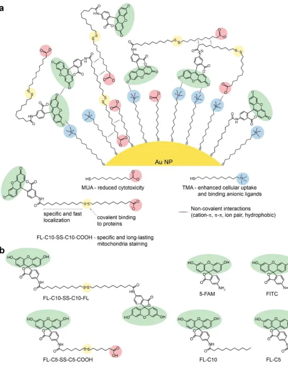
- Chemical synthesis of disulfide-based and alkyl fluorescein derivatives
- Supramolecular assembly of mixed-charge fluorescein nanoparticles
- Selective and long-term staining of mitochondria
In fact, zeta potentials for fluorescence nanoparticles were slightly more positive than those for the corresponding standard mixed-charge nanoparticles (Figure 3.3d, Table 3.1) thus retaining the ability to accumulate in the mitochondria based on negative mitochondrial membrane potential. The total number of FL-C10-SS-C10-COOH ligands attached to the different types of fluorescence nanoparticles was then estimated by comparing the fluorescence intensities after etching of the cores (Figure 3.3b) with the calibration curves obtained were briefly mixed with the corresponding single- and mixed-charge nanoparticles by KCN treatment of known amounts of the free ligands. For these experiments, we chose to use 80:20 NPs because the corresponding [+/-]9:3/FL NPs had the largest number of FL ligands bound in ex vivo experiments (shown in Figure 3.3c).
The brightness of mitochondria labeling (quantified as fluorescence intensity in mitochondria) (Figure 3.6a) reflects the number of ligands per nanoparticle detected in ex vivo experiments (Figure 3.3c) with [+/-]9:3/FL nanoparticles yielding the brightest nano-dye, and at [+/-] to FL ratio of 60:1, even more than [+]/FL NPs. Surprisingly, even nanoparticles with net negative surface charge [+/-]3:9/FL NPs (violet, Figure 3.6a), albeit with less intense fluorescence, localized to mitochondria. To establish this selectivity, we compared the time course of co-localization of mixed charge [+/-]9:3/FL NPs to the mitochondria versus the LysoTracker-labeled acidic cellular compartments (Figure 3.6b,c).
We observed that ~66% of the fluorescence intensity of [+/-]9:3/FL NPs remains in the mitochondria after dissipation of the negative potential across the inner mitochondrial membrane with a protonophore carbonyl cyanide-4-(trifluoromethoxy)phenylhydrazone (FCCP). ) (Figure 3.14).
Discussion
- Proposed mechanism for targeting of fluorescent dyes to mitochondria with mixed-charge
In the absence of direct delivery, fluorescein disulfides produced in the cytoplasm will be reduced by abundant reducing glutathione and thiol oxidoreductases35,55. Similar methods can enhance the accumulation of small molecules/fluorophores with unstable disulfide bonds in the mitochondrial matrix. In the mitochondrial matrix, exposed reactive protein thiols (eg, cysteines) are 25-fold higher than glutathione concentrations, suggesting that protein-thiol interactions will predominate51.
At the higher pH of the mitochondrial matrix (pH 7.8-8.0) compared to the cytosol (pH 7.2), protein thiols (pKa 8.3) will be five times more reactive than anywhere else in the cell for processes requiring thiolate anions are needed. S-), including thiol-disulfide exchange. Deprotonated thiolate groups of exposed cysteine residues in the matrix attack fluorescein disulfides. or mixed FL-disulfide complexes) more readily than in the cytoplasm, resulting in covalent attachment of fluorescein moieties to mitochondrial protein cysteines via disulfide bonds (shown in Figure 3.12).

Conclusion
Much less intense, but still selective, staining with shorter disulfides (compare FL-C5-SS-C5-COOH, see Figure 3.4) suggests that longer (alkyl) linkers may be required for efficient translocation across the lipid bilayer, analogous to polydisulfide compounds50.
Materials and methods
Unless otherwise stated, cells were stained in their native media containing 50 nM FL NPs for 1 h at 37 °C. Then, media containing dye and NPs were removed and cells were imaged in their native media. All cells were imaged in their native media and 5% or 0% CO2 levels (as specified in the Cell Culture section).
Cells were pretreated with endocytosis inhibitors (as described above), followed by the addition of 50 nM [+/-]9:3/FL NPs. MCF10A cells were grown in glass-bottomed cell culture dishes in their native media for 24 h.
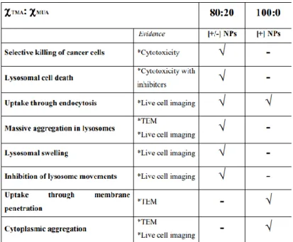
Together with colleges, we have demonstrated: (1) that gold nanoparticles functionalized with a mixture of positively [+] and pH-sensitive, negatively [-] charged ligands, so-called mixed charge. NPs with positive-to-negative surface ligand ratios around 80:20, but not purely cationic NPs, exhibit cancer-specific cytotoxicity mediated by destabilization of cancer lysosomes through gradual, pH-dependent assembly of [+/-] NPs into ordered supracrystals inside cancer lysosomes, which then leads to increased osmotic pressure, swelling of the lysosomes and leakage of their contents into the cytosol, triggering apoptosis-like cancer cell death; (2) a novel strategy for targeting, visualizing, and long-term tracking of mitochondria in living cells that does not involve TPP+ lipophilic cations by incorporating a fluorescein disulfide ligand with mixed-charge nanoparticles. Our system consists of fluorescein dye in the form of asymmetric alkane disulfides that are not covalently bound to mixed-charge nanoparticles.
For the future, we hope to expand our in vitro research with animal models (mice). Additionally, we consider using another nanomaterial instead of gold nanoparticles, such as polymer-based particles or dendrimers, to see how the mixed charge strategy has general value for pH-based organelle targeting.
Summary