To understand the role of TonEBP in obesity, I examined a series of mice with TonEBP haplodeficiency. To map the mechanisms of action of TonEBP in adipose tissue, I further studied the role of TonEBP in both adipocytes and macrophages. To investigate the role of TonEBP in macrophages, a line of mice with myeloid-specific deletion of TonEBP was generated.
TonEBP was rate-limiting, as increased TonEBP expression increased NFκB activity and decreased TonEBP expression decreased it without affecting NFκB itself. These data suggest that variations in TonEBP expression levels determine individual variations in insulin resistance and inflammation regulated by NFκB and PPARγ. To address the role of TonEBP in adipocytes, a second line of mice with an adipocyte-specific deletion of TonEBP was generated.
These data indicate that TonEBP in adipocytes promotes obesity and insulin resistance by suppressing Adrb3 expression and blocking WAT initiation. In conclusion, TonEBP in adipocytes and macrophages contributes to obesity and insulin resistance due to the suppression of WAT beiging and the promotion of inflammation, respectively.
Chapter1. Background
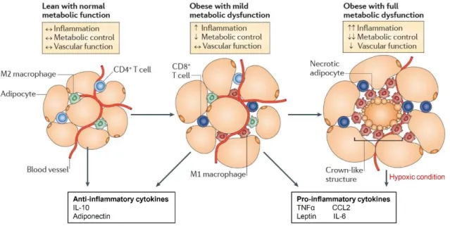
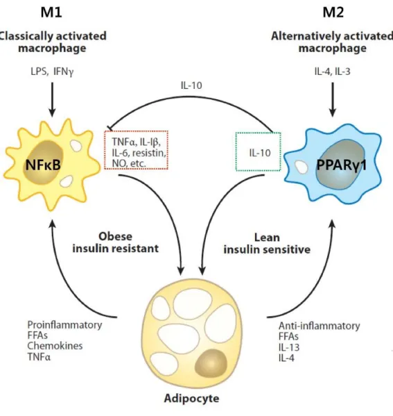
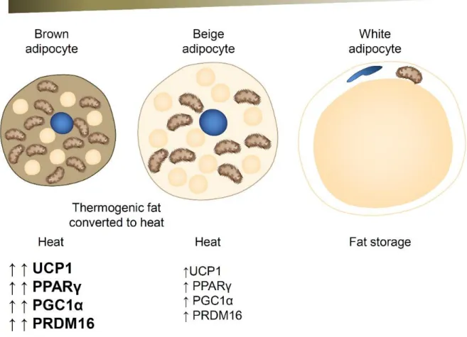
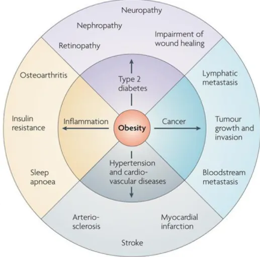
The obesity-infiltrated M1 macrophages secrete pro-inflammatory cytokines such as TNFα and promote insulin resistance in adipose tissue. Therefore, macrophage polarization in adipose tissue is critical for the development of insulin resistance in obesity. Adipocytes are classically divided into white adipose tissue (WAT), which stores energy by accumulating lipids, and brown adipose tissue (BAT), which regulates energy expenditure through non-shivering thermogenesis.
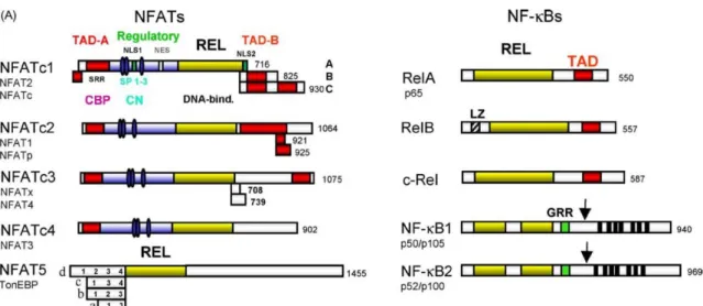
Recently, a new type of brown-like adipose tissue was discovered in humans, called 'beige' and 'brite' from brown in white [11] (Figs. 1-5). Adipose tissue bleaching causes adipocyte dysfunction, inflammation, and decreased thermogenesis, leading to obesity and type 2 diabetes. TonEBP was originally identified as the central DNA-binding transcription factor for cellular response to hypertonic stress by regulating genes such as BGT1, SMIT, AR and HSP Fig. 1-8).
These studies demonstrated that TonEBP has pleiotropic functions, including DNA-binding transcription factor, transcriptional co-activator, and transcriptional co-repressor in inflammation and adipocyte differentiation. Regulation of the hypertonic stress response and other cellular functions by the Rel-like transcription factor NFAT5.
Chapter2. Macrophage TonEBP promotes inflammation and insulin resistance by regulating M1/M2 polarization
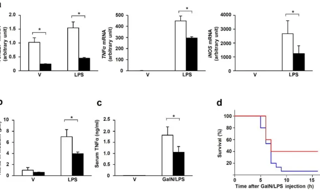
When stimulated with LPS, PECs from TonEBPfl/fl, LysM-cre mice showed significantly reduced expression of the NFκB-dependent proinflammatory genes TNFα and iNOS (Figure 2-1a) and NO production (Figure 2-1b) in response to LPS. In addition, the severity of sepsis, as measured by the resulting death, was reduced in TonEBPfl/fl, LysM-cre animals (Figure 2-1d). Expression of the NFκB target genes TNFα and IκBα in response to LPS was also reduced in these cells (Fig. 2-6c), consistent with reduced NFκB activity.
The TonEBPΔ/Δ MEF cells expressed the TonEBP Δ protein which migrated faster than the wild type TonEBP protein (Fig. 2-6d). In addition, serine 276 phosphorylation of p65 in response to LPS was normal in the TonEBPΔ/Δ MEF cells (Fig. 2-6f). The interaction increased in response to LPS (Fig. 2-8a) in correlation with increased nuclear localization of p65 (Fig. 2-6e).
The inability of the TonEBP Δ protein to interact with p65 and recruit p300 explains the reduced NFκB activity in the TonEBPΔ/Δ MEF cells (Fig. 2-6b). This was further supported by the observation that increased expression of p300 did not lead to increased κB-driven luciferase expression in the TonEBPΔ/Δ MEF cells (Fig. 2-8b). Cerulenin inhibited NO production in response to LPS in a dose-dependent manner without impairing cell viability (Fig. 2-12b).
Decreased NO production was associated with decreased NFκB activity based on decreased expression of κB-driven luciferase (Fig. 2. 12c) and decreased expression of NFκB target genes (Fig. Very interestingly, decreased NFκB activity was not associated with changes in nuclear localization, DNA binding, or p65 phosphorylation (Fig. 2-15). Consistent with increased IL-10 secretion, TonEBP knockdown increased STAT3 phosphorylation in response to LPS (Fig. 2. 17c) and enhanced LPS-induced SOCS3 expression (Fig.
Macrophages differentiated from monocytes responded to LPS by increasing the expression of TONEBP, IL-10, and anti-inflammatory regulators, such as RAW264.7 cells (Figure 2-18). As expected given the decreased expression of TonEBP, β-lapacone increased IL-10 and CD206 mRNA expression while suppressing COX-2 and iNOS mRNA levels in response to LPS (Figure 2-19b). PPARγ1 without changes in STAT6 phosphorylation and expression and C/EBPβ expression in TonEBP-knockdown RAW264.7 cells (Figure 2-20a).
Interestingly, myeloid-specific TonEBP KO mice showed a similar increase in body weight compared to WT mice (Figure 2-21a). However, myeloid-specific TonEBP KO mice were protected from obesity-induced glucose intolerance and insulin resistance (Figure 2-21b-d).
Chapter3. Adipocyte TonEBP promotes obesity through the suppression of WAT beiging
In addition, HFD-fed mice showed lower mRNA expression of thermogenic genes PPARGC1A (encoding PPARγ coactivator 1α (PGC1-α)), Dio2, CPT1α, Cidea, PPARα, and Adrb3, and beige markers (UCP-1, CD137, and TMEM26 ), in iWAT (Fig. 3-2d and e). Haplodeficient mice on NC showed similar weight gain to WT mice (Fig. 3a), but those on HFD were resistant to weight gain (Fig. Accordingly, haplodeficient mice showed lower weights of inguinal, epididymal, and dorsal fat pads (Fig. 3-3e), suggesting resistance to diet-induced obesity.
We also observed that TonEBP haplodeficient mice on a Leprdb/db background were relatively protected from weight gain and adipose tissue gain compared to WT mice (Fig. 3-4a and b). A reduction in adipose tissue weight without a change in food intake (Fig. 3-3e) suggested a potential increased energy expenditure by TonEBP haplo-deficient mice. The measurement of oxygen consumption (VO2) and carbon dioxide production (VCO2) over 72 hours (including both light and dark phases) showed higher consumption in the haplodeficient mice (Fig. 3-5a and b).
Furthermore, TonEBP haplo-deficient Leprdb/db obese mice had lower blood glucose levels than WT mice (Fig. 3-8d). To confirm the effect of TonEBP deficiency on insulin sensitivity, we injected HFD-fed mice with insulin and found that Akt activation was significantly enhanced in the eWAT and liver of haplo-deficient mice (Fig. 3–8e) . Consistent with this, adiponectin mRNA expression was higher in HFD-fed haplo-deficient mice than in WT mice (Fig. 3-9a, right).
In addition, eWAT contained significantly fewer corona-like structures, lower macrophage infiltration, and lower expression of the inflammatory cytokine tumor necrosis factor α (TNFα) in HFD-fed haplodeficient mice (Fig. 3-9d and e). HFD-fed haplodeficient mice did not show elevated fasting serum cholesterol levels (total and low-density lipoprotein (LDL)) and were therefore lower than in WT mice (Figure 3-11). Consistent with the increased expression of a key lipolytic enzyme, HSL, in haplodeficient mice (discussed below), serum triglycerides were lower, but there was no difference in serum free fatty acids (FFA) (Figure 3-11). ).
Interestingly, we found that the HFD-mediated decrease in Adrb3 expression was restored in the iWAT of TonEBP haplodeficient mice (Fig. 3-12a). In addition, there was a trend toward increased expression of Adrb3 in iWAT of haplodeficient mice exposed to a cold environment (Fig. 3-12b). Consistent with the in vivo study, Adrb3 expression was increased in primary adipocytes isolated from haplodeficient mice (Fig. 3-12c).
TonEBP deficiency in iWAT and 3T3-L1 reduced DNA methylation in the B and C regions of the Adrb3 promoter, demonstrated by bisulfite sequencing (Fig. 3-14c and d). Consistent with this, miR-30b and miR-30c administration enhanced the mRNA expression of thermogenic genes (Fig. 3-19c).

Conclusion
Their white adipose tissue has elevated thermogenic activity and beige phenotype with multi-locular lipid droplets and UCP1 expression in iWAT. In summary, I have found that TonEBP regulates multiple processes including inflammation and thermogenesis and WAT formation. Increased expression of TonEBP in adipose tissue, including macrophages and fat of obese mice, promotes insulin resistance and obesity (see graphical summary below).
These findings raise the possibility that TonEBP is an attractive target for future therapeutics in the treatment of obesity and type 2 diabetes.
Acknowledgement (감사의 글)