Putative HIV-1 Reverse Transcriptase Inhibitors:
Design, Synthesis, In vitro Evaluation and In Silico Analysis
By
Preantha Poonan
A dissertation submitted in fulfilment of the academic
requirements for the degree of Master of Science in the School of Life Sciences, University of KwaZulu-Natal, Durban
January 2018
ABSTRACT
One of the most significant treatments for HIV-1 infection has been the combination of drugs targeting the HIV life cycle with the aim of preventing further destruction of the host immune system. This study addresses the design, synthesis, in vitro evaluation, and in silico analysis of putative HIV-1 reverse transcriptase (RT) inhibitors. The inhibitors comprise two structurally diverse components which are intended to bind separately to the enzyme allosteric site and to a location at, or close to, the polymerase active site. Therefore, the hydrophobic N-tritylated p- halo-DL-phenylalanine derivatives (fluoro, chloro, bromo, iodo) have been coupled to 8-(6- aminohexyl) amino-adenosine-3',5'-cyclic monophosphate through N-hydroxysuccinimide- carbodiimide chemistry.
Compounds were characterized by thin layer chromatography, UV spectroscopy, MALDI-TOF mass spectrometry and proton NMR spectrometry. A reverse transcriptase colorimetric assay kit, which features a sandwich ELISA protocol, based on biotin-avidin and digoxygenin-anti DIG interactions, was used for quantitative determination of the inhibitory effect of synthesized compounds on recombinant HIV-1 reverse transcriptase activity in vitro. Molecular docking simulations of the chimeric inhibitors within the allosteric site of HIV-1 RT, were performed using AutoDock Vina. The predicted binding associations were compared with laboratory findings on HIV-1 RT inhibition. Two dimensional representations of protein-ligand interactions were generated using LigPlot.
The non-halogenated N-trityl-L-phenylalanine-8-(6-aminohexyl)amino-adenosine-3',5'-cyclic monophosphate derivative (4a) inhibited RT activity down to 57 % at 10-4 M, while the N- trityl-para-fluoro-DL-phenylalanine-8-(6-aminohexyl)aminoadenosine-3′,5′-cyclic
monophosphate derivative (4b) was the strongest RT inhibitor reducing RT activity to 69 % at 10-7 M (IC50 = 29.2 μM). In the same assay, Nevirapine, a first-line anti-retroviral drug, showed a decline in RT activity down to 43% at 10-5 M (IC50 = 3.03 μM).
Ranking of inhibitors according to estimated docking energies obtained from in silico docking was in excellent agreement with potencies calculated from experimental studies. The docking
i
score of N-trityl-para-fluoro-DL-phenylalaline-8-(6-aminohexyl)amino-adenosine-3',5'-cyclic monophosphate was -8.8 kcal/mol, while that of Nevirapine was -9.9 kcal/mol. The benzene rings of the N-trityl-fluoro-DL-phenylalanine-8-(6-aminohexyl) amino-adenosine-3',5'-cyclic monophosphate derivative formed hydrophobic interactions with hydrophobic, non-aromatic amino acid residues Pro176 and Val179 in the allosteric site. Nevirapine, on the other hand showed strong van der Waals interactions with Val106 ,Val179 and Tyr188 due to the aromatic properties of the pyridine ring. Possible π-π stacking between phenyl rings of Nevirapine and Tyr 181/Tyr188 aromatic side chains may also be present. Other HIV-1 RT large subunit residues in the allosteric site common to the binding of Nevirapine and the active para-fluoro derivative include Lys101, Tyr318, Leu 100, Trp229 and Phe227. Apparent binding to the allosteric site suggests that compounds may be acting primarily as non-nucleoside reverse transcriptase inhibitors (NNRTIs).
ii
PREFACE
The experimental work described in this thesis was carried out in the Discipline of Biochemistry, School of Life Sciences, University of KwaZulu-Natal, Durban from April 2016 to September 2017, under the supervision of Professor Abindra S. Gupthar and Professor Mario Ariatti.
These studies represent original work by the author and have not otherwise been submitted in any form for any degree or diploma to any tertiary institution. Where use has been made of the work of others it is duly acknowledged in the text.
Supervisors:
Professor Abindra S. Gupthar
Professor Mario Ariatti
iii
DECLARATION
I, Preantha Poonan, declare that:
i. The research reported in this dissertation, except where otherwise indicated or acknowledged, is my original work;
ii. This dissertation has not been submitted in full or in part for any degree or examination to any other university;
iii. This dissertation does not contain other persons’ data, pictures, graphs or other information, unless specifically acknowledged as being sourced from other persons;
iv. This dissertation does not contain other persons’ writing, unless specifically acknowledged as being sourced from other researchers. Where other written sources have been quoted, then:
a)
b)
v. where I have used material for which publications followed, I have indicated in detail my role in the work;
vi. this dissertation is primarily a collection of material, prepared by myself, for journal manuscripts or presented as a poster and oral presentations at conferences. In some cases, additional material has been included;
vii. this dissertation does not contain text, graphics or tables copied and pasted from the Internet, unless specifically acknowledged, and the source being detailed in the dissertation and in the References sections;
viii. the turnitin plagiarism score (Appendix E) of the thesis compilation yielded 17 %.
their words have been re-written but the general information attributed to them has been referenced;
where their exact words have been used, their writing has been placed inside quotation marks, and referenced;
iv
Student signature Date
v
TABLE OF CONTENTS
ABSTRACT I
PREFACE III
DECLARATION IV
TABLE OF CONTENTS VI
LIST OF FIGURES X
LIST OF TABLES XIV
LIST OF ABBREVIATIONS XV
ACKNOWLEGDMENTS XVII
CHAPTER ONE INTRODUCTION
1.1 Human immunodeficiency virus 1
1.1.1 Structure of HIV 3
1.1.2 Life cycle of viral replication 5
1.1.2.1 Attachment, un coating and fusion 5
1.1.2.2 Reverse transcription 6
1.1.2.2.1 First strand synthesis (minus strand) 7 1.1.2.2.2 Second strand synthesis (positive strand) 8 1.1.2.3 Integration and reinfection 8 1.2 Genesis, structure and enzymatic functions of HIV-1 RT inhibitors 10
1.3 Early history of therapeutic interventions 13
1.3.1 Reverse transcriptase: the target for anti-viral drug therapy 13 1.4 Nucleoside/nucleotide reverse transcriptase inhibitors and their inhibitory
mechanism
14
1.4.1 Clinically approved NRTIs 16
vi
1.4.1.1 Azidothymidine (AZT) 16
1.4.1.2 Tenofovir (TDF) 17
1.4.1.3 Abacavir (ABC) 17
1.4.1.4 Stavudine (D4T) 17
1.4.1.5 Didanosine (DDI) 18
1.5 Non- Non-nucleoside reverse transcriptase inhibitors and their inhibitory mechanisms
18
1.5.1 Commonly used NNRTIs 21
1.5.1.1 Nevirapine (NVP) 21
1.5.1.2 Etravirine (ETR) 22
1.5.1.3 Delavirdine (DLV) 22
1.5.1.4 Efavirenz (EFV) 22
1.5.1.5 Riplavirine (RPV) 23
1.5.1.6 Azvudine 23
1.6 Combinations of NNRTIs 24
1.7 Mutations that cause HIV resistance 25
1.7.1 NRTI mutations 26
1.8 Chiral inhibitors 31
1.9 Characterization methods 33
1.9.1 Nuclear magnetic resonance spectrometry 33
1.9.2 Ultra violet (UV) spectroscopy 34
1.9.3 Mass spectrometry (MS) 34
1.9.4 Computational molecular modelling 35
1.9.5 Principle of reverse transcriptase assay 40
1.10 Rationale framing the entire study 43
1.11 Aims and objectives 44
CHAPTER TWO
MATERIALS AND METHODS
2.1 Materials 45
2.1.1 Chemicals and reagents 45
vii
2.2 Methods 46
2.2.1 Drug design and synthesis 46
2.2.2 Synthesis of N-trityl-L-phenylalanine 46
2.2.3 Synthesis of N-hydroxysuccinimide ester of N-trityl-L-phenylalanine 47 2.2.4 Synthesis of N-trityl-phenylalanyl-8-(6-aminoadenosine)3′,5′-cyclic
monophosphates
48 2.2.4.1 Synthesis of N-trityl-L-phenylalanyl-8-(6-aminoadenosine) 3′,5′-
cyclic monophosphate (4a)
48 2.2.4.2 Synthesis of N-trityl-F-DL-phenylalanyl-8-(6-aminoadenosine) 3′,5′-
cyclic monophosphate (4b)
48 2.2.4.3 Synthesis of N-trityl-Cl-DL-phenylalanyl-8-(6-aminoadenosine) 3′,5′-
cyclic monophosphate (4c)
48 2.2.4.4 Synthesis of N-trityl-Br-DL-phenylalanyl-8-(6-aminoadenosine) 3′,5′-
cyclic monophosphate (4d)
49 2.2.4.5 Synthesis of N-trityl-I-DL-phenylalanyl-8-(6-aminoadenosine) 3′,5′-
cyclic monophosphate (4e)
49 2.3 Thin layer chromatography (TLC)- detection of desired product using aluminium
backed sheets of silica gel 60F254
51 2.4 Isolation of N-trityl-phenylalanyl-8(6-aminohexyl)aminoadenosine- 3′,5′-cyclic
monophosphate by preparative TLC
51
2.5 Characterization methods 53
2.5.1 1HNMR spectrometry 53
2.5.2 Ultraviolet (UV) absorbance spectra of N-trityl-phenylalanyl-8-(6- aminohexyl)aminoadenosine-3′,5′-cyclic monophosphates
53
2.5.3 MALDI-TOF mass spectrometry 53
2.6 Colorimetric HIV-1 reverse transcriptase assay 54
2.6.1 Preparation of solutions required for the assay 54 2.6.1.1 Reaction mixture containing poly (A). oligo (dT)15 and nucleotides 54
2.6.1.2 Washing buffer (solution 6) 55
2.6.1.3 Anti-DIG-peroxidase stock solution (Solution 5) 55 2.6.1.4 Working solution of anti-DIG-peroxidase (Solution 5a) 55 2.6.1.5 2,2'-azino-bis (3-ethylbenzothiazoline-6-sulphonic acid) (ABTS)
substrate solution (Solution 7)
55 2.6.1.6 Stock recombination HIV-1 reverse transcriptase solution (Solution 1) 56 2.6.1.7 Ready to use solutions (as per kit) 56
viii
2.6.2 Stock test nucleotide solutions and Nevirapine 56 2.6.3 HIV-1 reverse transcriptase colorimetric ELISA protocol 57
2.7 Computational studies 59
CHAPTER THREE
RESULTS AND DISCUSSION
3.1 Thin layer chromatography 60
3.2 Melting points of N-trityl-phenylalanyl NHS esters 63
3.3 Spectral analysis of N-trityl-phenylalanyl amino adenosine-3′,5′-cyclic monophosphates
66
3.3.1 1HNMR spectrometry 66
3.3.2 UV spectroscopy 70
3.3.3 MALDI-TOF MS 72
3.3.4 RT colorimetric assay 72
3.3.5 Effects of organic solvents on HIV-1 RT activity 80
3.3.6 Computational studies 81
CHAPTER FOUR CONCLUSION
91
REFERENCES
93
APPENDIX A: Calculations 107
APPENDIX B: UV spectrum of tritanol 116
APPENDIX C: The proton 1HNMR spectra of N-trityl-phenylalanyl conjugates (4a-e) and 8-AHA-cAMP
117 APPENDIX D: The matrix assisted laser desorption ionisation-time of flight mass
spectrometry (MALDI-TOF) of N-trityl-phenylalanyl conjugates (4a-e)
122
APPENDIX E: Plagiarism report 125
ix
LIST OF FIGURES
Figure 1.1 People on antiretroviral treatment from 2010-2016 (UNIAIDS, 2016). 2
Figure 1.2 Mother to child transmission rate % from 2000-2013 (UNICEF annual report, 2013).
3
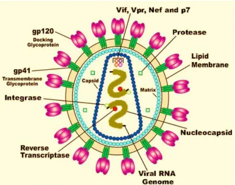
Figure 1.3 Diagram of HIV illustrating the viral RNA genome, nucleocapsid, lipid membrane, the gp120 docking glycoprotein, gp41 transmembrane glycoprotein, the viral enzymes (reverse transcriptase, integrase, protease) and viral proteins (Vif, Vpr, Nef, p7). https://www.google.co.za/search. Diagram of human immunodeficiency virus (HIV). Adapted from U.S. National institute of health (U.S. Department of health and human services).
4
Figure 1.4 HIV life cycle (De Clercq, 2009). 9
Figure 1.5 (A) Structure of HIV-1 RT and the different subdomains (Pata et al., 2004);
(B) Polymerase active site with the YMDD motif and divalent ions (Chong and Chu, 2004); (C) NNRTI-binding pocket, showing the residues at which NNRTI-resistance mutations occur (Sarafianos et al., 2010).
12
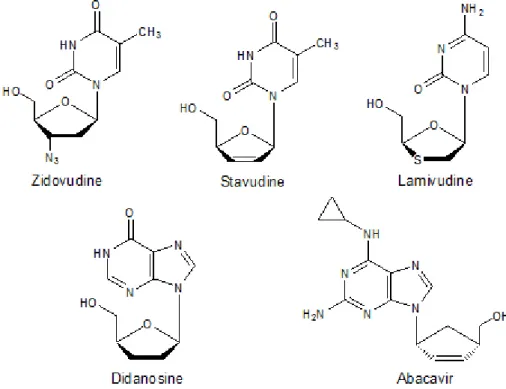
Figure 1.6 Clinically used NRTIs (Sarafianos et al., 2010). 15
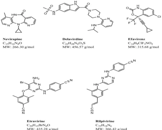
Figure 1.7 Structures of common NNRTIs (Usach et al., 2013). 21
Figure 1.8 Illustrating the strategic flexibility of NNRTIs to handle resistance mutations by conformational wiggling and positional jiggling
28
Figure 1.9 Illustrating different conformational modes in the binding pocket (A) butterfly-like model (B) Horseshoe model.
30
Figure1.10 Conformation of the subdomains of the HIV-1 RT subunit with NNRTI Riplivirine accommodated in the hydrophobic binding pocket. Trp24 on the finger subdomain and Lys287 on the subdomain are represented by green circles. Adapted from (Nizami et al., 2016).
30
x
Figure 1.11 Mechanism of work-flow in a MALDI-TOF MS (Singhal et al., 2015). 35
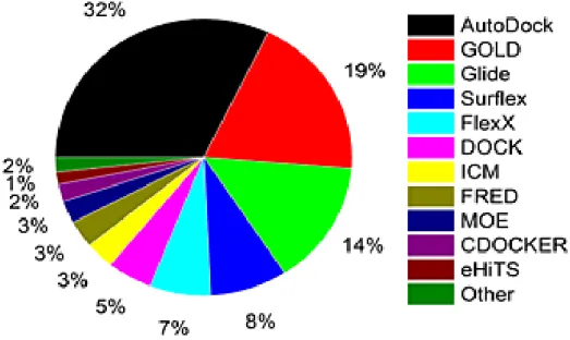
Figure 1.12 Various docking programmes (%) commonly used (Yuriev et al., 2013). 36
Figure 1.13 Chemical structures of (A) (6R)-6-acetoxydichotoma-3,14-diene-1,17-dial (ADD), (B) (6R)-6-hydroxydichotoma-3,14-diene-1,17-dial(HDD) and (C) diterpenes dolabelladienotriol (THD). Adapted from Miceli et al., (2013).
37
Figure 1.14 Docking complexes of (A) ADD, (B) THD and (C) HDD (orange)bound to HIV-1 RT enzyme (grey) showing hydrogen bonding (blue) and van der waals interactions (red) with specific NNRTI-BP residues. Adapted from Miceli et al., (2013).
38
Figure 1.15 A 4-thiazolidinone derivative (1656714) forms interactions with aspartate residues (Seniya et al., 2015).
39
Figure 1.16 2D Structure of (A) Alizarine derivative K-49 docked into wild type RT (E) Alizarine derivative K-49 showing possible binding interactions with Tyr188, Leu100, Val106, Phe227, Tyr318, Leu234 (Esposito et al., 2011).
39
Figure 1.17 Structures of: (A) digoxigenin-dUTP (DIG-dUTP) indicated in the orange box and (B) D-(+)-Biotin (only biologically active isomer of 8 possible isomers).
41
Figure 1.18 Colorimetric HIV-1 reverse transcriptase assay. 42
Figure 2.1 Reaction scheme for synthesis of N-trityl-phenylalanyl-8-(6- aminohexyl)aminoadenosine-3,5-cyclic monophosphates.
50
Figure 3.1 Thin layer chromatograms (A) N-hydroxysuccimide esters of N-trityl phenylalanine derivatives, H, F, Cl, Br, I (3a-e respectively). (B) Synthesis of p-fluoro conjugate 4b; 8-(6-aminohexyl) adenosine-3',5'-cyclic monophosphate (NUC), NHS ester of N-trityl-p-F-DL-phenylalanine (AA), reaction mixture (RM), N- hydroxysuccinimide (NHS) as well as the purified product.
61
Figure 3.2 (A) Numbering of 8-AHA-CAMP and (B) conjugate with hydrophobic trityl components via a 6-carbon spacer to form the N-trityl-phenylalanyl derivatives (4a-e).
64
Figure 3.3 UV absorbance spectra of N-trityl-phenylalanyl conjugates (4a-e). Base line(
). (4e) ( ). (4d) ( ). (4c) ( ). (4b) ( ). (4a) ( ).
69
xi
Figure 3.4 UV absorbance spectrum of 8-(6-aminohexyl) aminoadenosine-3′,5′-cyclic- monophosphate.
69
Figure 3.5 Detection of inhibitory effect on HIV-1 reverse transcriptase using the reverse transcriptase colorimetric assay kit. (A) Nevirapine (B) fluoro derivative 4b (C) phenylalanyl derivative 4a.
72
Figure 3.6 Relative reverse transcriptase activity (%) plotted against Molarity of inhibitor (A) Nevirapine, (B) 8-(6-aminohexyl) adenosine,3′,5′-cylic-monophosphate (C) 4a (D) 4b (E) 4c (F) 4d (G) 4e. RT mixed with lysis buffer and DMSO was used as the control (positive). Results were reported as mean +/- SD (n=3).
77
Figure 3.7 Effect of 10% DMSO on HIV-1 reverse transcriptase activity. 81
Figure 3.8 3D docking interaction of: (A) Nevirapine (B) 4a (C) 4b (D) 4c (E) 4d (F) 4e and HIV-1 reverse transcriptase allosteric site. Compounds are in stick representation while the RT allosteric site is in ribbon representation.
84
Figure 3.9 LigPlot predicted docking binding interactions between: (A) Nevirapine, (B) 4a (C) 4b (D) 4c (E) 4d (F) 4e and HIV-1 reverse transcriptase allosteric site. Hydrogen bonds are indicated by green dashed lines between atoms involved, while hydrophobic contacts are shown as an arc with red spikes pointing towards the atoms they contact.
88
Figure 4.1 UV spectrum of tritanol showing the log10ε at specific wavelengths. 116
Figure 4.2 1HNMR spectral analysis of 8(6-aminohexyl) aminoadenosine-3′,5′-cyclic- monophosphate and N-trityl-phenylalanyl derivatives (A) 8-AHA-cAMP (B) 4a (C) 4b (D) 4c (E) 4d (F) 4e.
118
Figure 4.3 Anomeric region of ribosyl moiety (C-1′) in preparative N-trityl-Cl-DL- phenylalanyl- 8(6-aminohexyl) aminoadenosine-3′,5′-cyclic-monophosphate.
120
Figure 4.4 1HNMR spectral analysis of the first preparative N-trityl-Cl-DL-phenylalanyl conjugate (4c1).
121
xii
Figure 4.5 MALDI-TOF mass spectra of N-trityl-phenylalanyl conjugates (4a-e) (A) 4a (B) 4b (C) 4c (D) 4d (E) 4e.
124
xiii
LIST OF TABLES
Table 1.1 Commonly used non-nucleoside reverse transcriptase inhibitors, chemical structures and their EC50 values (µM) against wild type and mutant HIV-1 RT. (Das et al., 2007). Amino acid codes: K= Lys, Y=Tyr, L=Leu, N=Asn, C=Cys.
29
Table 2.1 Summary of synthesis of N-trityl subsituited-phenylalanyl-8-(6- aminoadenosine-3′,5′-cyclic monophosphates.
52
Table 2.2 Components for colorimetric HIV-1 reverse transcriptase ELISA. 58
Table 3.1 Rf values of N-trityl-phenylalanyl derivatives in ethanol: H2O (2:1, v/v) solvent.
62
Table 3.2 Melting points of starting materials. 62
Table 3.3 1HNMR spectral analysis of N-trityl-phenylalanyl -8(-6-aminoadenosine) 3',5'-cyclic monophosphate and of 8-AHA-cAMP.
65
Table 3.4 UV spectral data of 8-(6-aminohexyl) adenosine-3′,5′-cyclic-monophoshate and of N-trityl-phenylalanyl conjugates of 8-(6-aminohexyl) adenosine-3′,5′-cyclic- monophoshate (4a-e).
71
Table 3.5 Calculated molecular weights (g/mol) and those obtained by MALDI-TOF MS analysis.
71
Table 3.6 The estimated IC50 values for Nevirapine and N-trityl-p-substituted- phenylalanine-8-(6-aminohexyl)amino-adenosine-3′,5′-cyclic monophosphates against HIV-1 RT in vitro.
78
Table 3.7 Dock binding energy (kcal/mol) of Nevirapine and test compounds with HIV- 1 RT allosteric pocket as predicted by AutoDock Vina.
83
xiv
LIST OF ABBREVIATIONS
ABC Abacavir
AIDS Acquired immune deficiency syndrome
Ala Alanine
Asp Asparagine
AZT Azidothymidine
DDI Didanosine
DLV Delavirdine
D4T Stavudine
EFV Efavirenz
ETR Etravirine
FDA Food and drug administration
Gly Glycine
Glu Glutamic acid
HAART Highly active antiretroviral therapy
His Histidine
HIV Human immunodeficiency virus
Ile Isoleucine
IN Integrase
Leu Leucine
Lys Lysine
M-MuLV RT Moloney murine leukemia virus MTCT Mother to child transmission
NC Nucleocapsid
Nef Negative regulatory factor
NNRTI Non-nucleoside reverse transcriptase inhibitor
NNRTI-BP Non-nucleoside reverse transcriptase inhibitor binding pocket NRTI Nucleoside reverse transcriptase inhibitor
N(t) RTI Nucleotide reverse transcriptase inhibitor
xv
NVP Nevirapine
PBS Primer binding site
PBMC Peripheral blood mononuclear cells
Phe Phenylalanine
Ppt Polypurine tract
PR Protease
RPV Rilpavirine
RSV Rous sarcoma virus
RT Reverse transcriptase
Ser Serine
TFV Tenofovir
Trp Tryptophan
Tyr Tyrosine
Val Valine
Vif Viral infectivity factor
Vpr Viral protein R
WHO World health organization
xvi
ACKNOWLEDGMENTS
First and foremost, I would like to thank God for his richest blessings throughout this year.
My sincere gratitude and appreciation to the following individuals:
My supervisors, Professor M Ariatti and Professor AS Gupthar for their utmost guidance and support throughout this project.
Mr. D. Jagjivan, Department of Chemistry, University of KwaZulu-Natal (UKZN), Westville Campus, for the generation of NMR spectra.
Prof G. Kruger and his team, Department of Pharmaceutical Sciences, University of KwaZulu- Natal (UKZN), Westville Campus, for the generation of MALDI-TOF spectra and computational work.
Dr A. Kumar of the Discipline of Microbiology (UKZN, Westville) for his assistance on the ultra violet spectrometer.
The National Research Foundation (NRF) and the University of KwaZulu-Natal (UKZN) for the scholarship that allowed me to complete this project.
And finally, a special thank you to my Father, Mother, Sister and Deshwyn for their love, support, guidance and encouragement throughout my studies.
xvii
CHAPTER ONE
INTRODUCTION AND LITERATURE SURVEY
1.1 Human immunodeficiency virus
A family of malignant human retroviruses, among them the human immunodeficiency virus, also known as HIV (Killian et al., 2011), is responsible for Aquired Immune Deficiency Syndrome (AIDS), a worldwide epidemic. HIV is a single stranded RNA, blood-borne virus that is most often transmitted via shared intravenous drug paraphernalia (Gostin, 1991; Gaskin et al., 2000) sexual intercourse with an infected partner, or mother-to-child transmission (MTCT), during birth process or breastfeeding. HIV retroviruses function by attaching themselves to a healthy host cell, fusing with the host cell membranes, integrating into the host cell nucleus and incorporating its viral DNA into a normal host cell genome (Perilla et al., 2016).
AIDS was first discovered in 1981 (Gottlieb et al., 1981) and two years later, HIV and human- lymphotropic virus type III were isolated as the causative agents of the disease (De Clercq, 2009). The ability of HIV to replicate rapidly in healthy cells and the errors made during the replication process, cause the virus to infect and evolve much faster in patients, making it a global health care crisis (UNIAIDS, 2013). At the end of 2014, approximately 36.9 million people were infected with HIV and 1.2 million people had died from HIV-related diseases worldwide (World Health Organization, 2015). However, according to recent statistics, approximately 36.7 million people were living with HIV in 2015 (UNAIDS, 2016).
HIV infections can’t be cured (Ensoli et al., 2014) and drug administration is a lifelong commitment to those suffering with the virus (Sarafinos et al., 2010). Therefore, various anti- viral drug therapies have been studied and designed over the past years to provide HIV positive patients with a good quality of life (Figure 1.1). This, however, is an ongoing process since
1
HIV-1 has the tendency to show resistance to administered drugs, as the major enzyme in RNA replication, reverse transcriptase, lacks the ability to proof read (Asahchop et al., 2012). Other issues such as toxicity and harmful side effects such as nausea, diarrhoea and muscle disease are also common factors that contribute to HIV resistance. Therefore, new anti-viral drugs with low toxicity and possibly fewer side effects are continuously being sought and studied. Other approaches, such as bone marrow transplantation, have led to limited success.
With help from various funding organizations, HIV/AIDS victims have gained access to various anti-retroviral treatments as shown in Figure 1.1. According to World Health Organization (WHO), low income countries such as South Africa, Botswana and Guyana have gained access to drug therapy which have assisted 80% of HIV infected pregnant women (World Health Organization, 2012).
Figure 1.1 People on antiretroviral treatment from 2010-2016 (UNIAIDS, 2016).
According to statistics published in the UNICEF annual reports, (2013), mother to child transmission has decreased markedly in the period 2000 to 2013 in some African countries.
Botswana was shown to have less than 5% mother to child transmission after breastfeeding, however more than 10 countries were shown to have more than 15% mother-to-child transmission (Figure 1.2). Highly Active Antiretroviral Therapy (HAART) has indeed caused a great decrease in HIV infections world-wide.
2
Figure 1.2
1.1.1 Structure of HIV
HIV-1 is a lentivirus belonging to a family of retroviruses. HIV is composed of an outer layer that contains the envelope glycoprotein gp160 (Figure 1.3). In its native form, the HIV trimeric envelope glycoprotein consists of gp120 and gp41 subunits (Lobritz et al., 2010). Both molecules work together through non-covalent interactions. The gp120 glycoproteins assist in viral replication by binding to the receptors on the target host cells while the gp41 proteins play a role in fusion of the host cell and viral membrane. Found below the outer shell of a mature virion, is a layer of matrix (p17) as well as a round-shaped core that is formed from the virus capsid (p24). This core serves as a protector as it shields the components found in the virion as well as the p6 protein that functions in late viral assembly. The components found within the virion are two copies of the positive sense genomic viral RNA and are shielded by the nucleocapsid (NC) from nuclease digestion. The core also contains three viral enzymes:
integrase (IN), reverse transcriptase (RT) and protease (PR) (Sundquist and Kräusslich, 2012).
PR, along with the viral proteins; negative regulatory factor (Nef), viral infectivity factor (Vif) and viral protein R (Vpr) are said to be located within the virion. Vpr plays a role in the replication and transcription of non-dividing cells (i) induces apoptosis of cells and (ii) causes death of cell cycle in proliferating cells (Bukrinsky and Adzhubei, 1999).
Mother to child transmission rate (%) from 2000-2013 (UNICEF annual report, 2013).
3
The HIV genome contains three genes namely, 5'gag-pol-env-3' that encode the viral enzymes as well as structural proteins. Gag precursor protein (p55) is 55 kD in length and is formed by the gag gene. After the budding process the enzyme, protease cleaves p55 into the p17 matrix, p6, p24 capsid and nucleocapsid. This causes conformational changes in the viral structure such that the p24 capsid encloses, surrounding the viral RNA while the p17 matrix remains intact (maturation).
Figure 1.3 . Diagram of HIV illustrating the viral RNA genome, nucleocapsid, lipid membrane, the gp120 docking glycoprotein, gp41 transmembrane glycoprotein, the viral enzymes (reverse transcriptase, integrase, protease) and viral proteins (Vif, Vpr, Nef, p7). https://www.google.co.za/search. Diagram of human immunodeficiency virus (HIV). Adapted from U.S. National Institute of Health (U.S. Department of health and human services).
4
1.1.2 Life cycle and viral replication
Most retroviruses, such as Rous sarcoma virus (RSV), tend to infect dividing cells during the process of mitosis. HIV, on the other hand, can infect non-dividing cells when certain endogenous or exogenous genotoxic agents damage DNA (Lyama and Wilson, 2013), leading to multiple human diseases. When a person is infected with HIV, the virus is said to remain latent for long periods of time. It then starts attacking specific CD4+ T cells that serve in cell- mediated responses. Since HIV is a single-stranded ribonucleic acid (RNA) virus, it cannot replicate on its own and requires the formation of double-stranded DNA to replicate. The process and synthesis of viral DNA is shown in Figure 1.4.
1.1.2.1 Attachment, un-coating and fusion
T lymphocytes play a crucial role in protecting the body’s immune system. They contain specialized antibody-like receptors on their surface that are used to recognize and detect harmful antigens on the surface of other infected cells. T cells have two important functions: (i) attacking infected, and (ii) regulating and directing immune system responses.
HIV is known to attack T helper cells, as they are essential in stimulating the activation of other important immune cells such as B cells. When HIV attacks these cells, the immune system cannot function properly and this causes the body to become prone to many other infections (Casiday and Frey, 2001). These cells are also called T helper cells because HIV uses the CD4 proteins found on the surface of the helper cells, to attach to and enter the cell. T helper cells also contribute to the activation of B cells and cytotoxic cells with chemical signals. Therefore, when HIV attacks T cells, it also prevents the activation of B cells and cytotoxic T cells, which leaves the immune system vulnerable to harmful foreign antigens.
5
The protein Nef (1.1.1), is expressed during the attachment step. Nef promotes the survival of infected cells by reducing the presence of essential complexes such as major histocompatibility complex (MHC I) and MHC II present on antigen-presenting cells (APCs) and target cells, and CD4 present on helper T cells (Das and Jameel, 2005, Das et al., 2005).
The HIV-1 envelope (Env) contains spikes protruding from its binding site surface which comprise trimers of non-covalently-linked heterodimers consisting of glycoprotein gp120 and the transmembrane glycoprotein (Figure 1.3) (Engelman and Cherepanov, 2012). The process of replication occurs when glycoproteins (gp120) on the HIV-1 envelope bind to the CD4+ receptors protruding from the surface of T helper cells (Engelman and Cherepanov, 2012). This interaction causes the formation of a bridging sheet and brings about a conformational change in the structure of both molecules, therefore exposing a site known as the chemokine co- receptor binding site.
This enables co-receptors such as CCR5 or CXCR4 to facilitate viral entry into the cell by membrane fusion (Mehellou and De Clercq, 2010) after exposure of the gp41 peptide, which is inserted into the host cell membrane (Wilen et al., 2012). This brings the host and viral membranes closer, permitting the fusion peptide of gp41 to fold at a hinge region, bringing a carboxy-terminal helical region (HR-C) and an amino-terminal helical region (HR-N) from the gp41 subunit together to form a six-helix bundle (6HB). Due to the proximity of HR-C to the viral membrane (caused by the glycoprotein, gp41) and the proximity of HR-N domain to the host membrane (caused by the gp41 peptide), the 6HB essentially links the two membranes causing a fused pore. The viral core is then released into the host cytoplasm (Wilen et al., 2012).
Once in the cytoplasm, host cell enzymes help in the removal of the viral capsid, resulting in structural change and the release of the viral RNA into the host cell. This process is known as un-coating.
1.1.2.2 Reverse transcription
Reverse transcription is the process of synthesizing a double-stranded DNA molecule from a single-stranded RNA template. It is called reverse transcription as it acts in the opposite direction to transcription. The process of reverse transcription was unaccepted at first as it contradicted the central dogma of molecular biology which states that DNA is the code which is transcribed into RNA which then carries the message to be translated into proteins. However,
6
due to the independent discovery of the enzyme reverse transcriptase in 1970, by Howard Temin and David Baltimore (Coffin and Fan, 2016) and its role in reverse transcription, the possibility that DNA could be copied from an RNA template in the reverse manner was accepted.
The formation of a complementary single-stranded DNA is carried out by the enzyme reverse transcriptase. The process of reverse transcription is essential as it involves the conversion of a single-stranded plus-sense RNA genome to a double-stranded cDNA which can be inserted or integrated into the host cell (Klickstein et al., 2001; Hu and Hughes, 2012). There are two enzymatic activities that are essential to carry out the process of reverse transcription. These include a DNA polymerase (reverse transcriptase) that can copy a DNA or RNA template, and an RNase H activity which helps degrade RNA during the synthesis of a provirus (Hu and Hughes, 2012). HIV-1 reverse transcriptase uses viral single-stranded RNA as a template, to catalyse the formation of a proviral DNA.
Viral genomic RNA is plus stranded and the synthesis of the first DNA strand, also known as (–) strand DNA, is synthesized by extending the 3′-end of a specific tRNAlys3 using the viral RNA as a 3′-5′ template (Betancor et al., 2015). Reverse transcriptase, like many other polymerases also requires a primer and template to initiate strand polymerization. The 3′ end of the cellular tRNAlys3 primer, is based paired to a complementary sequence of nucleotides at the 5′ end of the viral genome called the primer binding site (PBS). This site is approximately 180 nucleotides from the 5′ end of the viral genome.
1.1.2.2.1 First strand synthesis (Minus strand)
The enzyme reverse transcriptase attaches to the growing DNA strand and copies the 5′ end of the viral RNA genome to form a RNA-DNA hybrid duplex and with the help of RNases H, the viral RNA is degraded nucleolytically, exposing the newly synthesized single-stranded minus DNA. This creates direct repeats (long term terminal repeats) at the 5′ and 3′ ends of the viral RNA, which act as a bridge to allow the newly synthesized single-stranded minus DNA at the 5′ end to join with the complementary repeat sequence (R) at the 3′ of the viral RNA.
Retroviruses contain two copies of their RNA genome; the minus strand (also called the first
7
jump) (Sarafianos et al., 2010) undergoes a transfer which involves the repeat sequence (R sequence) at the 3′ end of one of the two RNAs.
Once the minus strand DNA joins to the R sequence, the synthesis of the DNA strand continues along the viral genome in the 5′ direction. As synthesis of minus strand DNA continues, RNase H continues to degrade the RNA strand. However, at the 3′ end of the viral genome, there is a sequence that is rich in purines. This sequence is called the polypurine tract or ppt and is found to show resistance to RNase H activity. This purine sequence allows for a short stretch of RNA to remain attached to the newly synthesizing cDNA and serves to start the synthesis of the second strand DNA (plus strand), by serving as the primer sequence.
1.1.2.2.2 Second strand synthesis (Positive strand)
HIV- 1 has two polypurine tracts (ppts), one at the 3′ end and one at the middle of the RNA genome (Hu and Hughes, 2012). When RT creates the plus-strand DNA that is initiated from the 3′ ppt, it copies the minus-strand DNA, as well as the first 18 nucleotides of the tRNAlys3
primer. This stops DNA synthesis. A study carried out by Swanstrom et al., (1981) with avian sarcoma-leukosis virus suggested that the ppt-primed plus strand DNA synthesis would stop when it comes across a modified Adenosine (A) that the enzyme reverse transcriptase cannot copy. The same applies to HIV. Once the 3′ terminal of the tRNA is copied into DNA, it becomes sensitive to RNase H. RT in HIV-1 is the only RT that cleaves the tRNA from the 3′
end, therefore leaving a ribo-A nucleotide at the 5′ end of the viral minus strand DNA.
1.1.2.3 Integration and re-infection
The newly synthesized double-stranded viral DNA in the core is in the host cell cytoplasm and migrates into the host cell nucleus, where it is integrated into the host cell genome as a provirus with the help of the enzyme, integrase (Craigie and Bushman, 2012). The provirus acts as a template to form messenger RNA (mRNA) and viral RNA during the process of transcription.
The mRNA then travels to the host cell cytoplasm, where it undergoes translation to form Gag and Gagpol poly-proteins (Sarafianos et al., 2010). With the help of host enzymes, the viral
8
RNA and Gag and Gagpol poly proteins migrate to the cell surface to form new virus particles, each containing two copies of the RNA genome and the essential proteins needed for re- infection.
Figure 1.4 Illustrating the stages in the HIV life cycle. Virus adsorption, virus-cell fusion, uncoating, reverse transcription, integration, transcription, translation and budding (De Clercq, 2009).
9
1.2 The genesis, structure, and enzymatic functions of HIV-1 reverse transcriptase
A unique characteristic of the RT enzyme is that it can utilize both DNA and RNA templates.
HIV-1 RT has three main functions: It can act as an RNA-dependant DNA polymerase to produce cDNA from an RNA template, it’s Ribonuclease H activity degrades the RNA template during the formation of cDNA and it acts as a DNA-dependent DNA polymerase synthesizing double-stranded DNA using cDNA as the template (Sarafianos et al., 2010). Most of RT’s functions are found in the same protein subunit although they are considered to be monomeric enzymes. However, HIV-1 RT is a heterodimer, consisting of two subunits, termed p66 and p51. The p66 subunit contains two domains: the DNA polymerase domain and the RNase H domain, which are located at different regions in the p66 subunit (Thammaporn et al., 2015).
The polymerase domain of the RT contains 4 subdomains: the connection, fingers, palm and thumb (Figure 1.5A). The connection domain connects the polymerase and RNase H domains as it acts as a bridge. These subdomains create a site for the binding of the primer, template, two divalent cations and dNTPs during the synthesis of DNA (Yokoyama et al., 2010). Hence it is called the polymerization active site. This site is composed of three key aspartic residues;
Asp185, Asp110 and Asp186, which are in the palm domain. The Asp186 and Asp185 comprise part of the YMDD motif (Tyr-Met-Asp-Asp) corresponding to the more general YXDD motif (X = Met, Val, Leu or Ala) of HIV-1 RT. Tyr183 and Asp185 play a role in the formation of a hydrogen bond with the 3′-hydroxyl group at the primer end as well as act as a base to undergo a nucleophilic attack on the α-phosphate group of an incoming nucleoside-5′- triphosphate (Figure 1.5 B). The overall structure of the RT is often described as a right hand, where the polymerization active site is found in the “palm sub-domain’’ between the ‘’fingers’’
and the ‘’thumb’’ and ‘’runs’’ through the connection and RNase H domains (Das and Arnold, 2014).
The second subunit, p51 is similar to the p66 subunit, however it lacks the C-terminal RNase H domain and is formed by HIV-1 protease mediated cleavage of the C-terminal RNase H domain of the p66 subunit. The amino acid sequence that forms the polymerase active site of p66 domain is the same as the polymerase active site in the p51 domain, which is however not
10
functional (Xia et al., 2007). Therefore, the function of the p51 domain is to basically provide structural support to the RT enzyme.
The second site to which RT inhibitors bind is called the non-nucleoside binding pocket (Figure 1.5 C). A non-nucleoside binding pocket is found in the palm subdomain of the p66 subunit and is approximately 10Å away from the aspartic acid catalytic triad in the polymerization active site (Santos et al., 2015). The binding pocket is located between β6-β10-β9 and β12- β13-β14 sheets of the palm subdomain. The allosteric binding pocket is known to be hydrophobic in its natural form, consisting of aromatic residues (Tyr181, Tyr188, Phe227, Trp229, Tyr233), along with hydrophilic residues such as Ser105, Lys101, Lys103, Asp192, Glu22 and Glu138 of p51 subunit (Sarafianos et al., 2010). The hydrophobic binding pocket allows the template strand to bind to reverse transcriptase enzyme as it exposes the 3′-OH end of the primer to the catalytic site. The “thumb’’ subdomain contributes to this exposure as it functions in the mobilization of the template and the primer to the polymerization active site when the reverse transcriptase enzyme forms a closed circle around the sub domains (Hu and Hughes, 2012). This brings the thumb and fingers to move closer to the palm subdomain and allows for binding of nucleic acids. It is also known to be more flexible than the ‘palm and fingers’ thus allowing for proper binding of strands (Kohlstaedt et al., 1992).
11
A
B C
Figure 1.5 (A) Structure of HIV-1 RT and the different subdomains (Pata et al., 2004);
(B) Polymerase active site with the YMDD motif and divalent ions. (Chong and Chu, 2004); (C) NNRTI-binding pocket, showing the residues at which NNRTI-resistance mutations occur (Sarafianos et al., 2010). Amino acid codes: F227=Phe, G190=Glu, K101, K103=Lys, L100, L234=Leu, P236, P95=Pro, V106, V106= Val, W229=Trp, Y181, Y188, Y318= Tyr.
12
1.3 Early history of therapeutic interventions
After the isolation of HIV-1 in 1983, extensive studies were carried out to control the spread of the chronic virus (Hoggs et al., 1999). The first anti-viral drug, Zidovudine (also known as AZT) was first synthesized in 1964 as an anti-cancer drug and thereafter became the first successful drug to be approved by the U.S. Food and Drug Administration (FDA) for extending the lives of those suffering with HIV infection up to 18 months in 1987 and later on became the preferred drug for the prevention of HIV infection (Corey et al., 2007). AZT at the triphosphate level was used to restore the immune system as it could enter the reverse transcriptase active site and block its activity in the HIV replication cycle (Furman, et al. 1986).
The discovery of AZT’s antiretroviral activity subsequently led to the development of other anti-retroviral drugs. However, one of the main limitations of AZT was that the HIV virus could easily show resistance to the drug within a short period of time as well as cause undesired side effects. For this reason, a second drug therapy, highly active antiretroviral therapy (HAART) was designed to overcome this problem (Asahchop et al., 2012).HAART was implemented after 1995 and has indeed improved the lives of HIV-infected patients, as this system brought about longer survival periods (Eswara Rao et al., 2015). The system of HAART consists of a mixture of 3 or more drugs usually from two different classes of anti-viral drugs, namely; nucleoside reverse transcriptase inhibitors (NRTI) and non-nucleoside reverse transcriptase inhibitors (NNRTI). Anti-retroviral treatment is very effective at preventing HIV from multiplying and spreading throughout the body. This prevention protects the immune system and thus allows the body to fight off other HIV-related opportunistic infections which eventually lead to AIDS.
1.3.1 Reverse transcriptase: The target for anti-retroviral drug therapy
Reverse transcriptase has long been a target for the development of anti-viral drug therapy, due to its major role in the replication process of HIV. HIV infections can’t be cured easily and therefore, the administration of drugs is a lifelong commitment for those infected with the virus (Sarafianos et al., 2010). Therefore, compounds should be easily administered and non-toxic.
There are different classes of drugs that intervene at different stages of the life cycle of HIV.
However, drugs that specifically target the DNA polymerization activity of the reverse 13
transcriptase enzyme are said to be the backbone of current HIV-1 strategies (Betancor et al., 2015). These drugs can be split into two groups, namely, (i) nucleoside/nucleotide reverse transcriptase inhibitors (NRTIs) and (ii) non-nucleoside reverse transcriptase inhibitors (NNRTIs). NTRI and NNRTIs that target the DNA polymerization active site of the reverse transcriptase enzyme are said to be the backbone of current HIV-1 treatment strategies (Betancor et al., 2015). Several drugs have been successfully implemented to inhibit RT. These include Azidothymidine, Nevirapine, Tenofovir, Abacavir, Stavudine, Didanosine, Etravirine, Delavirdine, Efavirenz and Rilpivirine. However, due to their toxic side effects and the resistance caused by viral mutations, their therapeutic effects are sometimes limited (Padariya et al., 2016).
1.4
Nucleoside reverse transcriptase inhibitors (NRTIs) were the first successful retroviral agents used against HIV and are thus, the oldest group of antiretroviral agents. NRTIs are analogues of naturally occurring nucleosides and are therefore inactive in their normal forms. To display their anti-viral activity an NRTI requires host cell entry and must undergo phosphorylation twice by initial conversion to its 5′-monophosphate (NMP), followed by pyro-phosphorylation to its 5′-triphosphate (NTP) form by host cell kinases in order to compete with naturally occurring deoxynucleotide triphosphates (Michailidis et al., 2009). NRTIs lack a 3′-OH group on the deoxyribose sugar moiety thus preventing the formation of a 3′,5′-phosphodiester bond between the NRTI and a naturally occurring 5′-nucleoside triphosphates (Sarafianos et al., 2010). When reverse transcription occurs in the presence of a NRTI, RT may bind a NRTI triphosphate instead of a naturally occurring nucleotide building block and this, in turn, prevents reverse transcription. NRTIs may compete with and block the addition of naturally occurring substrates as well as become incorporated into the growing DNA molecule. When the NRTI triphosphates are incorporated into the nascent DNA, they act as chain terminators (Goody et al., 1991) by preventing the process of elongation; hence a double-stranded DNA is not fully formed and cannot be incorporated into a new host cell.
Along with the amino acid residues previously mentioned, there are two Mg2+ ions that are also present in the polymerase active site that are approximately 3.6 Å apart from each other (Figure
Nucleoside/ nucleotide reverse transcriptase inhibitors and their inhibitory mechanism
14
1.5 B) (Goldschmidt et al., 2006). One of the ions, binds to the three phosphate groups of the incoming inhibitor as well as the Asp110 and Asp185 residues. This causes the Mg2+ to enter the catalytic site preventing exchange of free deoxynucleoside triphosphate. The second Mg2+
ion binds the three aspartate residues: Asp110, Asp185 and Asp186 as well as the α phosphate group of the incoming deoxynucleoside triphosphate (Goldschmidt et al., 2006).
Figure 1.6 Clinically used NRTIs (Sarafianos et al., 2010).
Nucleotide reverse transcriptase inhibitors (NtRTIs) generally display the same inhibitory mechanism. The only difference is that NtRTIs are nucleotide analogues and require further phosphorylation to be converted to their active triphosphate form as they already possess a phosphate group (De Clercq, 2009). NtRTIs are polar in nature as they contain a triphosphate group, a 5-carbon sugar and a nitrogenous base. Therefore, their polar property prevents their movement across the plasma membrane to enter the cell.
15
1.4.1 Clinically approved NRTIs
1.4.1.1 Azidothymidine (AZT)
AZT, also known as Zidovudine (Figure 1.6) is a potent inhibitor of reverse transcriptase, when it is converted to its 5′ triphosphate form. When reverse transcriptase utilizes AZT-5′- triphosphates to incorporate an AZT residue into a growing DNA strand, this serves as a chain terminator, therefore inhibiting reverse transcription (Corey et al., 2007). AZT possesses an azido group at the 3′-position on its 2′-deoxyribose sugar moiety, which prevents DNA chain extension using 2′-deoxynucleoside-5′-triphosphate building blocks. To display its anti-viral activity, AZT is first converted into its 5′- triphosphate form inside the cell. The triphosphate form of the drug cannot penetrate the cell membrane (Michailidis et al., 2009). Therefore, AZT in its monophosphate form also lowers the formation of 2′-deoxythymidine 5′-triphosphate (dTTP) by competitive inhibition. The mechanism and inhibitory effect of AZT was described in a study by Furman et al., (1987). In this study, the effect of AZT in its 5′- mono, di and triphosphate form on uninfected human fibroblasts and lymphocytes was investigated. It was shown that the inhibition of the growth of uninfected cells occurred at concentrations over 1mM and the conversion of AZT to its 5′-mono, di- and triphosphate forms was similar in HIV infected cells.
AZT is known to cause many harmful side effects including headaches and nausea (Santos et al., 2015). However, drug resistance allows for other opportunistic diseases to occur and to affect the patient. For this reason, AZT is mainly used in combination with other antiviral drugs. The combination of two or more anti-viral drugs in a patient’s body not only reduces viral replication, but also minimizes the chances of the virus showing resistance to the drugs.
Since AZT was the first successful anti-HIV drug, it was the most expensive medicine at the time, costing users $8,000-$10,000 per year. It was used exclusively until other anti-viral were developed. However, the quest for new more effective anti-viral with fewer side effects is an ongoing process.
16
1.4.1.2 Tenofovir (TFV)
Tenofovir, a NRTI was discovered in 1997 as a potent inhibitor of reverse transcriptase. In the same year, TFV was modified to Tenofovir disoproxil (TDF), making it the first oral prodrug of TFV (Wang et al., 2016) and becoming one of the most commonly used drugs for the treatment of HIV-1 infection. TDF is used in many fixed-dose combinations such as with Efavirenz and Rilpivirine and one of its side effects is renal toxicity (Wang et al., 2016). TFV is hydrophilic making it difficult to move across a hydrophobic membrane (Van Rompay et al., 2012). After following a two-step phosphorylation process, TFV is converted into its active form, Tenofovir diphosphate (TFV-DP) which shows anti-HIV activity (Biswas et al., 2014;
Wang et al., 2016).
1.4.1.3 Abacavir (ABC)
Abacavir (Figure 1.6) developed in 1998, is a carbocyclic 2′-deoxyguanosine nucleoside analogue (Adetokunboh et al., 2014) mainly used for the treatment of HIV positive children.
Analysis of the drug on peripheral blood mononuclear cells (PBMCs) revealed that it is considerably more potent than Didanosine (DDI) but as effective as AZT. Its low toxicity level and less harmful side effects such as; reduced hypersensitivity, fewer rashes and lower fever have allowed the drug to remain well tolerated long-term (Volberding et al., 2008). The drug is metabolically converted to its triphosphate form, carbovir triphosphate and competes with the natural substrates dGTP for incorporation in the growing DNA strand.
1.4.1.4 Stavudine (D4T)
Stavudine (Figure 1.6) was first approved by the FDA in 1994 and is effective when used in combination with other anti-viral drugs. In a study carried out by Kline et al., (1996), combination treatment of D4T and Didanosine (DDI) showed strong inhibition against HIV-1 infection in a small group of children. Three children with CD4 counts higher than 50 cells/mL showed a 20% increase in CD4 counts after being treated with the combination therapy for 12
17
weeks. One of the main side effects of D4T is peripheral neuropathy. This symptom can be tolerated for a short while, however, cannot be tolerated in the long term. Therefore, alternative antiviral drugs or combination of other antiviral drugs continues to expand (Volberding et al., 2008).
1.4.1.5 Didanosine (DDI)
Didanosine (Figure 1.6), a purine nucleoside became the second approved NRTI in 1991 (Brittain, 1993). This inhibitor acts against both HIV-1 and HIV-2. It requires intracellular phosphorylation by cellular kinases to initiate its inhibitory mechanism. Earlier studies have shown DDI to be an effective antiviral drug. However, when used in drug combination with AZT, it has been shown to be more effective. Side effects of Didanosine include diarrhoea, abdominal pain, dose-related peripheral neuropathy, vomiting and nausea.
1.5 Non-nucleoside reverse transcriptase inhibitors and their inhibitory mechanism
Non-nucleoside RT inhibitors (NNRTIs) are an important component of antiretroviral therapy.
NNRTIs and protease inhibitors are more potent inhibitors of viral replication than nucleoside RT inhibitors (NRTIs) and integrase inhibitors (Seckler et al., 2011). NNRTIs are structurally diverse antiviral drugs that are shown to be less effective to HIV-2. HIV-2 is less readily transmitted and is generally less pathogenic than HIV-1. Given the slow development of immunodeficiency and limited clinical experience with HIV-2, it is unclear whether antiretroviral therapy significantly slows progression. NNRTIs do not inhibit HIV-2 due to the residues at codon 181 and 188 (Tyr181 and Tyr188 in HIV-1; Ile181 and Leu188 in HIV-2) which prevent the drugs from binding to HIV-2 RT (Sluis-Crèmer and Tachedjian, 2008). For this reason, NNRTIs are described as selective inhibitors of HIV-1 reverse transcriptase (Famiglini and Silvestri, 2016). NNRTIs are a group of compounds that are known to act as allosteric inhibitors of RT, thus preventing DNA polymerization. There are 5 compounds that are commonly used against HIV-1 infection. These include: Nevirapine, Efavirenz, Delavirdine, Etravirine and Rilpivirine. NNRTIs differ from NRTIs as they do not mimic or
18
compete with naturally occurring substrates, instead they bind directly to the hydrophobic binding site/pocket in the palm subdomain of the p66 domain of the RT enzyme. This site is termed the non-nucleoside reverse transcriptase inhibitor binding pocket (NNRTI-BP) and is approximately 10Å from the polymerase active site (Arts and Hazuda, 2012).
NNRTIs block the process of reverse transcription by binding to the NNRTI-BP and altering the mobility of the DNA polymerase allosteric site, more specifically, the thumb subdomain of the RT. NNRTIs are also known to deform a region in the RT, known as the ‘primer grip region’. This region functions in correctly positioning the DNA primer in the polymerase active site located in the p66 subunit. Therefore, an alteration in the primer grip region tends to cause a change in template/primer conformation and position, thus blocking formation of a ternary complex. It is also known that the NNRTI binding pocket functions as a bridge between the thumb and palm subdomains. Therefore, any alteration to the binding pocket will affect the functions of the thumb and palm subdomains.
During favourable conditions of DNA synthesis, the RT fits a “closed” conformation bringing the fingers and thumb subdomains closer to the palm subdomain and thus allow for the binding of nucleic acids. However, in the presence of an NNRTI, an open conformation is created that restricts the thumb to a hyperextension position, which prevents the polymerization of DNA (Das et al., 2012). It restricts the movement of the thumb subdomain, which prevents the template or primer strands from binding to the polymerization active site. Thus, preventing strand elongation and termination of reverse transcriptase.
It was shown that in the absence of an inhibitor, the aromatic side chains of Tyr181 and Tyr188 in the non-nucleoside RT binding pocket are positioned towards the hydrophobic core.
However, in the presence of an inhibitor the two aromatic residues move away from the hydrophobic core thus accommodating space for the incoming inhibitor (Sluis-Cremer et al., 2005).
In a study by Das et al., (2012) the mechanism of binding interactions of the NNRTI, Nevirapine on RT-DNA was compared with binding interactions of AZTTP on RT-DNA.
Upon binding of Nevirapine to the non-nucleoside binding pocket, the two amino acid residues Tyr181 and Tyr188 rotamer conformations are switched off, while β12-β13-β14 part ways from the β6-β10-β9 sheet. The β6-β10-β19 sheet contains the polymerase “catalytic triad”
(Asp110, Asp185, and Asp186), while the β12-β13-β14 sheet contains the “primer grip” that keeps the primer strand in position for the incorporation of a nucleotide. With the aid of crystal
19
structures, the binding interaction of Nevirapine was shown to cause a shift in the primer gap of about 4Å. This caused the shifted primer grip to lift the primer away from the P-site leading to lost interactions with the Tyr183MDD motif (at the polymerase active site).
In a recent study by Lu et al., (2011) an analogue of the RT inhibitor Calanolide A, 10- chloromethyl-11-demethyl-12-oxo-calanolide A, also known as (F18) and the NNRTI, Nevirapine were bound by the HIV-1 wild type, Leu100 mutant and Tyr181 mutant RTs separately. Results indicated that Nevirapine showed better binding interactions with wild type RT compared to F18. This was probably due to the rigid structure of F18. The structure of Nevirapine, on the other hand, has aromatic rings that contribute to its hydrophobicity and this property is favourable in the NNRTI-BP and thus formed aromatic interactions with the aromatic side chains of Tyr188 and promoted RT inhibition. The structure of Leu100 mutant RT was altered when bound to F18. The NNRTI-BP is hydrophobic in nature and many hydrophobic compounds will be accommodated. However, in this study, the NNRTI-BP was shifted, indicating that the hydrophobic and aromatic side chains in this site must have shifted causing fewer interaction with the ligand and thus contributing to resistance. The Tyr181 mutant structure showed better spatial flexibility with F18 and resulted in excellent antiviral activity. The change from Tyr181 to a cysteine amino acid residue was shown to contribute to this result.
20
1.5.1 Commonly used NNRTIs
Figure 1.7 Structures of common NNRTIs (Usach et al., 2013).
1.5.1.1 Nevirapine (NVP)
Nevirapine (Figure 1.7) was approved by the FDA to be the 9th successful anti-viral drug against the enzyme reverse transcriptase in the year 1996. In a study done by Sluis-Cremer et al., (2004), a single dose of Nevirapine was shown to have prevented HIV-1 transmission from mother to child. However, the virus was shown to develop resistance to the drug when administered as an immunotherapy. However, Nevirapine when given in combination with one or more other drugs was shown to be more efficient. This was first validated by Montaner et al., (1998) where the combinations of Nevirapine and Zidovudine and Didanosine with Zidovudine were compared with the combinations of Nevirapine, Zidovudine and Didanosine.
The triple combination treatment brought about a 51% drop in HIV-1 RNA levels at week 52 in patients, while the duel combinations: Zidovudine and Didanosine, Nevirapine and
21
Zidovudine showed a drop of 12% and 0% in HIV-1 levels respectively (Montaner et al., 1998).
This showed that the outcome of combining three types of drugs was much more effective and superior to that of combing two types of drugs. Although Nevirapine is widely used in the treatment of HIV-1 RT, it is also noted that 5% of individuals treated with Nevirapine develop allergic reactions with symptoms of drug reactions that occur rarely (idiosyncratic drug toxicity) (Isogai and Hirayama, 2016).
1.5.1.2 Etravirine (ETR)
Etravirine (Figure 1.7) previously known as TMC125, is a diarylpyrimidine-based NNRTI that exhibits effective antiviral activity against wild type HIV-1 as well as some viruses that show resistance to some NNRTIs (Wainberg, 2012).
1.5.1.3 Delavirdine (DLV)
Delavirdine (Figure 1.7) belongs to the bis(heteroaryl) pyridinyl group of non-nucleoside reverse transcriptase inhibitors. This compound was first described in 1993, but due to its high toxicity levels and its inability to inhibit human DNA polymerases, it is rarely used in clinical treatment. The structure of delavirdine is extremely bulky and projects from the hydrophobic binding pocket in the reverse transcriptase enzyme (Esnouf et al.,1997).
1.5.1.4 Efavirenz (EFV)
Efavirenz (Figure 1.7) is an NNRTI and is a generally safe and highly effective antiretroviral drug. This drug is one of the most commonly prescribed antiviral drugs in the world (Kryst et al., 2015). However, it is also known to cause side effects such as anxiety, insomnia, dizziness and abnormal dreams (Highleyman, 2014). Efavirenz is known to possess a half-life of 40-55 hours (Gaida et al., 2015) and is primarily metabolised in the liver by the CYP450 enzyme
22
system. The specific isoform within the system most important for the metabolism of Efavirenz is CYP2B6. According to a recent study, 50% of all patients administered with Efavirenz experience at least some of the above-mentioned side effects. This, however only occurs during the first few days of intake and subsides after a few weeks.
In a study by Highleyman, (2014) (http://www.hivandhepatitis.com, Accessed 17/11/2017) the effect of Efavirenz given at different doses was investigated. In this study, two separate groups of participants were orally given 400 mg of Efavirenz and 600 mg of Efavirenz respectively (once daily), on a 48 week analysis. Results, showed equivalent effects between the two groups.
After 96 weeks, 90% of participants on each treatment had a reduced HIV RNA <200 copies/mL in an intent-to-treat analysis (ITT). Fewer side effects were also witnessed in participants administered with 400 mg Efavirenz.
ITT involves all the randomized patients in each group that undergo treatment irrespective of the treatment they had initially received, irrespective of withdrawal and irrespective of protocol deviations (Gupta, 2011).
1.5.1.5 Rilpivirine (RPV)
Rilpivirine (also known as TMC278) is a diaryl pyrimidine NNRTI (Figure 1.7). It is one of the few NNRTIs that show strong inhibitory action against wild type and mutant HIV-1 RT at doses of 25-75 mg/day (Das et al., 2007).
1.5.1.6 Azvudine
Azvudine, is a cystidine analogue which has shown good inhibition on HIV-1 RT, hepatitis B virus as well as hepatitis C virus. In a study by Wang et al., (2014), Azvudine exercised effective inhibition on HIV-1 with EC50 (concentration of a drug that gives a half-maximal response) values ranging from 0.03 to 6.92 nM.
23
1.6 Combination of NNRTIs
In a recent study by Getell, 2015 (https://www.aidsmap.com, Accessed 12/11/2017), Doravirine, belonging to the group NNRTIs, was shown to be as effective as the antiviral drug, Efavirenz, while displaying fewer side effects. In this study, two separate groups of participants were orally given Doravirine and Efavirenz respectively, once daily. Thereafter, 100 mg of Doravirine was administered along with Tenofovir to participants in group one and 600 mg of Efavirenz was given to participants in group two on a 24-week analysis daily. However, after 24 weeks, the overall treatment response showed that 88.9% of patients treated with Doravirine and 87.0% of those who were administered Efavirenz were shown to have a viral load count below 200 copies/ml while the CD4 cell counts were 154 and 146 cells/mm3, respectively. The similarity of viral load and CD4 counts in the two groups indicated that the two regimens were equally effective. However, Doravirine was shown to cause fewer side effects than Efavirenz.
In a study by Borges et al., (2016) NNRTIs were compared with protease inhibitors such as Ritonavir. In this study, clinical investigation using both inhibitors were conducted and showed equal outcomes. This was substantiated by calculating risk ratios or mean differences. In previous studies, NNRTIs exhibited much faster suppression effect on the virus while protease inhibitor, Ritonavir was shown to recover damaged CD4 cells (Ridder et al., 2008). In another study by Pozniak, (2000) patients were switched from Indinavir, a protease inhibitor to Efavirenz, a NNRTI because of its short-term toxicity and virologic failure on viral loads.
Other derivatives such as oxochromenyl xanthenone and indolyl xantheone were recently studied as anti-HIV reverse transcriptase inhibitors by Kasralikar et al., (2015). Chromene derivatives have been useful inhibitors as they possess anti-HIV pharmacological properties and have shown potent activity against wild type HIV-1 replication. Two DCP (3′ R,4′ R-di- O-(-)-camphanoyl-2-ethyl-2′-2′-dimethyldihydro-pyranol[2,3-f] chromone) analogs, 2,5- dimethyl DCP and 2-ethyl DCP have shown remarkable inhibitory effects on wild type HIV replication as well as on the drug-resistant strains, making chromene derivatives highly potent inhibitors. In this study, structure activity relationship (SAR) plays a major role in revealing the inhibitory mechanism (Kasralikar et al., 2015). A planar ring system on these inhibitory structures was shown to be a requirement for the anti-HIV activity against the wild type HIV strains as well as resistant HIV strains. Therefore, with the addition of an indole and coumarin ring onto the xanthenone core, a more planar structure was created. The use of xanthene
24
derivatives has been extensive due to their diverse properties which include anti-bacterial, anti- viral as well as anti-inflammatory activities. In this study, a one-pot three component reaction of salicylaldehyde, 1,3-cyclohexadione component and a 4 hydroxy chromene (as nucleophile) were used to prepare 4H-chromenes in the presence of 1-hexyl-3-methylimidazolium hydrogen sulfate ([Hmim]HSO4) as a catalyst, which was required in relatively small amounts (Kasralikar et al., 2015). Molecular docking studies were also performed to rationalize the structural activity relationship of the compounds as well as to determine the possible binding conformation between the designed compounds and how well they interact with the HIV enzyme. According to the study the most active compounds were the indolyl xanthenone compounds with docking scores of -12.487, -12.457 and -12.256 (kcal/mol) while the native compound was found to be -13.413 (kcal/mol). It was observed that the xanthenone ring structures interacted better with the hydrophobic binding pocket in the presence of hydrogen bonds. The indolyl xanthenone derivatives not only formed hydrogen bond interactions with the Lys101, they also formed π-π interactions in the hydrophobic binding pocket with the aromatic side chain of Trp229. Some compounds showed reduced binding activities due to the lack of hydrogen bond interaction with Lys101. It was also observed that compounds with hydrogen bonding with the side chain backbone of Lys101 as well as π-π interactions with the aromatic side chain of Trp229 display