Onderstepoort Journal o j
r
eterinary Science; and Animal Industry, Volume 11, Number 1, July, 1938.Printed in the Union of South Africa by the GoYernmcnt Printer, Pretoria.
South African Helminths.-Part V . Some Avian and Mammalian Helminths.
By R. J. ORTLEPP, Section of Parasitology, Onderstepoort.
b· the ensuing pages some helminths are described and discussed which were recovered from six species of birds and three species of mammals. Of particular interest is the multiple infection encoun- tered in the Reed Cormorants and Giant Bustard; in one case of the former seven different kinds of worms were present, namely Prosthogonimus cuneatus (Rud., 1809), ll aTvarcha sandgroundi Baer, 1932 and Parphostom;um ra(hatum Duj., 1845 (Trematoda);
Ligula intestina[is Linn., 1758, Paradileps·is delachauxi (Fuhrm., 1909) and Hymenolepis connonmti sp. n. (Cestoda); and the nema- tode Contmcaecum .. carlislei sp. nov. The Giant Bustard harboured eight different worm species, the majority of which appear to be new to science; these helminths were Schistometm conoide-is (Bloch, 1782), Schistometra sp., I dwgenes l.·01·i, sp. nov. and I d1:ogenes lwlbei sp.
nov. (Cestoda) and Subulu1·a otidis sp. nov., Ar:uaria senwi sp. nov., Histiocephalus clwriotidis sp. nov. and two immflture female speci- nJens of a HnuTonema sp. (Nematoda).
Class TREMATODA.
Prosthogonimus ctmeattts (Rud., 1809).
On opening up the cloaca of a Reed Cormorant under water a single specimen of this species floated out. into the water; in con- sequence it is not possible to say whether its locotion was the bursa fabricii or the cloaca.
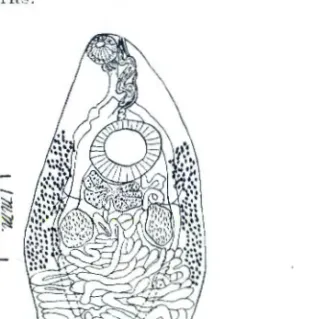
The species is tentatively referred to the above species because from the mounted specimen (Fig. 1) no character was observed by which it could be differentiated from Rudolph's species. In the unpressed condition the total length is 2 · 5 mm. and the maximum breadth 1·7 mm., the oral sucker has a diameter o£ 0·31 mm. and the ventral sucker measures 0 · 68 mm. across. The much convoluted uterus is packed with numerous eggs o£ which about half are brownish and appear to be mature.
SOUTH AFRICAN I-IEJ~NIINTHS.
Fig. 1.-PTosthoaonimtts cuneatus. Ventral view of toto mount.
As far as the writer could ascertain this is the first record of a member of this genus from ihe u nion.
Host: Microcarbo africana africanoides (A. Smith).
Licat'ion: Cloaca (? Bursa fabricii). Locality: Pretoria district.
Class CESTODA.
Ligula intestinalis Linneus, 175S
Two of three Reed Cormorants yielded four and two adult specimens respectively; they were allowed to die in cold \Yater and were then fixed in cold 70 per cent. alcohol-glycerine with slight stretching; the two largest specimens measured 225 and 240 mm. in length 'vith a maximum breadth of 5 and 5·5 mm. respectively.
The anterior portion for a length of 25 to 35 mm. is very muscular and its lateral margins are provicled with about 45 serrations on either side. 'l'hese help to keep the worm in position and this por- tion may be compared to a metascolex, in fhat, although a scolex is present, it and its bothriclial grooves are so small that it is doubtful whether they play any part as an organ for attachment. In trans- verse sections of the " metascolex " it is seen that the musculature is very well developed consisting of numerous bundles of longitu- dinal muscles arranged in 7 to 10 layers separated by sheets of transverse muscles; these sheets run more or less parellel to each other but they may join up to others immediately adjacent; the individual bundles are for the most part separated from each other by bundles o£ well developed dorso-ventral muscles. This extensive
JL J. ORTLEl'l'.
development of the~e Inu~de..; has causerl the medulla to be repre- sented bY onh a thin layer of parenchyllla. Genital organs are pre,;enL ~nly i;t the po~terior fifth of the " metascolex " and here two tocostomes, oue in front of the other, are present oppo;;ite ea<·h lateml serration. In the re111aining vortion of the strobila, "·hieh becomes naJTO\Yer posteriorly, the musculature is arranged some- what similarly exeepl tlmt it:; clegr.-e of deYelopment decreases, until in the posteriormost pmtion bundles appear to be ahsenl and only isola tell fibre:; are fo uncl.
Th genitalia are silllilar
to
those rleHcribecl by Baer (1!)3:3). The eggs are opt>rculate and mea:;urt> 0·002 to 0·06 by 0·0-:1:7 to 0·05 nun./1 ost: .1/ icracarbo afn.cana african old es (A. Smit b). Location: Small intestine.
T.oNdif.'J: Pretoria district.
'l'he writer is tentatively referring to this spe<:Ies :;ome plero- cercoid:s recoYererl from the body caYiLy of a fresh water fish, Eng rmtl·icypris wh itei- eollected from the Hartebeestpoort Dam by Mise; DOI'een Stewart of the Zoological Department, UniYersity of the \Vitwatersran<l and placed at the writer's disposal for determi- nation. These lan ae vary in length from 20 to 30 mm. \vith a maximum b1·eadth of 4 mm. DeYeloping genitalia are present in their posterior two-thirds.
Raillietinn (S.L.) thryonom.ysi sp. no\·.
This speries, collected from e1 Cane Hat, was represented by about half a dozen specimens all of w hicb were 1mfortun a tely immature; they were from 9·5 to lG mm. long with a maximum thickness of 0·5 to O·G mm. and consisted of a head, neck and from G4 to 114 segments.
A.
B.
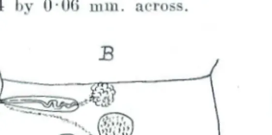
Fig. 2.- llail/ietina (S.L.) thryonomysi sp. noL A =scolex; B = hook,
'l'he scolex (Fig. 2A) is prominent and measures from 0·42 to 0 ·48 mm. across; the suckers are rounded and unarmed and have a diameter of 0 ·115 to 0 ·12 mm. The rostellum is retraeted in all cases and measures 0 · 18
to
0 · 192 mm. across; it r·arries a double row of about 100 t,vpieal hoo],s (Fig. 2B), the hooks of the anteriorsm;TH AFltTCAN IlEL)!l::ST.HS.
and posterior rows measuring respectively 0 · 04 and 0 · 036 mm. in length. The bnse of the rostellum i.s covered by innumerable small
;;pines; the fact thnt these spines and rostellar hooks are present on all the scolices makes the absence of spines from the suckers appear to be a normal occul'l'ence and not due to their loss. The neck is fairly long and may reach 1· 6 mm. in length; its breadth varies from 0·228 to 0·252 mm.
The g-enital pores are unilateral and situated in the anterior quarter of the segment. The cirrus sac is somewhat pear-shaped and reaches 0 ·12 mm. in length by 0 · 066 mm. thick; it crosses the ventral excretory canal (Fig. 3). There are about 50 to 60 rounded testes of which 15 to 20 are poral in position and the rest aporal; they are from 0 · 03 to 0 · 039 mm. in diameter; the area he tween these testicular groups is occupied by the somewhat centrally placed female glands, which in the material available, have not yet attained maturity. The oldest segment are 0 · 516 mm. broad by 0 ·168 mm.
long to 0·6 mn1. broad by 0·216 mm. long.
Fig. 3.-Raillietina (S.L.) tll1'YOnomysi sp. noY. Segment showing male genitalia.
A.tfin1.ties .- The fairly large rostellar hooks, the collar of rostellar spines, the unarmed suckers and unilateral genital pores suggest that these parasites belong to the genus H ovttuynia Fuhrmann, 1920. The absence of frayed endings to the handle of the hooks, the somewhat central position of the female glands, the presence of a cirrus sao which crosses the excretory canal an<l because of the laek of ripe segments shmTing the nature of the egg capsules hns c·ausecl the writer to refer his material to the genus Ra·ilhetina Fuhrmann 1920 (S.L).
Among the mamwalian forms of Davaineinae there is no species whieh combines the chnreters enumeraterl above, and although the material is immature the writer thinks that the characters sho,Yn are Rufficient to warrant the creation of a ne'" species for its reception.
Spec'ifi:c diagnosis.- Davaineinne having a fairly large head, unanuecl suckers, about 100 rostellar hooks arranged in h-o circles, anterior hooks 0·04 and posterior hooks 0·036 mm. long. Neck present, g-enital pores unilateral; cirrus sae crosses the ventral
66
excretory ('ctnal; about 50 to GO testes arranged m an uporal group ancl a pond group of 15 to 20 te~tes; bet11·een them !he centrally pbC'ecl female glmHls.
llost: 'l'hr.IJOnomys swinrlerionvs vorieyat.us Ptrs.
l~ocation: Small intestine.
Lor'olity: Zululand, Xlltal.
l'ypes in ihe Ondt~rstepoort R<>lminthological Collertion.
Ldioqene.s kori ,;p. nov.
Ahout .jQ specimen:; of thi:::; specie,; 11·ere coll<·e:ted from !he small int!'stine of a Giant Bustanl; the :1t·eplwlus strobila<> Y<li'.Y i.n leugth frolll 7 mm. to 60 mn1. with '' maximum thil'kness of' 0·72 mm.
to11·ards their posterior extn·mit..v. The strobilaf' arr thin and tleli- cate and in most cases are semitransparent. The anterior seg-ments, of whit·h at leafii eig·ht are inYolvetl, are n10difiecl to form a pst•u<loscolcx (Fig. -1-); i.n the:;e thP segments ;n·e trnt·-shaprd 11·ith
L~i~. 4.- ldiO(}elll'-' 1.-ori sp. no1·. J's(•udoscolicPs.
their posterior nwr<rin slnndin<r Ll\l·av in an angle frolll ihe axis of thr strobiLI and h;l~ing thi,; m;rgin ;leeply indented in itr; lllitldorsal ancl mi<h·enlral liues; these indentations are less marked towards the
po.~terior end of the pseudoscolex. In hYo strobilae, wbi(·h were c:omposecl of 120 ~~n 170 strobilae ead1, the genital organs appeared in the following· segments respectiYl'ly :- first genital primon1ia in :JOt h and 72nd segment; Lt oYaria n primordia in Both and lOOth :seg-n1ent; mature Sl'g-ments in !)8tl! to JO-±th and ]30th and J37th seg·ment8; 1st appearance of pnruierine organs in 112th an<l 1-Wth seg-n1eJ1ts and the last fell' segments of eal'lt strobila appearecl to ha1·e fully formed paruterine org·ans. these organ,; rrnching a length of 0 · 3 mm. by 0 · 19 mm. wide; no rgg~' ho,1·ewr. had, en terecl these organ::;.
67
SO CTH Al'JUC.-~=" ITEL~IIJ\THS.
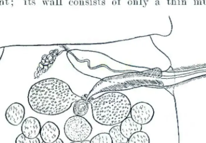
The genital poTes are uon-protuberant and are situated at the junction of the first and second body thirds. The <'irrus sac is large, long anc1 club-shaped (Fig. 5) anc1 generally it Pxtends obliguely im1·a.rds and forwards to reaeh the midline of fhe anterior margin of the segment; its 11·a 11 consists of only a thin muscular sheath.
I 0-2
lltl!VFig. 5.- lrliogenes koTi sp. no,·. :Uatu1·e segment.
and it:; length varies from 0 · 27 to 0 · 3 mm. by 0 · 07 to 0 · 078 mm.
in diameter. A burwh of retractor muscles are attaehed to its ante- rior apex. The cirrus is massive and when fully exertecl it 1·eaehes a length of 0 · 198 mm. and its base has a thickness of 0 · 039 mm. it is densely covered with prominent neec1le-like spines directed inwards; the Rpines being from 0 · 015 to 0 · 018 mm. lou g. \Vhen the cinus is retracted the vas deferens is thrown into loops inside the cirrus sac, but \Yhen the cirrus is exerted the vas deferens is more or less Rtraight; a vesicula seminalis interr1a appears to be absent.
'fhe testes, of which there are from 12 to 15 in each segment, are all situated behind the ovary; they are arranged in two layers in the medulla, and are roum1ed with a diameter of 0·06 mm. The vagina opens to the exterior immediately ventral of the cirrus pouch; it is practically straight "·ith only its inner tip bent haelnmrds; it is massive, having a maximum thickness of 0 · 041) mm. near its external orifice and being about 0·03 111111. thiek at its proximal end; the whole length of its lumen is lined by prominent inwardly directed spines. 'l'he two rounded ovarian lobes are placed one on either side of the midline in about the centre of the segment, the aporal lobe is generally slightly larger than the poral; their outline is generally smooth, hut in some cases their eclge appears Rlightly crenated; their diameters are about 0 · 09 and 0 ·11 mm .for the poral and aporal lobes respectively. A rounded and centrally placed receptaculum seminis is present. The vitelline gland lies imme- diately behind the receptaculum seminis; it is rounded to slightly oval, and its diameter is only slightly greater than that of the testes. The uterus first makes its appearance as a transverse tube behind the ovary; it rapidly becomes U -shaped but this shape is not apparent in older segments, because with the development of the
R. J. ORTLEPP.
parnterine org-an, thi.~ organ prt>:;sp,; on the anterior bee of the uterus cnusing it to appe:u fo t·on:;ist of t11·o i~obtetl bag::;, one on either :;ide of the ptnnterine organ; in no segment 11·ere there nuy eggs seen inside f lw p:1ruterine organ. As the oldest segmentR available cou- tained no mnture eggs, if \Yould nppear as i.£ they only r.ipen after tletachtnent of the :;t'glllenhi <liHl nlso only then !:'lltt>red the paruterine organ.
A.f/inities.- Tbree species of this genus haYe so far been desGribed from otidifonn birds; these are 1. gmnrhporus Cholod-
hoYSl\)-, 1900, 1. nuna Fuhrmann, 19:20 and 7. otidis Krahhe, 18G7.
The furnwr ~P!'t·ie~ cliffer:; front lhe 1niipr's in that it nurtu:tlly carries a s(·oh·x in UlE· adult, the Jllllnher of fe::;fcs is grealpr (30) and f he genital tluds open into a large nncl conspicuous rloat.;a.
Fuhrmann's :;per·ies diHerR in being much smaller (les:; fhan 10 mm. long), the p~eudoscolex is formed of the first fiye seglllent::;, ihe
it·.~tes are fe11·er (9-11) and the ,·agina is much longer, thinner and
!'ollPd. The abon tleseribed speries has its doseset n·latiYe in 1.
utid,.s, 11·hi!'h speeiPs di:ft('rR from it, ho11·eyer, h.1· ilR thinner :1nd
!'oiled Yag·in<~ 11·hiGh front ihe figureR <lYnilal>le, is abo unarnwd;
in that· the p::~e1tclos('olex i:; fonned of the first four segments and in that thf• einus sac i-, much shorter in length (0·15 mm.).
Sz)('cijic dia.r.rnosis.- ldioge11inae reachi11g a length of 60 llllll.;
::;l'olex absent in adult an tl rPpb!'ecl by a pseudos!'olex formed by at least the anterior eight segments. GPnital pores unilateral; cirrus large, up to 0·3 mm. long; cirrus massiYe and clenRely covered by largt> spines; 12 to 10 ronn(led testes in po:;terior half of segment.
Yagina mas:;iYe antl :;traight, its lumen linc><l by strong spines. 01·arian loht•s roun<lefl, C'C'Ufrally placed OJ! eifher side of micllint'. r·terus an iltYeited U, hut pressure of paruteniu<' organ on its nnterior fnre causes it laler to appenr as f11·o bags lateral of this org-an.
Host: Clwrlotis kori (Bm'Ch.). /,of'otion: ~lttall intestine.
f~ol'ltllty: X orthern TransYanl.
Types in tht> On<lPrstepoort Helminthologicnl Collection.
Scolex of Jdiogene., kori.
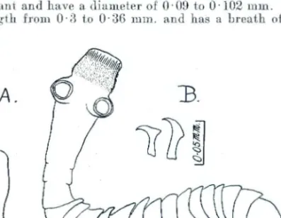
'l'lw 1niter is also tentati,·ely referring to fhe above spec1es :;u small strobilae can.1·ing Sl'olices, which ''"ere <~bo Jlresent am.ong the lttaterials of t·he aho,·e tlesrrihecl spc>cies. 'l'he long-Pst strobila (Fig. 611) is 1·S Jllllt. long, carries a scolex, 17 segment;; anll a eaudal appendage nearly half the lengfh of the enfire strobila; the other fhe strohibe are sitnilar, only smaller anll ha1·e less segnwnts. ln the fig1tred strobila it appears as if the ~rd lo 8tl1 segments a1·e b<·ginning to assunw the ~hape of the segments of the pReullosrolex, and if this is the case lhen t>ither the h l and 2ncl segments, 11·hich in th i::; strohlln are not yet fully dilineatecl. nre Rhed "·ith the S!'olex, or ebe the.1· onl~r .assume tl1e characteristic shape of those 0f the pseucloscolex at a later period.
SOUTH AFRICAN HEL)iiNTHS.
The scolex, which is very similar to that described for the next species, is roughly acorn-shaped, the acorn being- represented by the large rostellum which is separated from the scolex proper by a constriction. The rostellum has a diameter of 0·114 to 0·21mm. and carries a rlonble erown of 44 to 50 hanuner-shaped hooks (Fig. 6B) of which those of the anterior r-ow are 0 · 046 to 0 · 048 mm. long and those of the posterior row 0·036 mm. long. The axis of the blade o£
the longer hooks forms an obtuse angle with the axis of the handle, whereas in the smaller hooks these axes form roughly a right angle. rrhe whole rostellum' posterior of its hooks, is covered by innumerable minute spines. The scolex has .a diameter across the suckers of 0 · 24 to 0·3 mm. and the four some\Yhat spherieal suckers are unarmed anJ protuberant and have a diameter of 0·09 to 0·102nun. The neck varies in leng-th from 0 · :i to 0 · 36 mm. and has a breath of 0 ·102 to 0·19 nun.
A . "B.
I
027lt71?Fig. 6.- Idiogenes koTi sp. nov. A = young strobila with scolex; B = hooks.
The shape of the scolex, the presence of ha mm er-sh a peel rostellar hooks, and collar of minute spines round the rostellum, the protuberant suckers, presence of a caudal appendage, and differentia- tion of the anterior segments, all these charaeters are very similar to those of the larval stages of I. nana figured by Fuhrmann (1925);
also the scolex is very similar to that described for the next species and that figured by Cholodokovsky (1905) for I . .91·andipon1s. In consequence of these similarities the writer is fairly r·onfident that these small strobilae are very young stages of the species described above; this view is further supported by the fact that all the other cestode species harboured by this host had their scolices, so that these scolices had either to belong to the species described aboYe or belong to a different species of cestode with which this host had only recently become infected.
R. J. ORTLEPP.
Should further material prove that the vi.e>~c expressed above is correct, then the size and shape of the rostellar hooks would definitely sho,,- that this species is different to all the hitherto described species of this genus.
Idio.r;enes lwlbwi sp. noY.
From the same host "·hich harboured the preceding species about 60 specimens of the above species were recovered; on examination it was noted that a bout 20 of these still carried theiir heads, but most of them had partially or completely lost their hook ; those without heads sho"·ed no signs of pseudoscolex and a cursory examination easily showed that their scolices had been separated and lost during collection.
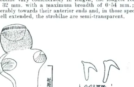
The strobilae vary eonsiderably in length, the longest reachmg a length of 32 mm. with a maximum breadth of 0·54 mm.; they taper considerably towards their anterior ends and, in those specimens which are well extended, the strobilae are semi-transparent.
0·02?ltJ!v. I
Fig. 8.-Icliogenes kolbei sp. nov. Hdoks.
Fig. 7.- 1 diooenes kolbei sp nov. Scolex.
The heacl (Fig. 7) is Yery similar in shape to that described for the precediug species; across the suckers its breadth varies from 0 · 22 to 0 · 27 mm.; the rostellum, when fully exerted is prominent, 0 ·12 to 0 ·16 mm. thick and carries a double crown of typical hammer- shaped hooks (Fig. 8); these hooks appear to vary in number trorn 120 to 140 alld the larger ones measure 0·015 to 0·016 mm. in length and the smaller ones 0·012 to 0·013 mm. The suckers are relatively small, unarmed and rounded and have a diameter of 0 · 06 to 0 · 078.
As in the preceding species the base of the rostellum is covered by i.nnumera hle minute spines. A short neck is present, from 0 ·15 to 0·21 111111. long by 0·114 to 0·132 111111. broad.
SOUTH AFRICAN HELMINTHS.
'rhe longest strobila (32 mm.) consisted of 153 segments, but the most segments (183) were encountered in a strobila 31 mm. long. In two strobilae 31 and 24 mm. long with 176 and 150 segments each the organs appeared in the following segments respectively: first appearance of genital primordium 58th and 70th segment; first appearance of ovary 100th and 92nd segment; mature segments 125 to 133 and 112 to 121; first appearance of paruterine organ 141st and 132nd segment; mature eggs in 17lst to 176th segments and 146th to 150th segments. No strobila carried mature segments in which the eggs had entered the paruterine organ.
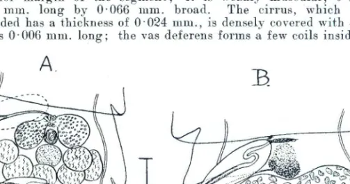
The unilateral genital pores are situated in the anterior half of the segment, generally at about the junction of the first and second thirds; as a rule they are very prominent (Fig. 9A). 'rhe cirrus sac is large and extends obliquely forwards to about the middle of the anterior margin of the segment; it is weakly muscular, 0·19 to 0·21 mm. long by 0·066 mm. broad. The cirrus, which when extended has a thickness of 0 · 024 mm., is densely covered with small spines 0 · 006 mm. long; the vas deferens forms a few coils inside the
B
Fig. 9.-ldiogenes kolbei sp. nov. A = mature segments (the male duct has been omitted in the anterior and the female duct has been omitted in the posterior segment); B = nearly ripe segment.
e1rrus and on emerging from it is thrown into numerous closely packed loops. A vesicula seminalis interna is absent. The testes are large and round and become mature before the ovary; they number from six to eight, have a diameter of about 0 · 06 mm.
and are situated in the posterior half of the segment behind the ovary and between the excretory canals. The vagina opens immediately ventral of the opening of the cirrus sac; it is more or less straight, not forming any loops anrl its terminal third has its lumen widened to 0·024 mm. and is lined by small spines. 'rhe ovary is more or les~5
centrally placed in the anterior half of the segment, an(l consists of two rounded to oval wings up to 0 ·12 mm. long by 0 · 06 mm. across;
a small shell gland is situated at the junction of the two ovarian
R. J. ORTLEPP.
lohe::;. 'l'he roundt>d receptaculum ~PmtnJs ts Riluatt>d between the OYarian ,,·iug-~ arHl has a diameter of about 0·0-!5 JJJnl. The ~-olk gland is g:en('rall~- m·.al, O·Q(i(i mm. by O·ll42 mm. an<l is generally centrally placed lwhind the receptaculum se111inis; ho,,·eyer, this latter organ may di~place it slig-htly to the right or lo the left of the mitlline. 'fht> uterus arises as an inverted U-shaped structure iu the a1·Pa occupit>cl b_,. thP oYary; as it eitlaq.!,·es it £lls up lliOSt of !he space beh,·een the pxcretory canals <llld the cirrus sac leaving the space lwt"·een its limbs Free \\'hPrP the rr!'eptncuh1n1 semini~ is found to pPr~ist (Fig-. Ull). Snon after the paruterine org-an Jllakes its appear- HII!'P a,; a clnrkl:v st:1ining- cell m~1ss in the mi<l<lle of tbe an(Prior half Of the Sf'g'lllPIIt; it gro\\·~ ha!'kward. antJ f'Olllpl'eSf\lllg ihe nnterio1· antl po..;terior ''"''lis of th<· uteruf\ 011 to il1P recPptacuhnn SPIIJJJUS t·ausP:; tlw uterus to loos<· itR U-shape an1l .assume the appt>arancP of hn1 isolatt>d latPral lJags. l n ihP hindmost segllJt•nts.
thfl eggs appear to l1<• mature hut no Pgp;s h:He enteretl Lhe Jlaruterine org-an; it \\·ould ihu~ ::tppP:tr that their entr:~nce only l:tkPs plare .aft-er thP end f't'f.('IIH'tJts ha\·e hern shec1. The eggs are thin-\\·alled and round, and h:tY(' a tliametPr of O·OJS to 0·042 mnt . .and the hPXal':tn1h hool\~ a rP tl· 011-1 nmt. long.
Af/initiPs.- 'l'his spet·iPs i~ easily tlifferenti:~ted from tht• prt>ce<l- lllg' spPt·it•s in that a psPnclo~t·olex is not forniPd, :1 sC'olPx being normally present in the atlult; the testes are 1'<·\\·er; the eitTUR and ,·agina :~re not ~o mnssiYP anrl their :;pi.nec; are !'onsidrrabl_,. :-nnnllPr.
The ah,;encP of a pseurloscolex distinguishes this spet'ies from L. otidis Krahlw, JK(i7 .antl /. nn-no Fuhrmnnn, Hl2:), anrl tlw smaller and gn•atPr nuntber of rostellar hooks, fe"·er tPstes .antl ahsencP of large genital atrium di:ffnenhates it from /. yronrli;10nts Cholo(lk, ]!)()!'").
,\JiCI'ijic dingnos'is.- T<liogPninae rpaehing :!2 mm. in lPngth by 0 · :1-t 111111. hro:H1. S!'olex JIOl'lltall.'· pn f\t'nt in thP a<lult; psentloscolex ah,;pnt. 1:!() to ].j.() rostellar hooks of t,\'jlil'al Rhnpe :ln<l about 0·0]3 and 0·0\(i llllll. long·. Jtostellum t·oYPrPtl hy iJJnllltJPrnhiP sntall spinPs. Uenital pores unilntPral. C'irrus ,;ar l:ng-P, reaching- 0·21 111111. in lPngth; cirrus t·ovf•rPtl "·ith small spinPs; Y:tgina ,;traig-lt1, ib rn<l third hns a "·iclenPrl lumen lined "·ith f\mall spines. 'J\·s1es SiX to PigJtt in JlOStPrior half of segntent. 0\·ar.\· J\Yo-Jobed; uterus r-~hapPd. Eggs I'OllllllPd, :111(1 do not PiliP!' p:nnterinf' org-an while segntent is :;till atl:~t·ltell io strobila.
lfosi: CluJI·iotis 7,01';. (Hueh .). Loco/ion: Small intrstine. Locality: X ort hPrn Tr:lns,·aal.
Types in the Understepoort Ilelminthological CollPd.ion.
ThP \\Titer ha::; ntuch pleaf\ure in naming this spP<:ies in honour of J\ir. F. F. Kolbe, H.Sc., ()ffil'er in ChargE> of the Zoological SmYey in the fiPlfl, '"ho 1\'il:i pPrsonally responsible for col1Pcting all the mah•rial from this host.
SOUTH Al,RICAN HELMINTHS.
SchistOTnetra connoideis (Bloch, 1782).
About a dozen full-gro\vn speeimens were reeovered from a Giant Bustard shot in the K orthern 'rransvaal. In addition about 50 immature specimens were present ranging in length from about 10 111111. to 60 mm. with a maximum breadth of 4 mn1. The longest mature specimens were from 150 to 175 mm. long with a maximum breadth of 9 nun.
This species 'ns :first recorded from this host by Beddard (1912) as Otid·itaenia ev.podotidis gen. and sp. nov. The writer fully agree with Skrjabin (1914) that this species is cospecific with Bloch's (1782) species (syn. S. togata Cholodk, 1912) as an examination of the writer's material shows. The suckers each carry two muscular appendages, each about 0·06 mm. long. The rostellum carries about 500 small and typical hammer-shaped hooks, in two regular circles; those of the anterior row are 0 · 01 mm. long and those of the posterior row · 0085 mm. long. The base of the rostellum is densely covered ·with minute spines. The ovary is small ancl
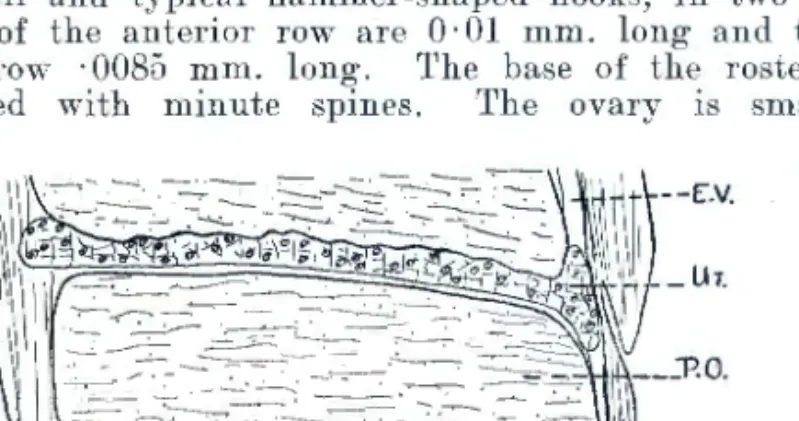
Fig. 10.-SchistometTa conoide·is. Horizontal section of hindmost segment. E.V.= excretory vessel; P.C.= paruterine capsules; P.O.= paruterine orgen; U.T.= uterus.
decidedly poral in position, being located just internal to the poral ventral excretory canal and anterior of the testes. The recepta- culum seminis is fusiform and increases in size posteriorwards; in mature segments it is about 0·18 mm. long by 0·06 mm. thick and in the hindmost segments 0·48 mm. long by 0·1 mm. thick. 'rhe uterus arises as a transverse canal immediately anterior of the testes and extends across the whole segment hebYeen the excretory canals; near the excretory canals it becomes enlarged; its cavity in ripe segments is traversed by trabeculae which divide it into cavities extending antero-posteriorly; these cavities are again sub-divided to form irregular <:avities each containing a single egg. In sections of the last few segments of three worms (Fig. 10) it was noted that the eggs had entered the paruterine organs in the last two segments of each worm. vVhen entering this organ three of four tlask-shaped paruterine capsules are formed for the reception of a considerable
74
R. J: ORTLEJ'P.
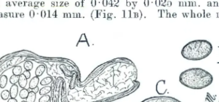
number of eggs. These capsules a rise from the posterior face of the paruterine organ and become connected with the uterus; after having receiYed their eggs they become severed from the uterus and pass forwards to lie separately in the posterior half of the paruterine organ. 'l'he capsules have a fibrous wall, and consist of a " body " and '' neck ", the latter generally kinked (Fig. llA) ; the whole capsule has an overall length of about 0·42 mm. of which 0 · 2 mm. represents the neck and the remainder forms the body measuring about 0 · 22 by 0 · 26 mm. The " body " is filled by a considerable number (roughly about 60) of oval and thin-walled eggs having an average size of 0 · 042 by 0 · 025 Inm. and the hexa- canth hooks measure 0·014 mm. (Fig. 11JJ). 'l'he whole mass of eo·gs
Fig. ll.-Schistom etTa conoideis. A = section of paruterine capsule with eggs; B = eggs hom paruterine capsule; C = eggs from uterus of same segment (B and C are equally enlarged).
is enclosed in a thin membrane separating them from the " bo(ly ., of the capsule. From the literature available the writer has not been ahle to find any definite reference to these paruterine capsules.
Beddard (1914) is unfortunately not clear on this point, in that when discussing the ultimate fate of the uterus he states (p. 218) " the eggs are at the very end grouped into many more or .less isolated but not \Yell marked causples "; whether he refers to the uterine or paruterine capsules is not clear, although on the previous page he states that there is a " tendency for tbe fibrous parenchyma to en- wrap the bundles of eggs " which would suggest that these egg bundles were lodged in the paruterine organ. His figure of a section of a ripe segment (Fig. 30) shows groups of eggs in the paruterine organ but these do not appear to be enclosed by fibrous capsules.
A remarkable feature of the eggs in the paruterine capsules is that they are Yery much smaller than the apparently ripe eggs still present in the uterus and the hexacanth hooks are also much smaller; eggs in the uterus of the same segment containing paruterine cap- sules were globular, had a thin and smooth shell, measured from 0 · 066 to 0 · 084 mm. in diameter and their hexacanth hooks were from 0·037 to 0·039 mm. long (Fig. llc). How this change takes place, especilly in the size of the hexacanth hooks, the ''Titer is unfortunately not able to say, as he rlid not observe any capsules into which the eggs were in the process of entering. The possibility
SOUTH Al'lll CAK 11 EL~fl.;\Tl-IS.
of t11·o types of eggs being present Ill the uterus is also exclude<l as a careful search of a number of sections through different uteri onl_,- shmn'd the presence of r·ound large eggs.
A curiolli; feature of the pan1terine caps1des 1s t:hat· ihe neck ('Olliai.ns a short coiled <luct, about 0 · 015 nml. 111 diameter and having its internal surface lined by short spines.
'['he annngentent of the tesit>s i~ typical for the genus consisting of <t ,-erti('al ~heet at the himl Pnd of the segment; i hen' are from 25 to :J-± testt·s in a transYersP ro11· and there are fiye o1· six of thesP rows one nbrn·e iht> other. A YeSi('uln seminalis is absent, hut the .-a:; deferPns is mu('h roile<l aftpr enterg·ing- fro111 the ('inus s;w 11·hi('h ren('he,; hut does (·ross thl· pxndory l'<lllnl: this btter organ is about 0·24 llltll. long- by 0·0!) to 0·01 nnn. thick (Skrjabin says 0·6 mnl. long h_y 0·2 to 0·2:) HUll. broad). In all the transYersP sections, obtained from rlifterPni parts nf hYo adult 11·orm,;, no trace of a dorsal excretory eanal was flee11. 'rhe genital dul·ts passed dorsal of the ,-eniral excretory canal and ner.-e.
Host: Cl10notis kori (Buurh.) Loration: Rmn ll intestine.
Lurn.lity: ::'\ortbern Transvaal.
Schistom.eira sp.
The 1n·iter is tentatiYely refening to this genus two ,·ety inuHature siTobilae ohtaineJ in associalion 11·ith the precelling- speeies. The strobilae are respeetively 10 :mel 1.5 mm. long-. The head is wry similar to that of S. 1.-orlwnw (htlt>pp ] ~):38; it~
rostellum is f'OYerecl by minute spines and also a double circle of ahmtt 000 hooks arranged in a z:ig-zag- aud having a break at each lateral corner; the hooks, ho11·eyer, are longer than in this specief:!
being 0·021. and 0·018 mm. long· for the anterior and posterior 1'0\Y respectiYely. Each sucker is provided with two muscular appendnges.
!lost: Choriotis l.:.ori (Buuch.) Location Small intestinf'.
0or:nlily: ~orthern Trnnsvaal.
HynwnolC'pis corm.oumti sp. nov.
Ahout a clozen spec·imens of thi,· deli('atc cestode ~~-ere collected front h1·o or three Reed Connornnis. Onlv h,-o 11·orm,; still had their solicPs present but a few atlditional • detachNl scolices \Yere recovered hom scrapings of the intestine.
'fhe total length renches 1.50 mm. with a maxinnun thickness of 0·6 mm. at the posterior end; the strobila is thin, especially in its anterior half whi('h has the appearance of a thread of white rotton.
76
lL J. ORTLEPP.
The head is small and only reacbt:>s 0 ·138 mnl. across; it is somewhat spindle-shaped. In all cases the rostellum is retracted and its sac extende<l haebn1rcls for a bout 0 ·13 llllll. posterior of the level of the suckers; it carries a single row of ten small hooks (Fig.
12A) 0 · 024 to 0 · 025 long, haYing a long handle and a blade only 0·006 mm. long. 'l'he suckers are slightly oval and small and measured 0 · 054 by 0 · 06 mm. across.
A
Fig. 12. -liymenolep·is cO?·nwTanti sp. noc. A = rostellar hook; n = segment with mature testes.
'l'he genital pores are unilateral and situated in the anterior quarter of the segment's margin. The cirrus sac is elongate and tubular, crosses the excretory canal and reaches the poral testes and sometimes extends beyond it; it is about 0 ·15 mm. long by 0 · 03 mm. in thickness, but in older segments it may even reach a length of 0·17 mm.
1 0·271£71?.
L<'ig. 13.- Ilymenolepis cormoranti sp. noL Segment with mature ovary.
'l'he three testes, which are rounded and have a diameter of U · 045 to 0 · 06 mm., a1·e ananged in the form of a triangle, one poral and two aporal (Fig. 12B); the anterior aporal testes it situated slightly more internal than that behind it. In segments, where the testes had attained maturity, the female glands are only represented by a darkly staining mass of eells; as the testes begin to disintegrate the ovary begins to assume a definite shape, and further back when the testes have completely disappeared the ovary and its associated glands become mature; in these segments (Fig. 13) the ovary is trilobed and measured from 0·17 to 0·2 mm. across by about 0·08 in length. The yolk gland lies immediately behind the ovary, it is rounded and has a diameter of about 0·05 mm.
SOUTH AFRICAN HELMINTHS.
A.Qinihes .-Baer (1933) identified fragments of a cestode obtained from the same host as H. phalacrocora:c (Woodland, 1929), although he noted that his material shm\·ed several differences from
\Vooclland's description based on material obtained from an Indian Large Cormorant (Pitalocrocorax carbo). Although W oodlancl found no hooks or rostellum on the one available scolex, it is not certain whether this species is normally unarmed as this scolex \Yas much distorted. Baer's material unfortunately contained no scolex. Apart from the armed scolex the \niter's specimens differ from both Woodland's and Baer' s specimens m that the testes are not found external of the exnetory vessels, in that the cirrus sac is a simple muscular tube and not the compli<.:ated structure described for W oodlann's species, and in that the ovary is trilobed; from Baer's specimen it differs in that the cirrus sac is much longer and extends almost to the middle of the segment, the peculiar structure of the vagina was not noted, and the testes are much smaller.
'rhe number, size and shape of the rostellar hooks easily dis- tinguishes this species from the species H. ficticia (Meggitt, 1929), H. magniuncinata Meggitt, 1927, H. panicirroscL, Meggitt, 1927, and H. 1nedic1~ (Stossich, 1890), from Pelicaniformes; H. parviun- cinata )feggitt, 1927, also from this group of birds, has the ::mme number of hooks (10) as in the writer's species, but they are much smaller (0·013 to 0·018 mm.) and their shape is also different.
Specific d·ia.gnosis.- Hymenolepididae attaining a length of 150 mm. or more by 0 · 06 mm. broad; scolex small, 0 ·188 mm. across;
rostellum with 10 hooks 0 · 024 to 0 · 025 mm. long having a long handle and small blade. Genital pores unilateral. Three testes, one poral and two aporal, disappear before appearance of female glands; cirrus long, crosses excretory canal and reaches poral teste:>.
Ovary trilobed, large; yolk gland round; uterus a transYerse bag, extends across excretory canal to edge of segment.
Host: Jlicrocarbo aj1·icana afn"canoides (A. Smith).
Location: Small intestine.
Localdy : Pretoria district, Transvaal.
'rypes in the Onderstepoort Helmintholog-ical Collection.
Class NEMATODA.
Cont?·ocaecum carlisle?~ sp. noY.
The Heed Cormorants from which this species "·as obtained \Yere shot by Mr. Carlisle of this Institute and placed at the writer's disposal for section: the writer has much pleasure in naming this species after :M:r. Carlisle in recognition of his services for having from time to time plaee<l materials for section at his disposal.
The above species \Tas represPnted by a bout two dozen speci- mens, represPnting fourth stage larYae, adolescent and mature worms. They \Yere found firmly attached to the inner lining of the oesophagus and stomach.
18
R. J. ORTLEPP.
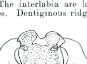
The worms are creamy yellow in colour and attenuated towards both extremities; the mature worms reach a length of 10 nun. by 0 · 5 mm. broad for the males and 27 mm. bv 1 Jlllll. thick for the females. The three lips are ;;eparatecl from the body by a constric- tion and each is somewhat rectangular, being slightly longer than broad and each is bilobed anteriorly having the lateral corner of each lobe drawn out into a point (Fig. 14); in the males they reach a length of 0 · 075 mm. and in the females 0 · 84 mm.; their pulps are provided with two swollen processes which extend one into each of the anterior lobes of the lips. The dorsal lip carries two single papillae but the sub-Yentral lips each cany a ventral double and a dorsal single papilla. The interlabia are large and curved, being almost as long as the lips. Dentiginous ridges are absent.
0·1 7/Zlll
Fig. 14.-Conh·acaecwn wTl-islei sp. nor. Dorsal lip.
The body is transversely striated, those striae ju~t behiurl the lips being more prominent and giving this portion of the body a serrated appearance. The tail is short and conical in both sexes, being about 0 · 25 mm. long in the male and 0 · 24 to 0 · 264 mm. long in the female. Lateral alae are absent.
'l'he oesophagus is 3 ·1 to 3 · 7 long and increase slightly in thick- ness posteriorly; in the male its anterior and posterior diametert:~
are 0 · 084 :md 0 ·12 mm. and in the female 0 ·108 and 0 ·192 mm.
respectively; its ventral caecum is 0 · 78 mm. long in the male and 0 · 86 mm. long in the female. The intestinal caecum is long and extend3 to almost 0 · 5 mm. behind the level of the nerve ring, being 2·76 mm. long in male 2·25 mm. long in the female. The nerve ring is 0 · 48 to 0 ·52 from the anterior end and the two small cervieal papillae are situated about 0·1 to 0·12 mm. behind the nerve ring.
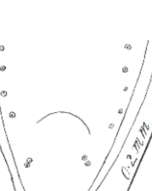
The tail of the male carries no alae; there are 6 pairs of post cloacal papillae (Fig. 15); the first two pairs are longer and ventro- lateral in position; they :ue situated dose together and only a careful examination reveals the fact that they are two separate papillae and not a single doubled one; the remaining four pairs are small, and situated in the posterior half of the tail, two pairs being lateral and two pairs ventral. The spicules are equal 2·2 to 2·4 nun.
long by 0 · 036 mm. broad at their proximal ends; they are alate in their posterior half. 'l'here are 30 or more pairs of precloacal papillae extending forwards on either side from the cloaca. A gubernaculum is absent.
SOl"TII .·IFRICAX HEL,li:\TlTS.
The Yuh-a is slightly protuberant and situated in the anterior
bo<l~ half; its position <liYicles the body roughly into the ratio of 2 : :J; the vagina is about 1·2 mm. long by 0·15 mm. thick and its lumen ha~ a diameter of O·OG llll1l.; it may extend transversely
acros~ the body and thell bend hacln1·arcls in a. right angle, or it may first pass obliquel~· fon1·ards for about 0·6 mm. an<l then bend baclnYarcls.
Fig. 15.- ContracaeCiun carli,/ei sp. nol'. Ventral view of male tail.
The llUlllt'rous egg~ are rounded and relativdy thin shelled;
thH are from 0 · 001 to 0: 05-.l: mm. in diameter and thPir sl1ells are 0·0.00 to 0·00-l: mm. thick.
A.Oinlties.- 1'he nature of the labial papillae and the arrange- ment of the male po~tcloacalpapillae shmn that this sper·ies is c·losely reb ted to C. TOrlhaini (Gedoelst., 1916) from 1'1otm Tujvs; the only di tl'ereuc·p being that in GedoP lst' s species the su bY en tral lips each carry only a double papilla and the first postcloacal papilla is <louble;
ho1n•.-er, more important· diffprences arc that in C. 1·odhalm; the spi('ules are nmch longer (3 · (i mm.), the .-uha is more anterior in
po~ihon (<livi<lt·s bO<l.V int·o r;d.io of 3 : 7), the 111ale tail is shorter (0·75 nlnt.), the fe1nale t<lil i~ longPr (0·:!36 nun.), <lllcl the k1~es of the lip~ are more cut-in tban in the 'nit·er's species.
'L'he ~ize and o;hape of thP spindes <llHl position of the vuh-a are
I'E'l'.\" similar to those of C. micro('eplwlmn CRud., 1809), but this
sp<·cies ha~ t\\'(> tlouhle p<tpillae to e;~ch dorsal lip and a single double papilla to e;~r·l1 sub-lateral lip; in adddion the arrangement of the post-cloa.('al papillae arE' also dlfrerent.
'l'ht' ~ize of th(' spicules tli.stinguislws the \\Titer's species fron1 the recently <l('scrihecl species C. toll]1tatum Yamaguti, 1935, C. milv'i Ya magu t i, 19Tj and C. lwgPdosluae Sanclgronn cl, 1933.
Specific rhngnosis .-Anas;~ kinac reaching a leng-th of 15 mm. fm thP male, allll 27 Jllln. for the females. Three lips somen·hat rect- angular and bilol>ecl anteriorly; inter-labia large and rnrverl;
dor~al lip wit-h bvo single p~1pillae and subYentral lips each with a double and a single papilla: Intestinal c·aeCIIJn long. Tail in both sexes about 0·26 mm. long. Spicules equal, alate, 2·2 to 2·4 mm.
long; gubernaculmn absent; 10 or more pairs of precloacal papillae 80
R. J. ORTLEPP.
and six pairs of postcloacal papillae of which the papillae of the 1st and 2nd pairs are close together but nor forming a double papillae.
Yulva cliYides body into ratio of 2 : 3. Eggs round and thin-shelled. llost: J[ictocarbo afr'icana africanoides (A. Smith).
Habitat: Oesophagus and stomach. Locality: Pretoria district, 'l'ransYaal.
Types in the Onderstepoort Helminthological Collectio11.
Ponocaecum spath'tdespiculum sp. nov.
The materials on n·hich the following description 1s based consists of two males and one female, all mature, from one bird host (unidentified), and two males and two females (all mature) and two immature females from another bird host (unidentified). 'fhey are creamy white in colour and are attenuated towa1~ds both extre- mities in both sexes. 'l'he males are from 32 to 44 mm. long with a maximum thicknt>ss in their mirldle of 1·17 mm. and the females are 34 to G5 mm. long 1Yith <1 maximum thickness behind the vulva of 1· 6 mm. 'fhe cut.icle is transYersely striated, and cervical and caudal alae are absent.
Pig. 16.- Porrocaennn spathulespiculmn sp. nov. Dorsal lip.
The three lips are rounded and slightly indented anteriorly and are deeply cut in at their bases (Fig. Hi) ; their length varies from 0·145 mm. to 0·16 mm.; the labial papillae are all simple, large and dome-shaped, hYo being present on the dorsal lip and one on each subventral lip. Each lip carries an internal dentigerous ridge whose denticles are largest towards the apex of each lip. The pulp of the dorsal shows no special peculiarities and simply follows the outline of the lip. The interlabia. are large and prominent and about hYo-thirds the length of the lips. 'l'he oesophagus, including its Yentriculus, is from one-tenth to one-twelfth of the total body length and increases slightly in thickness posteriorly, where it may reach a thickness of 0 · 45 mm.; its ventriculus is about 0 · 75 mm. long. The intestinal caecum is small and inconspicuous and only reaches 0 ·18 111111. in length. In a male 32 mm. long the nerve ring was 0 · 6 mm. from the anterior· end, and the small cervical papillae were situated 0 · 24 mm. behind the nerve nng-.
SOUTH AFRICAN HELMINTHS.
The vulva is non-protuberant and situated in the anterior body half. its position dividing- the body into the ratio of 8 : 11. The vagina is relatively short and soon lends in to a large egg reservoir passing posterior-wards; two uteri originate from its posterior end.
'rhe eggs are oval and have pitted shells; they are from 0 · 084 to 0 ·1 mm. long by 0 · 06 to 0 · 072 Inm. in thickness; their shells are from 0 · 003 to 0 · 004 mm. thick. The tail is somewhat stumpy and is from 0 · 74 to 0 · 84 mm. long (Fig. 17).
Fig. l7.- Porrocaec1t7n spathulespic,ulmn sp. no1·. Female tail.
The caudal extremity of the male is pointed and the tail 1s from 0 · 33 to 0 · 43 mm. long (Fig. 18A) ; there are five pairs of postcloacal papillae, that immediately behind the cloaca being double; the remaining four pairs are equidistant from each other and placed further back. There are 12 to 14 pairs of precloacal papillae. The two spiclues (Fig. 18B) are equal, robust, slig-htly arched and alate, the alae extending from just behind the head to the tip and giving the spicule the shape of a flour scoop; they are from 0 · 78 to 0 · 792 mm. long by 0 · 098 mm. broad at their head.
A gubernaculum is absent.
B
~
0·4 71?71?.I 0-2 77t 77v.
Fig. 18.- Porrocaecum spathulespic1tl1tm sp. nov. A =ventral Yiew of male tail; B = side view of spicule.
Affinities.- -The nearest relatives of the above described species appear to be P. angusticolle (Molin, 1860) and P. depTessum (Zeder, 1800), both of which possess a pair of double papillae immediately
82
lL J. ORTLEPP.
behind t.he cloaca and four additional pairs of single papillae; the former species differs from the writer's in that its interlabia are small, its spicules are longer and non-alate anll its intestinal caecum is much longer; Zeder's species differs from the writer's by the shape of the pulp of its dorsal lip. the connical tail appendage in the male and its small interlabia.
Specific diagnos·1s.-Auisakinae reaching a length of 44 mm. in the male and 65 mm. in the female. Lips somewhat rounded and deeply cut in at their bases; pulp of dorsal lip simple; dentigerou~:~
ridges present; two simple papillae and one simple papilla on the dorsal and subventral lips respectively; interlabia large. No cervical alae. Inte;;tinal caecum verv small. Yuh-a in anterior body half. Egg,; oval and pitted. Five pairs postcloacal and 12 to 14 pairs precloacal papillae in male; first postcloacal papillae double. Spiclues, equal, alate and scoop-shaped. Gubernaculum absent.
Host: Bird (unidentified).
Location : Small intestine.
Locality : TransYaal.
Types in the Onderstepoort Heminthological Collection.
Sul)11l11N1 otid·,;s sp. nov.
X umerou;:; speci rnens \Yere recovered from a single Giant Bustard.
Unfortunately all the females "·ere still immature, and consequently the measurements giYen below must be taken as applying only to young individuals which have not attained maturity.
'fhe body is \l·hitish in colour and is strongly archecl dorsahnn·cls at the anterior encl. The body is thickest at the level of the junction of the oesophagus and intestine; from this roint the body becomes thinner to"·ards both extremities, this attenuation being less marked to"·ards the anterior than towards the posterior end. In the females the body becomes very much attennated posteriorly to end in a long and finely pointed tail; in the male, as far as the cloaca, the attenuation is not so marked, but in the tail region the bocly becomes considerably thinner to terminate in an elongate tail spike. The body cuticle sho'l"l·s fine annulations and also carries two long lateral alae which extenrl clown the body for about two-fifths of its length; they are from 0 · 025 to 0 · 036 mm. high and are vertically stria teo. 'l'he mouth is simple and leads into a buccal capsule having thickened cuticular walls (Fig. 19); it has .a diameter of 0 · 021 to 0 · 024 mm. at its anterior end and 0 · 040 to 0 · 045 mm. at its posterior end, and its average depth is 0 · 03 mm. Protruding into its base are three prominent teeth lodged on the apices of the three oesophageal segments. There are two wart-like lateral papillae and four small and dome-shaped submedian papillae round the mouth. The oesophagus is typical in shape and is from 1 · 27 to 1 · 34 mm. long in the males and 1 · 44 to 1 ·54 mm. in the females.
SOUTH AFRICAN HEL~UNTHS.
The length of the females is 9 · 0 to 11· 6 mm. "·ith a maximum thickness of 0·264 nun. The tail (Fig. 20A) is long, slender and pointed and .averages 1·0 mm. in lengt.h. The vulva is inconspicuous and situated in the anterior body half, dividing the body roughly into the ratio of 4:5. 'rhe ovejector (Fig. 20n) is very short, its total length being from 0·516 to 0·544 mm.; its vestibule is bent sharply forwards and is from 0 ·17 to 0 ·17 Imn. long; the sphinder measures only 0 · 7 mm. in length and the trompe is from 0 · 26 to 0 · 3 mm.
long. As the material is still immature no eggs are present.
Fig. 19.-SubuluTa otid'is sp. nov. Anterior extremity, dorsal view.
~--- ~
A.0·5
71! 1/b.0·27ltllt.
Fig. 20.- SubuluTa ot'idis sp. nov. A = female tail; B = ovijector.
The males are from 7 · 7 to 9 mm. long .and have a maximum thickness of 0·24 mm. The caudal extremity (Fig. 21) is hooked ventralwards and is provided with very narrow alae. There are 10 pairs of caudal papillae of which two are precloacal, three circum- cloacal and five post cloacal on the anterior half of the tail; a pair of small caudal pores is situated just anterior of the last pair of caudal papillae. The tail is relatively long, being from 0·36 to 0·4 mm. in
84