PREVALENCE OF FUNGI IN THE SEEDS AND SEEDLINGS OF BLACKBERRY
Shr":'!:"':;!~ t.~'::'!:tl!alUnlVeiSlly LilJl.Jry
...,~." n":J._ o00
k_.:: ::;:,,,,,Pll .....;;>. .. :/ ...:;J . .;:) ..:-..
Slgn~Dale:q9 11U13
A THESIS BY
NUSRAT JAHAN MASTER OF SCIENCE
IN
PLANT PATHOLOGY
-
. SHER-E-BANGLA AGRICULTURAL UNIVERSITY
;""1-' ..~,__
SHER-E-BANGLA NA.GAR, DHAKA-1207K~~~
BANGLADESH'":<
oaf,
JUNE,2006.---~----
....f .. 111~lnlnm ~III00111111111·
1
37330 . .' ~.1.,_i_ .. - ..__. .._.---... _ _..:._.. :~j
'. t
".~.~
PREVALENCE OF FUNGI IN THE SEEDS AND SEEDLINGS OF BLACKBERRY
By
NUSRA T JAHAN Registration No. 01520·
A Thesis
Submitted to the Faculty of Agriculture, Sher-E-Bangla Agricultural University, Dhaka,
In partial fulfillment of the requirements for the degree of
MASTER OF SCIENCE IN
PLANT
PATHOLOGYSEMESTER: JANUARY - JUNE, 2006 Approved by:
(M. Salahuddin M.Chowdhury) Assistant Professor
Department of Plant Pathology Sher-e-Bangla Agricultural University
Supervisor
(Dr. Md. Rafiqul Islam) Associate Professor & Chairman
Department of Plant Pathology Sher-e-Bangla Agricultural University
Co-supervisor
(Dr. Md. Rafiqul Islam) Chairman
Examination Committee
CERTIFICATE
This is to certify that the thesis entitled, "PREVALENCE OF FUNGI IN THE SEEDS AND SEEDLINGS OF BLACKBERRY" submitted to the Faculty of Agriculture, Sher-e-Bangla Agricultural University, Dhaka" in partial fulfillment of the requirements for the degree of MASTER OF SCIENCE in PLANT PATHOLOGY, embodies the result ofa piece of bonafide research work carried out by Nusrat Jahan, Registration No. 01520, under my supervision and guidance. No part of the thesis has been submitted for any other degree in any other institutes.
I further certify that any help or sources of information, received during the.
course of this investigation have been duly acknowledged.
Dated:
Dhaka, Bangladesh
(M. Salah uddin M. Chowdhury) Assistant Professor
Department of Plant Pathology Sher-e-Bangla Agricultural University
Supervisor
,,'
Dedicated' to
:My
(8e{ovea CParents
_"
\
ACKNOWLEDGEMENT
The author wish to acknowledge the immeasurable grace and profound kindness of Almighty Allah the supreme Ruler of the Universe, Who her enabled him to carry out this research work and prepare the thesis.
The author feel proud to expresses her heart-felt gratitude, immense indebtedness and sincere appreciation to her respected teacher and supervisor M. Salahuddin M. Chowdhury, Assistant Professor, Department of Plant Pathology, Sher-e-Bangla Agricultural University, Dhaka, for his scholastic guidance, valuable suggestions, constant encouragement, affectionate feelings, patience and advice extended throughout the research period and for completion of the thesis.
It is a great pleasure for the author to extend his deep sense of gratitude and indebtedness to her honorable teacher and co- supervisor (Dr. Md. Rafiqul Islam), Associate Professor and Chairman, Sher-e-Bangla Agricultural University, Dhaka, for his creative suggestions, constructive criticism and sincere co- operation in completing thesis.
The
cuthor
desires to express his respect and deepest sense of gratitude to all the respectable teachers of the Department of Plant Pathology, Sher-e-Bangla Agricultural University, Dhokc for their valuable suggestions and kind co-operation during the period of the study.1
The author pleased to convey cordial thanks to Dr. Ismail Hossain, Professor Department of Plant Pathology, Bangladesh Agricultural University, Mymensingh, for his kind help and valuable advice during the research period and for preparation of the thesis.
Cordial appreciation and thanks are extended to Md. Fajar Ali, Lab.
Assistant of Plant Pathology Laboratory of Bangladesh Agricultural University, Mymensingh, for his help and co-operation during the research work.
The author sincerely desire to expresses his heartiest gratitude to his friend and colleague Abu Noman Faruq Ahmmed and Najmun Nahar Tonu for their inspiration. and best co-operation during the research period and preparation of the thesis.
Sincere gratitude is also extended to all
of
his friends and well wishers specially ShakiI Mahmud Khan end Zioul Haque for their help and inspiration during the study.Lastly the author expresses his indebtedness to beloved parents, brothers, uncle, aunt and relatives for their blessings, love and affection.
The author
11
PREVALENCE OF FUNGI IN THE SEEDS AND SEEDLINGS OF BLACKBERRY
ABSTRACT
Prevalence of fungi in seeds of blackberry plant collected from six different sources under Netrakona and Dhaka district were recorded.
Seven different fungi, representing six genera, were detected in the seeds of blackberry. The seven seed borne fungi observed on blackberry seed , were Pestalotia psidii, Curvularia lunata, Fusarium equiseti, Aspergillus flavus, Aspergillus niger, Rhizopus sp. and Penicillium sp. All these seven
fungi on the seeds of blackberry appear to be new records for Bangladesh. Prevalence of all these fungi varied significantly (P=1 %) with respect to location. Pathogenicity test reveals that Curvularia lunata, Pestalotia psidii and Fusarium equiseti are pathogenic to blackberry tested in five selected nurseries in two different districts. In survey, four fungi species such as Puccinia psidii, Pestalotia psidii, Fusarium equiseti and Curvularia lunata were isolated from the leaves and stems of blackberry.
III
CONTENTS
SL.No CHAPTER Page
ACKNOWLEDGEMENT I
ABSTRACT Iii
CONTENTS. Iv
LIST OF TABLES Xi
LIST OF PHOTOGRAPHS Xii
1. INTRODUCTION 1-4
2. REVIEW OF LITERATURE 5-12
3.
MATHERIALS AND METHODS 13-263.1. Experimental site and period 13
3.2. Collection of seed sample 14
.3.3. Inspection of dry seed 15
3.4. Incubation test 15
3.4.1. Blotter method 15
3.4.2. Sterilization of incubating papers and dishes 18
3.4.3 Pre treatment of seeds 18
3.4.4. Agar plate technique 18
3.5. Isolation' and identification of fungi 19
3.6 Germination test 19
3.7. Pathogenicity test 20
3.7.1 Pathogenicity of the causal organism 20
3.7.2 Fungi included 20
3.7.3 Inoculation of leaves 21
3.7.3.1 Raising of seedling 21
3.7.3.2 Techniques used 21
3.7.3.3 Isolation and purification of fungi 22
3.8 Method of survey 22
3.8.1 Symptom observation 23
IV
3.8.2
Collection, isolation, purification and identification of25
causal organisms
3.8.2.1
Collection of diseased specimen25
3.8.2.2
Isolation of causal organisms25
3.8.2.3
Purification and identification of causal organisms25
3.9
Statistical analysis26
4. RESULTS 27-46
4.1.
Inspection of dry seeds27
4.2.
Germination test27
'4.3.
Prevalence of fungi29
4.3.1.
Total seed borne fungal infection29 4.3.2.
Fungi identified and their frequency of occurrence in30
Netrakona District
4.3.3.
Fungi identified and their frequency of occurrence in37
the seed of Dhaka district
4.3.4.
Prevalence of fungi in blotter paper37 4.3.5
Prevalence of fungi in PDA media38
4.4.
Pathogenicity42
4.4.1.
Leaves inoculation42
4.5.
Occurrence of foliage diseases in nurseries46
5. DISCUSSION 50-54
6. SUMMARY AND CONCLUSION 55-56
7. REFERENCES 57-64
v
LIST OF TABLE
SL. NO. TITLE
1 Dry inspection of the selected seed of blackberry 2 Germination percentage of the selected seeds
observed in different tests.
3 Prevalence of Seed borne infections of individualy fungi detected in blackberry seeds collected from six different sources.
4 Frequency of occurrence of fungi observed. in the seeds ofNetrakona.
5 Frequency of occurrence of fungi observed in the seeds of Dhaka
6 Prevalence of fungi in blotter method with different treatments
7 . Prevalence of fungi in PDA media with different treatments
8 Pathogenicity of Fusarium equiseti on two months old seedlings of blackberry as determined by leaf inoculation
9 Pathogenicity of Pestalotia psidii on two months old seedlings of blackberry as determined by leaf inoculation.
10 Pathogenicity of Curvularia lunata on two months old seedlings of Blackberry as determined ~y leaf inoculation
11 Occurrence and intensity of foliage diseases of seedling raised in three nurseries ofNetrakona
12 Occurrence and intensity of foliage diseases of seedling raised in three nurseries of My men singh
VI
PAGE
28 28
35
36 39
40
41 44
44
45
49
49
LIST OF PHOTOGRAPH
SL.NO TITLE PAGE
1 A. Healthy seeds of blackberry 16
B. Infected, shriveled and dead seeds of blackberry
2 A, B. Dishes ready to examine under a stereo- 17 microscope
3 A. A nursery survey inNetrakona (Sotabdy nursery) 24 B. A nursery survey in Mymensingh (Tulip nursery)
4 A. Pure culture of Curvularia lunata of blackberry 31 seeds on PDA (40x)
B. Pure culture ofAspergillus niger of blackberry seeds on PDA (40x)
C. Pure culture ofAspergillus flavus ofblackberry seeds on PDA (40x)
5 A. Pure culture of Fuserium equiseti of blackberry 32 seeds on PDA (40x)
B. Pure culture of Pestci/otia psidii of blackberry seeds on PDA (40x)
C. Mature fruiting bodies of Pestalotia psidii of blackberry seeds ofPDA (40x)
6 Conidia of Curvularia lunata seen under microscope 33 (400x)
7 Conidia of Pestalotia psidii seen under microscope 33 (400x)
8 A. Conidiophore, vesicle and conidia ofAspergillus 34 niger (400x)
B. Conidiophore, vesicle and conidia ofRhizopus sp.
(400x)
9 A. Leaf blight symptoms developed after two months 43 old leaves of blackberry by Curvularia lunata.
Band C. Leaf spot symptom developed after two months old leaves of blackberry by Pestalotia psidii and Fusarium equiseti.
10 A. Symptom of leaf blight diseases 47
B. Growth of Penicillium sp. on blackberry seed incubated on blotter (4Ox)
C. Symptom of leaf rust disease
VB
..
~.
~.,P~II
1. INTRODUCTION
Blackberry (Syzygium cuminii) belongs to the family Myrtaceae and is a native fruit of India, Burma, Srilanka, and Bangladesh. The tall tree with evergreen foliage is an excellent roadside tree and varies often used as wind break, It is widely grown both innorth and south part of Bangladesh. Small . dark purple colored fruits with sub-arid spicy flavor are eaten fresh.
There is a standard variety in blackberry. In north India, a type Known as
"Ra Jamun" with big (2-5 cm long fruits) fruits are normally grown. A small (1.5-2.0 em long and 1-1.5 cm. diameter) fruited type is also grown for late harvesting. A wide range of variability exists in blackberry and survey conducted (Keskar et al. 1989) in Pune and Abmednagar districts in Maharashtra state revealed. The variation in fruit weight (3.5 to 16.5 g ), pulp contents (54-85%), TSS (4.5 - 17%) and acidity (0.16 to 0.55%). Some promising lines have been selected. Blackberry has got various industrial uses too.
Bangladesh has the lowest per capita arable land due to its high population density. This limited land is inadequate for fulfillment of the requirements for the people. Therefore, much emphasis was not given on fruits production which is an important source of nutrition. The minimum dietary requirement of fruits per capita is 85 gm, whereas the availability is only 30-35 gm.
Fruits are important source of vitamins and minerals. The availability and consumption of fruits in Bangladesh are much less than it should be. Fruits are very costly and therefore, the majority of people cannot afford to buy them. As a result there has been a widespread malnutrition inthe country. It may be over come by eating indigenous country fruits like blackberry, bel, amloki, guava, kamranga, jujube etc. Itis less costly but rich of nutrition.
Bangladesh has been exporting fresh fruits since several decades. However, in the more recent years, export promotional activities for fresh fruits have received greater attention. The fresh fruits productions which were only 1631 metric tones inyear 2002-2003 rose to 1772 metric tones during 2003- 2004. According to Bangladesh Bourne of Statistics 2003-2004, per acre yield rate was 3306 kg.
It is said that a country· needs at least 25% forest cover to maintain its ecological balance. But we have only 160/0 forest land. Among them actual free coverage of only 5.4% area (Amin, 1994).Itis undoubtedly insufficient for ecological requirements. Blackberry may be introduced as agro forest tree. Being Bangladesh overwhelming with it booming population the forest area could only be increased through social afforestration, homestead gardening as well as intercropping.
2
Seed is the most important input for crop production. In modem agriculture seed health is a recognized factor for increased production. Pathogen free healthy seeds are considered as vital input for desired plant population and a good harvest. Many plant pathogens 'are seed borne which can cause enormous crop losses. Out of 16% annual crop losses due to plant disease at least 10% losses are incurred due to seed borne disease (Fakir, 1983).
Along with blackberry, pineapple, turmeric, zinger etc, can be cultivated as a intercrop with blackberry without the addition of cultivated .area, cultivation of blackberry at pond side and homestead areas might be important for increasing forest area minimizing the ecol~gical imbalance as well as meeting the demand of food crisis. The woods are used to make furniture, spokes for wheels, arms for easy chairs, 'knees for all kinds of boats, beams for construction, frames for musical instruments (violins, guitars etc.) and packing cases. It is also popular for general turnery. It is not durable in the ground and is prone to attack by dry-wood termites. The tree grows rapidly after cutting to a stump and consequently yields a continuous supply of small wood for fuel. Blackberry and rose apple wood makes very good charcoal (Morton, J. 1987).
During the past two decades, research on diseases of fruits crops has made considerable progress. However, tremendous amounts of losses in yield and
3
quality of fruits caused every year dew to diseases incited by different plant pathogen viz. fungi, bacteria etc. In Bangladesh considerable amount of work has been conducted on determining the prevalence of seed born pathogen of cereals, vegetables and some fruit seeds but no work has been conducted on blackbeny seeds and seedlings. The present study has been undertaken with the fol1owing objectives:
Objectives:
1. To study the prevalence of fungi in the seeds of black beny.
2. To determine the pathogenicity of the fungi associated with the seeds of blackberry.
3. To study the seedling disease (s) of blackberry.
4
2. REVIEW OF LITERATURE
Blackberry is a popular fruit in our ·country. Millions of fanner plant blackberry in their homestead garden. The tropical weather is suitable for blackberry production. But the production qualities of blackberry are hampered due to different pathogens and diseases, especially, the seed borne ones. Pathogens attack seeds and leaves of blackberry. However, to the best of our knowledge, no work has been done in our country and least work has been conducted inabroad on the detection of seed borne fungi of blackberry.
Therefore, reviews on similar work on blackberry and its relatives under same family pertinent to the present problem are presented below:
Diseases of blackberry
Gupta et al. (1955) stated that B. theobromae is one of the most serious pathogen of mulberry plant. It cause die-back and attack many plantation crops like pear, rose apple, mango, blackberry etc.
Edward et al. (1964) stated Syzygium cuminii.Psidium cattieianum,Chinese guava and wild Philippine guava were found to be resistant to Fusarium wilt of guava.
5
Morton (1987) reported that Syzygium cuminii is susceptible to several diseases like white spongy spot, leaf spot, leaf blight and lesions on leaves.
Ramaswamy et al. (1988) reported that Pestalotia psidii is the causal agent of guava canker. The pathogen is also isolated from infected coconut palm, mango, Jackfruit, blackberry and litchi ininoculation tests.
Ramaswamy et al. (1998) isolated Pestalotia psidii from infected guava, coconut, palm, mango, eucalyptus, achrous sapota, litchi. Inoculation tests confirmed P.psidii the pathogen.
Lyman et al. (2004) reported that fungus Cercosporella rubi is an important blackberry pathogen in the southeastern United States. This pathogen severely reduces fruit production and its management has been erratic due to a limited understanding of the host-pathogen relationship.
Diseases plants under same family of blackberry
Chattopadhyay et al. (1955) isolated both Rhizoctonia solani and Fusarium solani from diseased guava plants. The disease was considered a serious problem in commercial plantings. Other .guava diseases reported from India were Fusarium wilt caused by Fusarium oxysporium f.sp. psidii and a condition characterized by intraveinal .chlorosis and die-back of leaders associated with zinc deficiency.
6
Mathur (1956) reported one deficiency disease and a number of fungal diseases on common guava grown as a fruit crop inIndia. Wilting was first observed in Allahabad in 1935 and subsequently caused extensive damage in Uttar pradesh. Both Cephalosporium sp. and Fusarium were initially isolated from diseased plants, the latter was identified as Fusarium oxysporiem f.sp. psidii. A serious fruit canker caused by Pestalotia psidii was reported very common in several state of India. Canker or scab caused by Glomerella psidii was also reported ..
Glasscock and Rosser (1958) observed that the seedling of Eucalyptus Perrineana and E.gunnij became severely affected by a species of oidium in July. Various kinds of Eucalyptus leaf spots due to Cercospora epicocooides, MycosphaerelJa molleriona and Readeriella onirabilis; tar spots due to Phytisma eucalypti and rust caused by Puccinia psidii were reported.
Edward (1960) reported that F. oxysporum sp. psidii exists in a variety of clonal forms that differ in pathogenicity and in mo~hological and cultural characteristics. No correlation was observed between the cultural characters and pathogenicity.
Edward (1961) reported that wilt disease of guava caused by F. oxysporium f.sp. psidii was the most serious of the diseases to which guava is
7
susceptible. However, guava relatives, includingPsidium cattleinaum var.
lucidum and Syzygium cuminii reportedly had never been observed to be attacked by the disease. Inoculation experiments subsequently demonstrated that seedlings of Syzygium cuminii were immune.
Kapoor and Tandon (1970) isolated the fungus Curvularia tuberculata was reportedly newly from guava fruits stored in local markets in India.
Previously Curvularia tuberculata had been reported as causing die-back disease of citrus in India. The disease on guava caused by the fungus appeared as a circular yellow spot, which later changed to brown and followed by decay.
Rahman and Zethner (1971) found considerable fungal damage of seeds of forest tress occurs in Bangladesh. They obtained 36 isolates of fungi from the seeds of eleven different forest tree species. Genera of fungi identified by them were Acremonium sp., Penicillium sp., Fusarium sp. and Candida sp.
on Syzygium grandies.
Galli et al. (l980a) reported disease of Psidium guajava caused by Puccinia guajava, Puccinia psidii, Colletotrichum gloeosporidies, Sphaceloma psidii, Pseudomonas sp.,Phyllorsticta guajava.
Galli (1980b) described diseases of Psidium guajava with emphasis on guava rust (Puccinia psidii) in Brazil. He also mentioned fruit diseases
8
caused by the fungi Phyllosticta sp., Colletotrichum gloeosporidies, Sphaceloma psidii, Pseudomonas sp.,Phyllorsticta guajavae
Pandey et al. (1984) studied the fungi colonizing the phyUoplane (leaf surface) and internal tissue of guava leaves from bud stageto senescence in summer, rainy and winter seasons in India. The total numbers of fungi were highest in the rainy season and lowest during. the summer. Fungi were categorized into 3 groups, those exclusively seasonal Pestalotia psidii, Fusarium oxysporum f.sp. Psidii and Colletotrichum g/oeosporioides were' isolated from the foliage in different seasons in different levels of dominance. The number of phylloplane micro flora increased with leaf age.
Adisa (1985) conducted a survey on guava fruit rot diseases in 16 locations of Nigeria. Two types of rots, soft rot and dry rot were recorded. Aspergillus niger, Rhizoctonia so/ani, Colletotrichum gloeosporioides, Botryodiplodia theobromae, Erwinia sp., Rhizopus stolonifer, Rhizopus oryzae were established as soft rot organisms. While Penicillium sp., Fusarium equiseti and F. oxysporium were established as dry rot organisms.
Kuthubutheen et al. (J 988) encountered specres of Collectotrichum, Pestalotiopsis, Fusarium, Botryodiplodia on healthy leaves of several fruit trees and these fungi were all capable of lesion formation and subsequent leaf damage.
9
Pandey (1990) studied the mycoflora of common guava. He found the leaves were colonized by a limited range of fungi such· as, Alternaria alternata, Aspergillus niger. pink yeasts and white yeasts. Four consistently pathogenic species:· Colletotrichun gloeosporioides, Fusarium oxysporium f. sp.psidii, Pestalotia psidii, and phoma psidii were consistently present during all seasons.
Jan et al. (1991) isolated the Macrophonina phaseolina (Tassi) Goid, On guava fruists from orchards. Pathogenicity was confirmed by inoculations of guava fruits.
Hossain et al. (1992) surveyed the prevalence of guava fruit anthracnose in 3 major guava producing areas of Bangladesh during 1987-1988. All plants and 90-100% of fruits surveyed were severely diseased. Pestalotiopsis psidii, Glomerella cingulata, and Botryodiplodia theobromae were isolated
from infected fruits and pathogenicity was confirmed.
Hossain and Meals (1992) monitored the prevalence of guava anthracnose in Chittagong, Barisal and Mymensingh during 1987-1988 and found 100%
plant infection and 90-100% fruit infection.
Dwivedi et al. (1994) reported that disease incidence in guava orchards ranged from 3.9 to 30%. Symptoms included defoliation, die-back and bark cracking. Among the associated pathogens F. solani, F. oxysporum and F.
10
oxysporum f.sp. psidiis predominated In all seasons. Accompanying saprophytes included Alternaria alternata, A. flavus, A. niger, Penicillium citrinum, Trichoderma harzianum in summer, while A. luchuemis, Humicola sp. and F. solani were dominant in rainy and winter seasons. Wilted twigs yielded isolates of Cunningham ella, Fusarium sp and Macrophomina phaseolina, while roots yielded F. so/ani, F. longipus, F. monilifarmae, F.
oxysporum f sp. psidii and M phaseolina. The maximum soil temperature (43°C) was recorded in the summer while the organic matter content of the soil was highest (2.1 %) in the rainy season.
Alahakoon and Brown (1994) isolated Colletotrichum gloeosporioides from 23 fruit crops in Srilanka. This was the first record of G. eingulata on durian, mangostein, pini jambu (Syzygium jambos), trees inSrilanka, Symptoms of infection by G. cingulata on fruit tree seedlings especially mango and rambutan which can cause up to 40% loss of planting stock were previously believed to be a physiological disorder.
Smith et al. (1998) observed die-back of eucalyptus species, clones and hybrids during survey of forest plantations in the Mpumalanga and kwazulu- Natal provinces, South Africa. This symptom was often associated with environmental stress (drought, frost and· hot winds). Botryodiplodia dothidea was frequently isolated from twigs showing die-back symptoms. In some
II
cases, C. gloeosporioides was also isolated. Artificial inoculations of 3 years old clones ofE. grandis with both fungi resulted inlesion development.
Coutinho et al. (1998) describe the symptoms of the disease. He stated the disease first begin as. tiny bright yellow powdery eruptions in a circular pattern on the leaf or stem surface. These infection loci or spots expand and become necrotic, and spread over the entire leaf, stem or shoot. Leaves and stems can be deformed by the disease, and growing tips can die back if the infection is severe.
. 12
~J
_I~ ~-~
RF7_
3. MATERIALS AND METHODS
3.1. Experimental site and period
The laboratory experiments on the detection of fungi on blackberry seeds were conducted in the Plant Pathology laboratory' of Sher-e-Bangla Agricultural University, Dhaka, and· Plant Pathology Laboratory, Bangladesh Agricultural University, Mymensingh during the period May to August, 2006.
. '
Two places were selected for survey to determine the disease of blackberry.
The places were Mymensingh sadar in the district of Mymensingh and Netrakona sadar in the district of Netrakona. Five nurseries were selected.
from the two places for survey. The nurseries are-
Nursery and total number of plants served
District Nursery Total no. of plants
surveyed
Netrakona 1. Bono Bevagh nursery 2910
2. Sotabdhy nursery
2574
3. Mohammed nursery
1500
Mymensingh 1. Maroun nursery
1500
2. Tulip nursery
1000
Total 9984
13
3.2. Collection of seed sample
Seeds of blackberry species were collected from Netrakona and Dhaka districts. Three sources were included from each district for seed collection.
In Dhaka, DAB Horticulture center, Asadgate, karwan hazer and Tongi hazer were selected for seeds collection. And in Netrakona seeds were collected from the Government nursery, Choto bazer and Modonpur.
Totally 800 seed samples from the two selected seed sources. Seeds were collected during May- June, 2005 and 2006. After collection of seeds the samples were plated in the laboratory for the study.
The seed sources are presented bellow
District Source
Netrakona 1. Government nursery
2. Choto hazer 3. Modonpur
Dhaka 1. Karwan bazer
2. Tongi hazer 3. Asadgate nursery
14
3.3. Inspection of dry seed
Dry Inspection of seeds were done, 4 hundred seeds of each district were taken randomly and grouped into two categories viz. I. healthy seeds II.
Discolored and calculated the percentages of each group (photo.-l).
'3.4. Incubation test 3.4.1. Blotter method
The seed samples were tested by blotter method for the presence of the seed- borne infection of fungi following the International Rules for seed health,
.
testing (ISTA, 1999). In this method, three pieces of whatman no. 1 filter paper were soaked in sterile water and placed at 9 em petri dishes. In another method, the seeds were treated with Mercuric chloride and Alcohol for 30 seconds then wash three times under running fresh water and then plated on the wet filter paper in the petridishes. Seeds were plated at the rate of 9 seeds per petridish. In this way 36 seeds in 4 replicates were plated per sample.
The Petridishes, with the seed plated were incubated at 22 ± 2°c under 12/12 hours alternating cycles of NUV and darkness in the incubation room of the seed pathology laboratory for 20 days. After incubation, the plates were examined under stereomicroscope (photo.-2) ...
15 .
Photo. 1. A. Healthy seeds of blackberry B. Infected, shriveled and dead
seeds of blackberry
16
\
Photo. 2. A~B. Dishes ready to examine under a stereo-microscope
17
3.4.2. Sterilization of incubating papers and dishes
Often contaminants interfere with the detection of seed bOJ;11epathogen in blotter technique. Filter paper and petridishes were sterilized by autoclave.
-
Whatman no. 1 filter paper and glass petridish were packed in sterile condition for future use. Germination percentage and presence of major
.
.
pathogens were recorded following the procedures described in 3:4.1 ..
3.4.3. Pre treatment of seeds
Seeds were pretreated by soaking in 0.05% Hg2ch for 30 second. And in . another experiment the seeds were pretreated by 70% ethyl alcohol. Seed borne infection by the major seed borne pathogen and germination were noted following the procedures described insection 3.4.1.
3.4.4. Agar plate technique
In agar plate technique, Potato Dextrose Agar (Potato 200gms, Agar- 15 gm and H20 1000 ml) having pH 6.5 was used. About 15 ml of the media were poured in each sterilized glass petridish. Controlled seeds were plated. Then, Hg2ch 0.05% and alcohol 70% were used to pretreated the seeds. Seeds were. .
dipped in each concentration of chemicals for 30 seconds. The pretreated seeds were plated in the petri dishes. All the petridishes were kept in the glass chamber for avoiding contamination. .
18
3.5. Isolation and identification of fungi
The fungi associated with seeds, seed coat and cotyledons were isolated in pure form .and grown on the acidified PDA. Seed borne infections of fungi observed under the stereo microscope' and identified by measuring their growth characters on the incubated seeds. The fungi were identified to
·species level, wherever possible, following the keys of Malone and Muskette (1964), RamN ath et al. (I970), Booth (1971), Ellis (1971), Barnette and Barry (1972), Mathur and Kangsdal (2003). Identification of pathogen was done by preparing slides and examined them under the compound microscope.
3.6. Germination test
The germination test was carried out by blotter method, plastic tray and agar
. .
plate method. In blotter method, two layer of whatmen paper 1 was used. 9 seeds were plated in each petridish. In case of agar plate method, PDA media was prepared and then media and glass petridishes were sterilized in the autoclave. 15 ml PDA media poured in each the petri dishes. Then 9 seeds were plated in each dish. Germination test was also determined insoil on plastic tray measuring 1 x 2 ft. The soil was collected from the field laboratory of the department of plant pathology. SAU, Dhaka. 50 seeds were sown in each tray. 3 plastic trays were used for this purpose. Data on
19
t
germination test were recorded after 7, 15 and 21 days of sowing. After 21 days of sowing, data of germination test was taken for final count.
3.7. Pathogenicity test
3.7.1. Pathogenicity of the causal organism
Pathogenicity of causal organism was conducted inseed pathology laboratory of department of plant pathology, Bangladesh Agricultural University, Mymensingh. Young leaves of the blackberry seedlings included for pathogenicity test of the fungus.
3.7.2. Fungi included
Pathogenicity test was conducted with the three fungi isolated from seeds of blackberry, collected from two locations. The fungi used for inoculation were Curvularia lunata, Pestalotia psidii and Fusarium equiseti.
The fungi Aspergillus niger, Aspergillus jlavus, Penicillium sp. and Rhizopus sp. were not included in the pathogenicity test because they are predominantly saprophytic and storage fungi. The three fungi were included for the pathogenicity test because they were frequently associated with incubated seeds in the blotter test.
20
r
3.7.3. Inoculation of leaves 3.7.3.1. Raising of seedling
Leaves of two months old seedlings of blackberry were inoculated by the test fungi in order to determine the pathogenic potentiality of these fungi.
Seedlings were grown"in earthen pot filled with soil. Four seeds were sown per pot. There were eight replications. ~t two months of rising seedling one were uprooted and the rest were kept for experiment. Watering and necessary aftercares were taken throughout the growing period of the seedlings during the experiments.
3.7.3.2. Techniques used
Leaves of two months old plant (at the stage of five to six leaves) was chosen for inoculation in pathogenicity test. The leaves were washed with sterile water and then taken in glass house. Inocula of the test pathogen were prepared for inoculation. During inoculation selected parts of leaves were first 5 pricked with fine sterile needle. And then 5 mm block of 7-8 days young culture of the test pathogen was attached in the pricked zone. Thi:m the spot was wrapped with wet cotton pad. The inoculation was made with injury. For development of leaf spot, some leaves were inoculated at a point 1 em away from the tip. 2 plants per pot were inoculated and one plant kept
21
for control. The experiment was conducted in the glass house of the seed pathology laboratory in BAD, Mymensingh.
After inoculation, both the inoculated and control pots were kept at chamber. After 48 hours, the wet cotton pads were removed. During incubation, the leaves were examined for disease development from time to time.
3.7.3.3. Isolation and purification of fungi
The three fungi namely Curvularia lunata, Fusarium equiseti and Pestalotia psidii were included for the test. These fungi were obtained from the infected leaves after inoculation. The fungi growing. on: the leaves were transferred to PDA plates. Pure culture of each of the test fungi was prepared by single hyphal tip transfer method as follow~d by Riker and Riker (1921).
The transferred single hyphal tip of the each test fungi was allowed to grow on PDA at 22±2°C for seven days.
3.8. Method of survey
Each of the selected nurseries was inspected at seven days interval for three months. This was done in the months of June, July and August in the year of 2006. In each nursery 1000 seedlings of blackberry were randomly selected for observation. The total number of affected plants in each nursery
22
by each disease was recorded. Since leaf diseases were found in a few numbers through the seedlings.
In the field different diseases were recognized by visual observation of the symptoms. Then the samples of diseased specimens were brought to the laboratory for isolation of the causal organism and confirmation of the
diseases (Photo.-3).
3.8.1. Symptom observation
In the five selected nursery more or less similar kinds of diseases were observed viz. leaf spot, leaf blight and leaf rust.
The characteristics symptoms of leaf spot of blackberry were examined in details on infected leaf with naked eyes. The symptoms were also studied by hand lens or under a low power stereo-binocular microscope to observe the presence of signs of the pathogen.
23
Photo. 3. A. A nursery survey in Netrakona (Sotabdy nursery) B. A nursery survey inMymensingh (Tulip nursery)
24
3.8.2. Collection, isolation, purification and identification of causal organisms
3.8.2.1. Collection of diseased specimen
Diseased leaves were collected from the infected plants representing
~ the different area of survey. The specimens were preserved .inthe laboratory
-s
-o following standard procedure until isolation was made.<:)
3.8.2.2. Isolation of causal organisms
f
Isolation of causal organism was done by tissue planting method.t -
\0
Diseased samples were collected for isolation of the causal fungus. The tissue approximately 1.5 em length and 1em in width were cut out of the infected leaf area. Then the inocula were washed thrice in sterile water. The inocula were then placed on acidified potato dextrose agar media. The Petri- dishes containing the inocula were incubated at room temperature (24±2°C)
~ for several days and examined daily for any fungal growth. -
a
3.8.2.3. Purification and identification of causal organisms('{)
~
N--- The fungi which grew out from the inocula were transferred to fresh 0)
culture plates. The sub-cultures were made into PDA and purified the pathogen by transferring hyphal tip. Morphological characters of each colony were studied on PDA. The fungus was identified by observing
~ 25
colony characters, linear growth, color. and speculation (Arx, 1970;
Kulshrerthe et al. 1976; Sutton, 1980).
3.9. Statistical analysis
The data were analyzed following the Completely Randomized Design (eRD). The mean differences for efficiency of the treatments were judged by least significant difference (LSD) test.
26
~4
_~c::J m~b
r:::::1_
4. RESULTS
4.1. Inspection of dry seeds
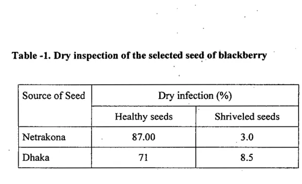
The results of dry inspection of seeds, that were collected from two different sources viz.' Dhaka and Netrakona districts. The results of dry inspection of seeds are presented in Table- 1.
It was observed that the two categories of seeds viz. i. apparently healthy seeds ii.Discolored and shriveled seeds ranged from 3'% and 8.50/0 in Netrakona and Dhaka district, respectively. The percentage of apparently healthy seeds was the highest in Netrakona (87%) and lowest in Dhaka (71%) districts.
4.2. Germination test
The germination test was carried out by blotter incubation test, agar test and pot soil test. The germination of blackberry seeds was the highest in pot soil which was 75% and 70% respectively in Netrakona and Dhaka districts. The lowest germination was recorded in,agar media 25% and 22%, in Netrakona and Dhaka, respectively (Table-Z).
27
Table -1. Dry inspection of the selected seed of blackberry
Source of Seed Dry infection (%)
Healthy seeds Shriveled seeds
Netrakona 87.00 3.0
Dhaka 71 8.5
Table-2. Germination percentage of the selected seeds observed in different tests.
Source Germination %
Pot soil Agar Blotter
Netrakona 75 25 68
Dhaka 72 22 65
28
4.3. Prevalence of fungi
4.3.1. Total seed borne fungal infection
In blotter method, out of 800 seeds, a total 183 seeds were found infected by fungal pathogens. The prevalence of total seed borne fungal infections varied significantly in respect of sources of collection of six samples from different location.
In general, more seed borne fungal infections 50% were observed in seeds collected from Dhaka district compared to Netrakona 30%.
In Netrakona, three sources were selected for seed collection. Among them the highest numbers of Aspergillus jlavus were observed in seeds collected from forest nursery (7%) and the lowest was Curvularia lunata (0.0%). The highest Rhizopus sp. (8.1 %) was recorded in the seeds collected from Choto bazer and Penicillium sp. was the lowest (0.0%) in number. The presence of Pestalotia psidii was also lowest (0.0%) in the seeds collected from Choto bazer. The infections of fungi in the seeds of Modonpur were Aspergillus niger (6.9%), Aspergillus flavus (6.4%), Rhizopus sp. (6.1%),
Curvularia lunata (3%), Pestalotia psidii (2.2%) and Penicillium sp. (3%).
In Dhaka, accept Aspergillus niger, the highest number seed born.e fungal infections were recorded in the seeds collected from karwan bazer. The highest Aspergillus niger (8%) found in the blackberry seeds collected from
29
Tongi hazer. Aspergillus
flavus
(6.9%) Aspergillus niger (6.5%), Fusarium equiseti (l~), Rhizopus sp. (2%), Pestalotia psidii (3.2%), Penicillum sp.(0.0%) and Curvularia lunata (0%), respectively observed in the seeds collected from Asadgate (Table-S).
4.3.2. Fungi identified and their frequency of occurrence in Netrakona District
Out of 74 seed borne fungal infections, 6 species of fungi representing, 5 genera were identified. The identified fungi were Pestalotia psidii (Photo.-5B, 5C, 7) Fusarium equiseti (Photo.-5A, 8A), Aspergillus jlavus(Photo-4C), Aspergillus niger (Photo.-4B,8B), Curvularia lunata
(Photo.-4A,6), Rhizopus sp. (Photo.-8C). Penicillium sp. (Photo. - 9B). Of all these fungi, most predominant fungi were Aspergillus niger (29.72%), followed by Aspergillus f1avus (25.6%), Rhizopus sp. (21.62%) and Fusarium equiseti (10.81%).
The Curvularia lunata had the lowest (4.05%) occurrence and the Aspergillus niger was the highest (29.72%) once. Penicillium sp. was not
found inthe seeds of blackberry in Netrakona (Table-4).
30
Photo 4. A. Pure culture ofCurvularia lunata of blackberry seeds on PDA (40x) B. Pure culture-of Aspergillus niger of blackberry seeds-on PDA (40x)
C.Pure culture ofAspergillusflavus of black perry seeds on PDA (40x)
31
Photo. 5.A.Pure culture ofFusarium equiseti of blackberry seeds of POA (40x) B. Pure culture ofPestalotia psidii of blackberry seeds of POA (40x)
C.Mature fruiting bodies ofPestalotia psidii of blackberry seeds of PDA (4Ox)
32
(
Photo. 6. Conidia of Curvularia lunata seen under microscope (400x)
.>"
-,
L) •
.f
'_
-
.~" .. '6.<~~.'
.. I ,-,
~': e
-.,_
Photo. 7. Conidia of Pestalotla psldii seen under microscope (400x)
33
, • I· .
•
,
•
•
,
I
.r-- •• ,.
Photo. 8. A. Conidiophore, vesicle and conidia ofAspergillus niger (400x) B. Sporangiophores, columella and sporangiosphores ofRhizopus sp.
(400x)
34
Table-3. Prevalence of Seed borne infections of individually fungi detected in blackberry seeds collected from six different sources.
District Source %of Seed borne infection
A. A. niger F. Rhizopus Penicillium C
P. psldi!
flavus equiseli sp. sp; lunata
Netrakona Government 7.000a 7.700a S.OOOa 2.00d O.100a O.OOOd 1.500b
nursery
Choto Bazcr 6.IOOc 6.800b 0.400d 8.IOOa O.OOOe 2.400c O.OOOd
Modonpur 6.400bc 6.900b 2.900b 6.104b O.300b 3.090a 2.200a
Dhaka Karwan 7.200a 7.100b 5.200a 8.300a 6.000a 2.200e 2.270a bazer
6.800ab 8.000a 5.100a 4.S00e 4.800a 1.700b 1.400c Tongi bazer
6.900a 6.Sb I.OOOe 2.000d O.OOOe O.OOd 3.210a Asadgate
nursery
LSD 0.445 0.3929 0.305 0.4050 0.1196 0.235 0.135
cv(%) 7.38% 6.05% 10.62% 8.73% 7.39% 14.22% 6.79%
Means bearing the same letter (s) in a column did not differ significantly at
1% level by DMRT.
35
Table- 4. Frequency of occurrence of fungi observed in the seeds of Netrakona.
Fungi No. of infection % of total No. ofSam~le Infections" infections
Aspergillus flavus 19 25.6 3
A. niger 22 29.72 3
Rhizopus sp. 16 21.62 3
Fusarium equiseti 8 10.81 2
Pestalotia psidii 6 8.10 2
Curvularia lunata 3 4.05 2
Total 74
s-rotal number of seed borne fungal infections recorded of the seeds of Netrakona.
"Total no. of seed samples were 3
36
4.3.3. Fungi identified and their frequency of occurrence in the seed of Dhaka district
Out of 109 seed borne fungal infections recorded on 400 blackberry seeds that represent 3 locations of Dhaka districts, seven different fungi were identified. The identified fungi, in order of prevalence were Aspergillus niger, Aspergillus flavus, Rhizopus sp., Fusarium equisiti, Penecillium sp., Pestalotia psidii and curvularia lunata. Of these, the most predominant seed borne fungus was Aspergillus niger; constituting 29.35% of the total fungal infections. Out of the 3 seed samples tested., 3 samples were found to be infected by 5 fungi viz. Aspergillus flavus, Aspergillus niger, Rhizopus sp, Fusarium equiseti and Pestalotia psidii (Table-5).
4.3.4. Prevalence of fungi in blotter paper
Among the three treatments, the prevalence of fungi was the highest in control seeds (8%), followed by seeds treated with alcohol and mercuric chloride. In control seeds, the occurrence of Rhizopus sp.. (8%) was the highest followed by, Fusarim equiseti (5.62%), Aspergillus flavus (5.27%), Aspergillus niger (3.87%), Penicillium sp. (3%), Pestalotia psidii (2.62%) and Curvularia lunata (2%). Incase of alcohol treatment, the
incidence of Rhizopus sp. (5.7%) was also the highest, followed by Fusarium equiseti (4%), Aspergillus flavus (4.8%), Aspergillus niger
37
(3.2%), Penicillium sp. (2.1 %) Pestalotia psidii (2%) and Curvularia lunata (1.5%). Incase of Hg2ch treated seeds, the incidence of Aspergillus jlavus was the highest (3.8%), followed by. Fusarium equiseti (3.2%), Rhizopus sp. (3%), Aspergillus niger (2.25%), Pestalotia psidii
. ,
(1.250/0), Penicillium sp. (0.9%) and Curvularia lunata (0.8%) (Table-e).
4.3.5. Prevalence of fungi in PDA media
The incidence' of fungi was relatively lower inPDA media. The occurrence of Aspergillus jlavus was the highest (6.62%) in PDA media in control seeds followed by Fusarium equiseti (6%), Rhizopus sp. (6.1%), Aspergillus niger (4.8%) and Penicillium sp. (1.9%). In case of Hg2c12
treated seed, the incidence of Aspergillus flavus was the highest (3.1%) while Fusarium equiseti was the lowest (1.1 %). The alcohol treated seeds showed medium incidence. Where, Rhizopus
sp,
caused the highest incidence (5%) followed by Aspergillus flavus (4.02%), Fusarium equiseti (3.8%) and Aspergillus niger (3.7%). No incidence of Curvularia lunata and Pestalotia psidii was observed (Table-7).38
Table- 5. Frequency of occurrence of fungi observed in the seeds of Dhaka.
Fungi No. of 0/0 of total No. of Sample
infection infections a infections b
Aspergillus
flavus
20 18.34 3Aspergillus niger 32 29.35 3
Rhizopus sp. 24 22.01 3
Fusarium equiseti 11 .10.09 3
Penicillium sp. 11 10.09 2
Pestalotia psidii 7 6.42 3
Curvularia lunata 4 3.66 2
109
"Total number of seed borne fungal infections recorded of the seeds of Netrakona.
~otal no. of seed samples were 3
39
Table=-S, Prevalence of fungi in blotter method with different treatments
Treatment Fusarium equiseti Aspergillus A. niger Curvularla Pastalotia Penicicillium Rhizopus sp.
flavus lunata psidii sp.
Control S.62Sa 5.27Sa 3.875a 2.000a 2.62Sa 3.000a 8.000a
H~d2(%) 3.250b 3.814b 2.250b O.87Sb 1.250c 0.900c 3.020c
A1cohol (%) 4.000b 4.875ab 3.2S0a 1.500a 2.000b 2.1l0b S.727b
LSD 0.5. 1.049 1.214 0.959 0.594 0.417 0.592 0.357
40
Table -7.Prevalence of fungi in PDA media with different treatments
Treatment Fusarium equiseti Aspergillus A. niger Curvularla Pastalotia Penicicillium Rhizopus sp.
flavus lunata psidii sp.
Control 6.000a 6.625a 4.875a
-- -
1.980a 6.147aH~d2(%) 1.125b 3.100b 2.750c
-- --
O.OOOb 2.992cAlcohol (%) 3.875b 4.025b 3.750b
-- --
O.OOOb 5.000b,LSD 0.5 2.026 1.968 0.935
-- --
0.998 0.20841
4.4. Pathogenicity
4.4.1. Leaves inoculation
The three fungi Fusarium equiseti, Pesta/otia psidii and Curvularia lunata tested for pathogenicity on blackberry. In case of infection caused by Fusarium equiseti raddish lesions were developed in .infected leaves later the leaves tum black and died out (Table- 8). On the leaves of two months old blackberry seedlings, PestaIotia psidii caused leaf spots. The centers of the spots were whitish with brown border. The fungus caused 10 spots on 5 leaves in 4 seedlings out of 8 inoculated plants. A few larger spots were found to coalesce to form bigger irregular lesions resulted in blight symptom. Infected cotyledon leaves with blight symptom ultimately dropped off (Table-9). Curvularia lunata caused characteristic dark brown to black colored lesions on the tip of the inoculated leaves of blackberry (Table-l 0).
The fungus caused shriveling of the leaves and irregular dark brown lesions.
The fungus caused altogether 3 lesions on 3 leaves 'in 2 plants out of the 8 inoculated plants (Photo.-9A, B, and C).
42
\
/
Photo. 9. A. Leaf blight symptoms developed after two months old leaves of blackberry by Curvularia lunata.
B and C. Leaf spot symptom developed after two months old leaves of Blackberry byPestalotia psidii andFusarium equiseti.
43
OJ
Table-S, Pathogenicity ofFusarium equiseti on two months old seedlings of blackberry as determined by leaves inoculation.
Fruit Total no. No. of No. of . No. of
of plants infected infected spots! Remarks Plants
inoculated plants leaves lesions
In severe cases reddish lesions developed.
Blackberry 8 2 3 3 Infected leaves
become dried. It turned black and died out.
Table- 9.Pathogenicity of Pestalotia psldii on ~o months old seedlings of blackberry as determined by leaf inoculation.
Fruit Total no. No. of No. of
No. of
of plants infected infected Remarks
Plants spots/lesions
inoculated . plants leaves
In severe cases blight symptom developed.
Blackberry 8 4 5 10 Infected
cotyledon acyleaves got fully blighted and dropped off.
44
Table-tO. Pathogenicity of Curvularia lunata on two months old seedlings of blackberry as determined byleaf inoculation.
Fruit Total no. No. of No. of No. of
of plants infected infected spotsl Remarks Plants
inoculated plants leaves lesions
Insevere cases blight symptom developed.
Blackberry 8 2 '3 3 Infected
cotyledon leaves got fully
blighted and dropped off.
45
4.5. Occurrence of foliage diseases in nurseries
Totally five nurseries were surved in Netrakona and, Mymensingh districts. Three nurseries in Netrakona, such as 1: Bonobevagh nursery 2.
Sotabdy nursery 3. Mohammed nursery. InMymensingh, 1. Mamun nursery and 2.Tulip nursery were surveyed. During the two months period of observation on foliage infection in three months old seedlings, the following diseases have been recorded:
1. Disease- Leaf blight of blackberry Pathogen- Pestalotia psidii
Symptoms- Irregular light brown spots with white centre surrounded by brownish border; the white area covered with black spore (photo-
lOA)
2. Disease - Red rust of blackberry Pathogen -Puccinia psidii
Symptoms - leaf blade covered with brownish superficial fungal growth on the dorsal surface; lesions of various sizes scattered on the surface (Photo.-l OC).
3. Disease - Leaf blight of blackberry Pathogen - Curvu/aria lunata
Symptoms - Withering of leaves started from the tip; the blighted area turned dark brown to black color; the border with healthy pinkish part.
46
A:
,i "r' . " I ,,;\ f\
,~4
JB
.i5..~-:.~:-_ ." ,c
Photo. 10. A. Symptom of leaf blight diseases
B. Growth ofPenicillium sp. on bJackbeny seed incubated on blotter (4Ox) C. Symptom of leaf rust disease
47
4. Leaf spot of blackberry Pathogen - Fusarium sp.
Symptoms -. Irregular reddish brown spots of different sizes were shown on the leaf surface.
Intensity of disease incidence was determined by counting the number of plants showing leaf infection in the selected nurseries in two locations.
Red rust of blackberry caused byPuccinia psidii was the least frequent.
Totally 9484 seedlings were survey in five nurseries. The germination was 75%. The disease severities were different in the nurseries. It depends on microclimate and locations.
Only three diseases were noted during the period of survey. The disease were leaf spot, leaf blight and leaf rust which occurred in all the seasons and in all the age of the trees. InNetrakona, the highest incidence (40%) of leaf blight disease' was found in Mohammad nursery caused by Pestalotia psidii. The incidence of leaf rust diseases caused by Puccinia psidii was the lowest (10%) (Table- 11). The highest infection was 80% due
to Pestalotia psidii recorded in the nursery of Mymensingh (Table- 12). The fungi isolated were Fusarium equiseti, Curvularia lunata, Pestalotia psidii and Puccinia psidii.
48
Table-ll. Occurrence and intensity of foliage diseases of seedling raised in three nurseries of Netrakona •.
Nurseries Diseases Causal organism 0/0 seedling infected Bono-bevagh leaf spot Fusarium equiseti 27.00
nursery leaf rust Puccinia psidii 20.00
leaf blight Petalotia psidii 33.50 Sotabdy leaf spot Fusarium equiseti 14.50
nursery leaf rust Puccinia psidii 10.00
leaf blight Curvularia lunata 28.00 leaf bight Pestalotia psidii 30.50 Mohammed leaf spot Fusarium equiseti 17.00
nursery leaf rust Puccinia psidii 11.70
]eafblight . Pestalotia psidii 40.00 Table- 12. Occurrence and intensity of foliage diseases of seedling raised in
three nurseries of Mymensingh.
Nurseries Diseases Causal organism
% seedling infected
Mamun leaf spot Curvularia lunata 21.80
nursery
leaf rust Puccinia psidi 8.00 leaf blight Fusarium equiseti 15.00 Tulip nursery
leaf blight Pestalotia psidii 80.00
49
5. DISCUSSION
Eight hundred seeds of blackberry were collected from two districts, Dhaka and Netrakona, and the seeds were tested for the prevalence of seed borne fungi. A total 183 seed borne fungal infections were recorded from 800 seeds of blackberry. The present study reveals that around 23% of the seeds were infected by fungi. Or in other words, at least one seed out of every four had fungal infection. This indicates that the .seeds of the blackberry plants under study had considerable amount of infection by pathogens.
Seven species of fungi were detected from the seeds of blackberry. No fungi have been reported from the seeds of this fruit species earlier in the country.
Thus, all these seven fungi recorded on the seeds of blackberry appears to be new records for Bangladesh.
In present study Aspergillus flavus, Aspergillus niger, Fusarium equiseti, Rhizopus sp., Curvularia lunata, Pestalotia psidii, Penicillium sp. has been recorded from the blackberry seeds. Rahman and Zethner (1971) isolated Acremonium sp., Candida sp., Penicillium sp. And Fusarium sp. from the seeds of Syzygium grandies which is close relative of blackberry. The study of prevalence of seed borne fungal pathogens on the blackberry seeds are very limited in abroad also. So, there is no confirm evidence of occurrence
50
of pathogen Aspergillus flavus, Aspergillus niger, Curvularia lunata, Pestalotia psidii, Rhizopus sp., Penicillium sp and Fusarium equiseti in blackberry seed available that can present in the thesis paper. The prevalence of the fungal infection varied with respect of fruit species and sources of seed collection. However, there is no report of variation in the prevalence of fungi in seeds of blackberry and no study has been made on this subject.
Such variation of occurrence of seed borne fungi has been demonstrated ina number of crops like rice, kaon, mustard, blackgram, wheat, jute and chilli by different research workers (Hossain et al. 1977; Barma and Fakir, 1981;
Dey and Fakir, 1988; kabir and Fakir, 1988; Rahman et al. 1988; Fakir and Islam, 1990; Basak et al. 1991 and Fakir andHalder, 1993). Variation in the occurrence of seed borne fungal infections on the seeds of blackberry could be due to the storage practice and climatic condition
of
the seed collected areas.Three fungi, Fusarium equisiti, Pestalotia psidii and Curvularia lunata were tested for pathogenicity on two months old· seedlings of blackberry. From pathogenicity test, it appears that Fusarium equiseti, Pestalotia psidii Curvularia lunata are pathogenic and caused spot and blighted symptoms on the leaves of blackberry. Morton (1987) reported several diseases like white spongy spot, leaf spot and leaf blight lesions on leaves of Syzygium cuminii.
Kuthubutheen et al. (I988) encountered species of Colletotrichum, Pestaloliopsis, Fusarium, Botryodiplodia on leaves of several fruits trees
5]
and stated that these fungi were all capable .of lesion formation and subsequent leaf damage. Pandey (1984 and 1990) found leaves of guava, which is a close relative of blackberry under same family Myrtaceae, were colonized by Colletotrichun gloeosporioides, Fusarium oxysporium f. sp.
psidii, Pestalotia psidii, andphoma psidii and these four were 'consistently pathogenic. Dwivedi et al. (1994) also reported that wilted twigs of guava yielded isolates of Fusarium sp. However, Edward (1961) reported that guava relative, Syzygium cuminii had never been observed to be attacked by wilt disease. As it is a very new finding in our country so further investigation ill future by researchers are required to' confirm the pathogenicity of theFusarium equiseti fungi.
Pestalotia psidii caused irregular small'spots (2-4 mm) of the leaves of seedlings of blackberry. Ramaswamy et al. (19~8 and 1998) isolated Pestalotia psidii from blackberry and he confirmed the pathogenicity of the pathogen to blackberry by inoculation test. They also reported that Pestalotia psidii is the causal agent of guava canker and this report is in accordance with the findings of Pandey (1990).
Curvularia lunata caused large lesions (3-7 mm) in leave's of seedling of blackberry. In severe case, blight symptom developed and often the infected cotyledonary leaves dropped off. No evidence of such pathogenicity could be presented due to lack of literature on blackberry but Kapoor et al. (1970)
52
isolated the fungus Curvularia tuberculata from guava which belongs to same family of blackberry. Previously Curvularia tuberculata had been reported as causing die-back disease of citrus in India.
In survey, 9984 seedlings were observed in five selected nursenes m Mymensing and Netrakona districts. At the time of survey, in addition to the
. .
three fungus viz., Pestalotia psidii, Curvularia lunata and Fusarium equiseti, Puccinia psidii has been encountered in the leaves of blackberry seedlings. This fungus is also recorded from the leaves of Syzygium jambos (White, 1990 and Galli, et al.1990), guava and eucalyptus (Galli, et ai,
1990). Itmay be mentioned here that these are close relatives to blackberry and all are belongs to the same family ofMyrtaceae.
Rust symptoms was first appeared as tiny, bright yellow, powdery eruptions that developed into circular, pustules on the stem and foliage. These pustules later expanded, coalesced, and became necrotic, spreading over the entire leaf and stem surfaces, and then leaves and stems were deformed and tip dieback ensued. Coutinho et al. (1998) describe the symptoms of the disease as first begin of tiny bright yellow powdery eruptions in a circular pattern on the leaf or stem surface. These infection loci or spots expand and become necrotic, and spread over the entire leaf, stem or shoot. Leaves and stems can be deformed by the disease, and growing tips can die back if the infection is severe.
53
As the study was limited to two districts only, further studies with more representative seed samples from different blackberry species, obtained from different parts of the country should be undertaken in order to depict the exact picture regarding the prevalence of. fungi and the pathogenicity of the fungi under study in this experiment.
54
6. SUMMARY AND CONCLUSION·
Prevalence and pathogenicity of fungi associated with the seeds of blackberry, collected form 6 sources under two districts of Dhaka and Netrakona, were studied inplant pathology lab, Sher-e- Bangia Agricultural.
University, Dhaka.
183 seed-borne fungal infections were recorded from 800 seeds of blackberry. In general, more seed-borne fungal infections were detected in seeds obtained from Dhaka district compared to Netrakona.
Of the total seed borne fungal infections recorded from the seeds of blackberry, seven fungi representing 6 genera were identified. The fungi identified were Aspergillus flavus, Aspergillus niger, Fusarium equiseti, Pestalotia psidii, Curvularia lunata, Rhizopus sp., Peniecillium sp.
The seven fungi recorded on the seeds of the one fruit species (blackbeny) appear to be new records of seed-borne. fungi of blackberry in Bangladesh.
Pathogenicity test conducted with Curvularia lunata, Fusarium equiseti and Pestalotia psidii and the fungi were found to be pathogenic to blackberry. As it is a very new finding, further investigations in future by researchers are required to confirm the pathogenicity of the above mentioned fungi. The
55
fungi produced dark brown to black spots and blight symptom on leaves of blackberry .
In survey, study four fungi Curvularia lunata, Fusarium equiseti, Pestalotia psidii and Puccinia