Angucyclinones from an Indonesian
Streptomyces
sp.
Serge Fotso,†Taifo Mahmud,†T. Mark Zabriskie,†Dwi Andreas Santosa,‡,§Sulastri,‡and Philip. J. Proteau*,† Department of Pharmaceutical Sciences, College of Pharmacy, 203 Pharmacy Building, Oregon State UniVersity, CorVallis, Oregon 97331-3507, Indonesian Center for BiodiVersity and Biotechnology, ICBB-Complex, Jl. Cilubang Nagrak No. 62, Situgede, Bogor 16115, Indonesia, and Department of Soil and Land Resources, Faculty of Agriculture, Bogor Agricultural UniVersity, Jl. Meranti, Kampus IPB Darmaga, Bogor 16680, Indonesia
ReceiVed August 7, 2007
Six new angucyclinone polyketides named panglimycins A-F were isolated together with the three known metabolites (+)-fujianmycin A, (+)-ochromycinone, and emycin C from the bioassay-guided fractionation of the extract of the IndonesianStreptomycesstrain ICBB8230. The new compounds are highly oxygenated angucyclinones that appear to be biosynthetically derived from ochromycinone or fujianmycin. Their structures were determined by X-ray crystal analysis, interpretation of 1D- and 2D-NMR spectra, and comparison of the data with those of structurally related known natural products. Despite structural similarities to angucyclinones with antibiotic activities, the panglimycins did not exhibit any growth inhibition when tested against several bacteria and fungi.
We recently began a screening program to discover new antibiotics from microorganisms isolated from the Black Water Ecosystem in Kalimantan, Indonesia. During our initial survey of
Streptomyces spp., the antibiotic activity of the crude extract of the isolate ICBB8230 against Escherichia coli,Bacillus subtilis,
Pseudomonas aeruginosa, andCandida albicansdrew our attention. Chromatographic separation of the extract and analysis using an antibacterial agar diffusion assay led to the known angucyclinone antibiotics (+)-fujianmycin A1and (+)-ochromycinone.2,3During
the processing of the extract to find the antibiotic compounds, it was noticed that the organism also produced additional angucycli-none metabolites. This family of antibiotics is characterized by a tetracyclic framework assembled in an angular manner, leading to the benz[a]anthracene system. These polyketide natural products have attracted much attention due to their biological activity, interesting chemical structures, and biosynthesis.4Further workup
of the extract led to the isolation of the new angucyclinones panglimycins A (1a), B (1b), C (1c), D (2a), E (2b), and F (3) and the known compound emycin C.5The name panglimycin signifies
the isolation of the producing strain from soil of the Black Water River, Pangkoh Lima. The known compounds were identified by substructure searches in AntiBase.6Here we report the isolation,
structure elucidation, and biological activity testing of the new compounds.
Results and Discussion
A culture of the strain ICBB8230, identified as aStreptomyces
sp., in M2medium delivered a greenish culture broth, which was
processed as indicated in the experimental section. Two crude extracts were obtained, one from the mycelium and one from the culture broth. They were fractionated using various chromatographic techniques.
Compound1awas isolated as colorless crystals. An AntiBase search provided elmycin A (4) as a likely possibility, but the1H
NMR shifts differed slightly from those published.7The13C NMR
shifts for 4were reported in CDCl3, but 1awas not soluble in
CDCl3, so a13C NMR spectrum was obtained in DMSO-d6. The
necessity to use a different NMR solvent, as well as shift differences, suggested that1awas not identical to4. In order to further assess the structure of1a, an X-ray crystal structure was
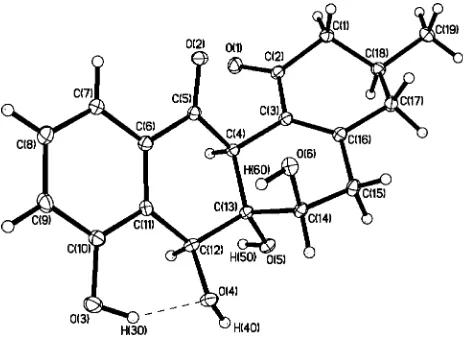
obtained. The relative configuration of1ais shown in Figure 1 and differs from that of48at C-6a and C-12a, indicating that1ais
a stereoisomer of elmycin A, which we have named panglimycin A. Although the absolute configuration was not determined experimentally, we propose the 3S,6R,6aS,7S,12aSconfiguration based on the likely derivation of compound 1a from (+ )-ochromycinone (3Sconfiguration),9which was also isolated from
the ICBB8230 strain.
Compound1bwas obtained as a yellowish powder. Extensive NMR (1H,13C, DEPT, COSY, HSQC, HMBC) and MS data were
used to establish the structure. The 1H-1H-COSY spectrum
indicated three coupled spin systems, along with two methines at
δ5.50 and 3.90 that appeared as singlets. The first spin system involved two doublets of doublets atδ7.31 and 7.00 (J)7.8, 1.1 Hz) and an apparent triplet atδ7.20 (J)7.8 Hz), which can be attributed to a 1,2,3-trisubstituted aromatic system. The second spin system consists of a methylene (δ3.08, 2.26) adjacent to a methine (δ4.47) attached to an oxygenated carbon. The final spin system has a methylene (δ2.56) coupled to a methine atδ2.18 bearing a methyl group (δ1.23) and also attached to another methine (δ4.11) on a carbon bearing an oxygen. The13C NMR spectrum indicated
19 carbon resonances as required by the high-resolution mass. There were two carbonyls atδ200.5 and 196.3, three sp2methines, five
sp3 methines including three bearing oxygen, one methyl, two
methylene carbons, and five sp2and one sp3quaternary carbons.
The (+)-ESIMS indicated the pseudomolecular ion atm/z361 [M + H]+, and the HRESIMS delivered the molecular formula * Corresponding author. Tel: (541) 737-5776. Fax: (541) 737-3999.
E-mail: [email protected].
†Oregon State University.
‡Indonesian Center for Biodiversity and Biotechnology. §Bogor Agricultural University.
Figure 1.ORTEP representation of panglimycin A (1a).
C19H20O7. The general NMR characteristics indicated that1bwas
quite similar in structure to1a.
In the HMBC spectrum, H-9 (δ7.00) and H-10 (δ7.20) showed cross-peaks to C-8 (δ157.6), suggesting an oxygenated carbon in the aromatic ring (Figure 2). The proton atδ7.31 (H-11) correlated to the carbonyl atδ196.3, suggesting a carbonyl adjacent to the trisubstituted aromatic system. The singlet at δ5.50 (H-7) had correlations to the carbons atδ157.6 (C-8), 128.1 (C-7a), and 77.7 (C-6a), establishing a secondary alcohol at C-7. Further correlations were seen between the three methines H-7 (δ5.50), H-6 (δ4.47), and H-12a (δ3.90) to the quaternary sp3carbon atδ77.7 and also
between both methine H-7 and H-12a to the carbon C-6 (δ66.2). On the other hand, the methine atδ3.90 and the methylene H2-2
(δ2.56) showed correlations to carbons atδ200.5 (C-1) and 129.4 (C-12b), while the methyl doublet correlated to the methylene carbon C-2 (δ45.4) and both methine carbons atδ76.6 (C-4) and C-3 (δ40.5), confirming the third spin system from the1H-1
H-COSY. On the basis of these correlations and comparison with1a, the structure of 1b was determined and named panglimycin B. Panglimycin B (1b) is a 4-hydroxy analogue of1a. As shown in Table 1, the key differences in13C NMR shifts between1aand1b
are at C-4 and nearby carbons.
The similarities in chemical shifts to1asuggest that the same relative configuration at the ring junction (C-6a/C-12a) is shared by1b. Thetransrelative configuration at C-3 and C-4 of 1bis proposed on the basis of the NOESY spectrum, which revealed an important correlation between the H-4 and CH3-3 protons (Figure
3), and on the large coupling constant between H-3 and H-4 (JH-3/H-4)9.0 Hz), which represents a diaxial-like relationship for
these protons.1This is the same relative configuration seen in (+
)-fujianmycin A,1which is also present in this extract and is a possible
biosynthetic precursor to 1b. The presence of a strong NOESY correlation between H-7 and H-12a is also supportive of the proposed ring junction configuration. These observations led to the proposed relative configuration of 1bas shown in Figure 2. An attempt to determine the absolute configuration of 1b was done using the Mosher method, but the derivatization reaction led only to a complex mixture of decomposition products.
Compound1cappeared as a yellow oil, and it exhibited similar
1H and13C NMR spectra to1b, with the same 1,2,3-trisubstituted
aromatic region, the methines atδ5.42 and 3.90, and the methyl doublet atδ1.13. The major differences in the1H NMR spectrum
for1cwere the absence of the protons atδ4.47 (H-6) and 4.11 (H-4) and a more complex region in the range ofδ2.58–2.00. The molecular formula was deduced from the (+)-HRESIMS to be C19H20O5, which requires 10 unsaturations as in1b, but two fewer
oxygen atoms are present in1c. The13C NMR spectrum of1clacks
the carbon resonances that appear in1batδ76.6 (C-4) and 66.2 (C-6) and instead has two methylene carbon signals atδ40.2 and 23.7. Extensive interpretation of the HMBC spectrum correlations, combined with comparison of data with those of 1b, established that1cwas missing the oxygens from C-4 and C-6. The resulting compound1cwas named panglimycin C.
Compound2awas obtained as a yellowish powder like1b. The proton NMR spectrum of2ais also similar to those of1a,1b, and
1cwith the trisubstituted aromatic system and the methyl doublet in the aliphatic region. The main difference between the2aand1b
spectra is that the H-5/H-6 spin system has been replaced by two coupled protons at δ3.97 and 3.94. The molecular weight was deduced from the (+)-ESIMS to be 358. The HRESIMS gave the molecular formula C19H18O7, requiring 11 unsaturations instead of
10 as in1band1c, suggesting the presence of an additional double bond or ring. The presence of two new sp3methine carbon signals
atδ57.1 and 48.4 and the lack of additional sp2carbon signals
compared to1bsuggested the presence of an epoxide in view of the chemical shifts of the methine carbons. The location of the epoxide was deduced from the HMBC spectrum, which indicated important correlations of the methine proton atδ5.56 (H-7) to the carbon atδ57.1 and of the methine proton atδ4.35 (H-4) to the carbon atδ48.4. These correlations indicated that the epoxide lies between C-4 and C-7, securing the structure of compound2a, which was named panglimycin D.
Figure 2.Panglimycins A-C (1a-c), selected HMBC correlations in panglimycin B, and the structure of elmycin A (4).
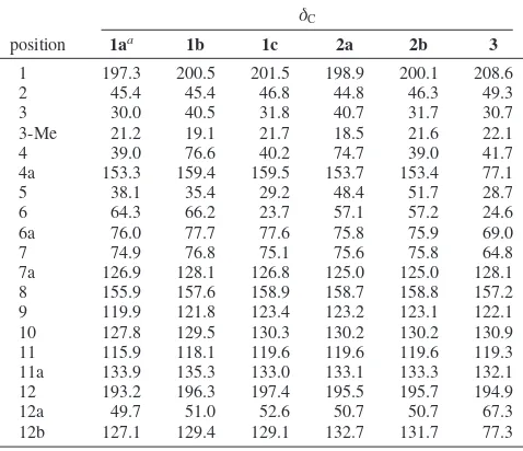
Table 1. 13C NMR (75 MHz) Data for Compounds 1a-c, 2a, 2b, and3in MeOH-d4
δC
position 1aa 1b 1c 2a 2b 3
1 197.3 200.5 201.5 198.9 200.1 208.6
2 45.4 45.4 46.8 44.8 46.3 49.3
3 30.0 40.5 31.8 40.7 31.7 30.7
3-Me 21.2 19.1 21.7 18.5 21.6 22.1
4 39.0 76.6 40.2 74.7 39.0 41.7
4a 153.3 159.4 159.5 153.7 153.4 77.1
5 38.1 35.4 29.2 48.4 51.7 28.7
6 64.3 66.2 23.7 57.1 57.2 24.6
6a 76.0 77.7 77.6 75.8 75.9 69.0
7 74.9 76.8 75.1 75.6 75.8 64.8
7a 126.9 128.1 126.8 125.0 125.0 128.1 8 155.9 157.6 158.9 158.7 158.8 157.2 9 119.9 121.8 123.4 123.2 123.1 122.1 10 127.8 129.5 130.3 130.2 130.2 130.9 11 115.9 118.1 119.6 119.6 119.6 119.3 11a 133.9 135.3 133.0 133.1 133.3 132.1 12 193.2 196.3 197.4 195.5 195.7 194.9
12a 49.7 51.0 52.6 50.7 50.7 67.3
12b 127.1 129.4 129.1 132.7 131.7 77.3
aDMSO-d6.
Figure 3. Observed NOESY correlations in panglimycin B (1b) and proposed relative configuration.
Figure 4.Structures of panglimycins D (2a) and E (2b) and key HMBC correlations for panglimycin D.
Due to the close proximity of the chemical shifts for the epoxide protons and H-12a in MeOH-d4, a NOESY spectrum was recorded
in acetone-d6to aid in determining the relative configuration of
compound2a. The NOESY spectrum exhibited cross-peaks between H-7 and H-12a and between H-4 and the C-3 methyl protons, which is consistent with the relative configuration observed for pangli-mycin B (1b), but no correlations were seen to the epoxide protons. Furthermore, preliminary molecular modeling of 2a(Chem3D) suggested that a H-5 to H-4 correlation, if observed, could be possible for both configurations of the epoxide. Therefore, the relative configuration of the epoxide remains unassigned. The cou-pling constant of 8.5 Hz from H-3 to H-4 also supports thetrans
relative configuration at C-3/C-4.1
Compound2bwas also obtained as a yellowish oil. Compound
2bwas found to have MS and NMR data similar to elmycin B, but as seen with1a, the NMR shifts were not an exact match.7Further,
the optical rotation of2bwas-31°, while the literature value for elmycin B was+28.5°. A comparison of the13C NMR shifts for 2band2aillustrates that the only major differences between the two lie near C-4, the site of the hydroxyl in2a. This suggests that
2b is in the same stereochemical series as is2aand that it is a stereoisomer of elmycin B, which we have named panglimycin E (2b).
Compound 3 was obtained as a colorless oil. The molecular formula C19H20O7obtained from HRESIMS requires 11
unsatura-tions as in2a. The1H-1H-COSY spectrum indicated the presence
of the trisubstituted aromatic system and two additional fragments; one consisted of CH2CH2 and the second fragment included a
methyl doublet atδ1.03, a methine atδ3.97, and two methylene groups (δ2.60, 2.22;δ1.97, 1.61) that were assembled in the same manner as H-2 to H-4 in 1a, 1c, and2b. The shifts of the H-4 protons, however, were upfield relative to the H-4 protons in1a,
1c, and2b, suggesting a change in the hybridization at C-4a. The
13C NMR spectrum of3indicated 19 carbon resonances, comprised
of two carbonyls atδ208.6 and 194.9, three sp2methines, three
sp2quaternary carbons, two sp3methines, four methylenes, one
methyl, and four quaternary sp3carbons. The downfield shift of
the C-1 carbonyl, the reduction in the number of sp2quaternary
carbons, and the addition of new carbon resonances between 70 and 80 ppm suggested the possible dihydroxylation of the isolated double bond between C-4a and C-12b. The lack of a C-4a/C-12b double bond would require an additional ring in3.
The HMBC spectrum revealed correlations between H-2, H-4, and H-5 and the carbons atδ77.3/77.1, which correspond to C-12b and C-4a, confirming the oxygenation of the C-4a/C-12b double bond. The H-7 proton correlates to the methylene carbon atδ24.6, the quaternary carbons atδ67.3 and 69.0, and the carbon at δ
77.3, suggesting that C-6a and C-12a also bear oxygen groups. Furthermore the correlation of H-5 and H-6 to the quaternary carbon atδ69.0 defines this carbon as C-6a. On the basis of the need for an additional ring in3and the presence of epoxides in a number of known angucyclinones, a reasonable conclusion is that an epoxide lies somewhere on the C-4a/C-12b/C-12a/C-6a backbone. The chemical shifts of these four carbons (77.1, 77.3, 67.3, and 69.0, respectively) and comparisons with NMR data for the related compounds simocyclinone D810and elmycin C11suggest that the
epoxide is formed between C-6a and C-12a, but further confirmation was necessary. When the 1H NMR spectrum was recorded in
DMSO-d6, all four exchangeable protons present in 3 were
observed. The phenolic hydroxy proton appeared at 10 ppm and the C-7 hydroxy proton was revealed by coupling with H-7, which left two hydroxy proton resonances (4.53 and 4.39 ppm) to be assigned. The HMBC spectrum of3 in DMSO-d6provided the
necessary correlations. The 4.53 ppm proton was correlated with carbons C-12a, C-12b/C-4a, and C-1, while the 4.39 ppm proton correlated to carbons at C-12b/C-4a and C-4. These correlations place the hydroxy groups at C-4a and C-12b, and therefore, the epoxide spans C-6a to C-12a, securing the structure of panglimycin F (3). The structure of panglimycin F is very similar to that of elmycin C11and simocyclinone A
1,12the only differences in the
planar structures being the presence of an additional C5-C6 double bond in elmycin C and a C2-C3 double bond in simocycli-none A1.
Biological Activities. Panglimycins A-E are related to the known antibiotics elmycins A (4) and B, which exhibited
antibacte-Table 2. 1H NMR (300 MHz) Data for Panglimycins A-C (1a-c)
δH(Jin Hz)
position 1aa 1bb 1cb
2 2.10–2.40, m 2.56, m 2.15–2.58, m
3 2.10–2.40, m 2.18, m 2.15–2.58, m
3-Me 1.07, d (5.7) 1.23, d (6.5) 1.13, d(5.9)
4 2.10–2.40, m 4.11, d (9.0) 2.15–2.58, m
5 2.59, dd (19.7, 4.2) 2.00, d (19.7) 3.08, dd (20.2, 4.6) 2.26, d (20.2) 2.12, m 1.38, m
6 4.16, br t (3.2) 4.47, br d (3.9) 2.15–2.58, m
7 5.38, s 5.50, s 5.42, s
9 6.96, dd (7.2, 2.0) 7.00, dd (7.8, 1.1) 7.05, dd (7.7, 1.3)
10 7.14, m 7.20, dd (7.8, 7.8) 7.25, ddd (7.7, 7.7, 0.8)
11 7.14, m 7.31, dd (7.8, 1.1) 7.39, dd (7.7, 1.3)
12a 3.74, s 3.90, s 3.90, br d (1.9)
aDMSO-d6additional signals atδ5.14 (C-6-OH),δ4.68 (C-7-OH).bMeOH-d4.
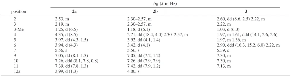
Table 3. 1H NMR (300 MHz) Data for Panglimycins D-F (2a,2b, and3) in MeOH-d4
δH(Jin Hz)
position 2a 2b 3
2 2.53, m 2.30–2.57, m 2.60, dd (8.6, 2.5) 2.22, m
3 2.19, m 2.30–2.57, m 2.22, m
3-Me 1.25, d (6.5) 1.18, d (6.1) 1.03, d (6.0)
4 4.35, d (8.5) 2.71, dd (18.4, 4.0) 2.30–2.57, m 1.97, m 1.61, ddd (14.1, 2.6, 2.6)
5 3.97, dd (4.3, 1.5) 3.92, dd (4.1, 1.4) 1.97, m 1.36, m
6 3.94, d (4.3) 3.42, d (4.1) 2.90, ddd (16.3, 15.2, 6.0) 2.22, m
7 5.56, s 5.56, s 5.39, s
9 7.05, dd (8.1, 1.3) 7.05, dd (7.2, 1.2) 7.30, m
10 7.26, ddd (8.1, 7.8, 0.8) 7.26, dd (7.9, 7.9) 7.30, m
11 7.39, dd (7.8, 1.3) 7.42, dd (7.9, 1.2) 7.13, m
rial and antifungal activities, as well as potent action against protozoa, especially Trichomonas Vaginalis.7 In contrast, the
panglimycins did not show antibiotic activity. Compounds1a-c,
2a,b, and3were tested against the bacteriaStaphylococcus aureus, Bacillus subtilis,Escherichia coli, andPseudomonas aeruginosa, the fungusMucor miehei, and the yeastCandida albicansin the agar diffusion test, at concentrations ranging from 20 to 120µg/ disk, but no growth inhibition was observed. In contrast, both ochromycinone (17 µg/disk) and fujianmicin A (18µg/disk), as positive controls, showed clear zones of inhibition againstB. subtilis
and P. aeruginosa. This difference in biological activity may be attributed to the opposite configuration at the C-6a/C-12a ring junction compared to elmycin A, which results in significantly different overall molecular shapes for the panglimycins.
Experimental Section
General Experimental Procedures. Optical rotations were mea-sured on a Jasco P1010 polarimeter. NMR spectra were meamea-sured on a Bruker Unity 300 MHz spectrometer. The spectra were referenced to the solvent line methyl at 3.30 ppm for1H NMR spectra and to the center line of the MeOH-d4septet at 49.15 ppm for13C NMR spectra. ESIMS data were recorded on a ThermoFinnigan LCQ Advantage system. HRESI mass spectra were recorded on a Waters/Micromass LCT spectrometer. HREIMS and HRCIMS were measured on a JEOL HMS-600H MS route magnetic sector instrument. IR spectra were recorded on a Nicolet Nexus 470 FT-IR spectrometer. UV–vis spectra were recorded on a Beckmann DU 640 B spectrophotometer. Prepara-tive HPLC was performed using an RP18 column (Phenomenex, RP 100-C18, 5µm) with the detector set at 254 nm. Flash chromatography was carried out on Si gel (230–400 mesh). Thin-layer chromatography was performed on aluminum sheets with Si gel 60 F254(EMD chemicals Inc.). Size exclusion chromatography was done on Sephadex LH-20 (Pharmacia). M2medium: 4 g of glucose, 4 g of yeast extract, and 10 g of malt extract were dissolved in 1000 mL of deionized H2O, the pH was adjusted to 7.8 with 2 N NaOH, and the medium was sterilized at 121°C for 35 min.
Organism Collection and Identification. Samples were taken from soil of the Black Water River, Pangkoh Lima, Malibu Village, Gandang Subdistrict, Pulang Pisau Regency, Central Kalimantan Province, Indonesia. This river is part of the unique Black Water ecosystem in Kalimantan. The ecosystem lies in the remote area about 150 km from the coast of South Kalimantan. Soil samples were stored at room temperature.
The selective agar plate medium was obtained by the addition of antifungal antibiotics and bacterial inhibitors (20 mg/L trimethoprim, 50 mg/L griseofulvin, 50 mg/L nystatin) into the YM medium (4 g/L glucose, 10 g/L malt extract, 4 g/L yeast extract) adjusted to pH 7. The antibiotics were dissolved in sterile water and added to the autoclaved agar medium prior to the pouring of the plates. Five grams of soil was inoculated into a 15 mL test tube containing 9 mL of sterilized H2O containing 0.85% NaCl, followed by serial dilutions. The soil suspension from the 10-3dilution (0.1 mL) was spread on selective agar plates. The plates were incubated at 30°C until the colonies appeared (4–7 days). The colonies with different characteristics were transferred repeatedly to selective agar plates until pure cultures were obtained. Each colony was checked by microscopy to differentiate between actinomycetes and other microorganisms. TheStreptomyces sp. ICBB8230 culture was deposited at ICBB-CC (Indonesian Center for Biodiversity and Biotechnology Culture Collection of Microorgan-isms) as 0.5 mL of a 20% glycerol stock stored at-20°C.
The 16S rRNA gene sequence ofStreptomycessp. ICBB8230 was found to be identical over the sequenced region to that ofStreptomyces sp. CNR875 PL04, isolated from marine sediment collected near Palau (see Supporting Information for details).
Fermentation and Isolation. Streptomyces sp. ICBB8230 was cultivated on a 5 L scale using 1 L Erlenmeyer flasks containing 250 mL of M2medium at 28°C for 4 days on a rotary shaker (300 rpm). The greenish culture broth was mixed with ca. 2.0 kg of Celite and filtered under vacuum. The filtered medium was passed through a HP-20 Diaion column (3×17 cm), and the resin was washed with distilled
H2O and eluted with MeOH. The mycelium was extracted sequentially with EtOAc and then MeOH. Both extracts were evaporated to dryness separately. The combined mycelium extract (500 mg) was
chromato-graphed on Sephadex LH-20 (50% MeOH/CH2Cl2), resulting in six fractions, I-VI. Fraction IV exhibited the main biological activity against E. coli, Ps. aeruginosa, and C. albicans. Fraction IV was chromatographed on Sephadex LH-20 (MeOH) and delivered four subfractions, IVA-IVD. Preparative TLC (PTLC; 5% MeOH/CH2Cl2) of the active subfractions IVB and IVC gave ochromycinone (30 mg), fujianmycin A (50 mg), and emycin C (1.5 mg). The crude extract from the medium (2 g) was first separated on Sephadex LH-20 (MeOH), yielding six fractions, I-VI. Fractions I and II contained only fatty acids and were discarded. Fraction IV was first separated on preparative HPLC using a gradient from 20% to 100% MeOH in H2O and delivered panglimycins A (1a, 5 mg) and E (2b, 4 mg) and a mixture of two compounds. The mixture was further purified by PTLC (8% MeOH/ CH2Cl2) and led to panglimycins C (1c, 2.5 mg) and F (3, 3 mg). The purification of fraction V twice over Sephadex LH-20 (MeOH) delivered two subfractions, VA and VB. The subfraction VA was washed with CH2Cl2and gave a mixture of ochromycinone and fujianmycin A and a CH2Cl2-insoluble yellowish powder, panglimycin B (1b, 102 mg). The subfraction VB was purified by PTLC and gave panglimycin B (1b, 7 mg) and panglimycin D (2a, 110 mg).
Panglimycin A (1a): colorless crystals (50% MeOH/CH2Cl2); [R]23 D +83 (c0.06, MeOH); UV (MeOH)λmax(logǫ) 311 (2.92), 247 (3.59), 209 (3.82) nm; IR (neat)νmax3299, 2915, 2847, 1695, 1652, 1634, 1587, 1559, 1463, 1293, 1055, 1003, 938, 839, 793 cm-1;1H and13C NMR data, see Tables 1 and 2; (+)-HRESIMSm/z345.1327 (calcd for C19H21O6, 345.1338) and 367.1165 (calcd for C19H20O6Na, 367.1358).
Panglimycin B (1b): light yellow solid; [R]27
D -100 (c 0.06, MeOH); UV (MeOH)λmax(logǫ) 321 (2.92), 262 (3.50), 222 (4.18) nm; IR (neat)νmax3347, 2958, 2922, 2854, 1657, 1588, 1464, 1390, 1297, 1263, 1085, 1062, 1023, 996, 939, 796, 737 cm-1;1H and13C
νmax3330, 2955, 2926, 1670, 1653, 1583, 1458, 1393, 1282, 1077, 797, 1385, 1107 cm-1; NMR data, see Tables 1 and 2; (+)-ESIMS m/z(%) 329 ([M+H]+
, 95), 679 ([2M+Na]+
, 100); HREIMSm/z 328.1298 (calcd for C19H20O5, 328.1311).
Panglimycin D (2a): light yellow solid; [R]27
D-30 (c0.1, MeOH); UV (MeOH)λmax (logǫ) 324 (2.90), 261 (3.80), 242 (3.77) nm; IR (neat)νmax 3212, 2955, 1685, 1656, 1634, 1580, 1463, 1291, 1271, 1234, 1152, 1083, 892, 786 cm-1; NMR data, see Tables 1 and 3; (+ )-ESIMSm/z(%) 381 ([M+Na]+, 18), 739 ([2M+Na]+, 100); (+ )-HRESIMSm/z381.0978 (calcd for C19H18O7Na, 381.0950).
Panglimycin E (2b): yellowish oil; [R]23
D-31 (c0.1, MeOH); UV (MeOH)λmax(logǫ) 315 (3.81), 254 (4.39), 228 (sh), 207 (4.45) nm; IR (neat)νmax3360, 2949, 1676, 1608, 1580, 1463, 1383, 1296, 1278, 1243, 1092, 1030, 900, 789 cm-1; NMR data, see Tables 1 and 3; (+)-ESIMSm/z)343 ([M+H]+
, 10), 365 ([M+Na]+ , 15), 707 ([2M + Na]+, 100); (+)-HRESIMS 343.1157 (calcd for C19H19O6, 343.1182) and 365.0988 (calcd for C19H18O6Na, 365.1001).
Panglimycin F (3): colorless oil; [R]27
D+150 (c0.09, MeOH); UV (MeOH)λmax (logǫ) 332 (3.42), 272 (3.94), 247 (4.13) nm; IR (neat)νmax 3360, 2956, 2917, 2849, 1698, 1662, 1640, 1456, 1266, 1221, 1157, 1089, 1071, 738 cm-1; NMR data in MeOH-d4, see Tables 1 and 3;1H NMR (DMSO-d6, 300 MHz)δ10.10 (1H, br s, C-8 OH), 27.2 (CH2, C-6), 23.0 (CH2, C-5), 21.2 (CH3, C-3 CH3) (*assignments interchangeable); (+)-ESIMSm/z(%) 383 ([M+Na]+
, 100), 743 ([2M + Na]+, 55); (+)-HRESIMSm/z 383.1115 (calcd for C19H20O7Na, 383.1107).
)4,Dcalcd)1.498 g cm-3,µ)0.112 mm-1, 10 222 reflections measured, 3529 reflections independent (Rint)0.0146),Rw)0.0294,Rw)0.0794. X-ray diffraction experiments for panglimycin A were carried out on a Bruker Smart Apex CCD diffractometer at 173 K using Mo KR radiation (λ ) 0.71070 Å). Absorption corrections were done by SADABS.13The structure was solved using direct methods and refined with full-matrix least-squares methods based on F2. Non-hydrogen atoms were refined with anisotropic thermal parameters. The H atoms were located by difference Fourier synthesis and refined with isotropic thermal parameters. The absolute structure of the compound has not been determined based on the X-ray diffraction data; the anomalous scattering power is too small. All calculations were performed using the SHELXTL (v. 6.10) package.14Crystallographic data have been deposited with the Cambridge Crystallographic Data Centre (deposit No. CCDC 666383). Copies of the data can be obtained, free of charge, on application to the Director, CCDC, 12 Union Road, Cambridge CB2 1EZ, UK (fax:+44-(0)223-336033 or e-mail: [email protected]).
Acknowledgment. We thank Dr. L. Zakharov, OSU Chemistry Department, for X-ray crystallography, J. Turner and Dr. L. Zhang for microbiology and technical assistance, and D. Ragland for 16S rDNA analysis. High-resolution mass spectra were obtained by J. Morré at the mass spectrometry facility of the Environmental Health Sciences Center at Oregon State University, which is supported in part by a grant from NIEHS (ES00210). This work was supported by the OSU College of Pharmacy Research and Scholarship Fund.
Supporting Information Available:1H and13C NMR spectra of 1a-c,2a,2b, and3, COSY, HSQC, and HMBC spectra for1band
2a, details of the 16S rDNA analysis, and a Crystallographic Informa-tion File (CIF) for1a. This material is available free of charge at http:// pubs.acs.org.
References and Notes
(1) Rickards, R. W.; Wu, J. P.J. Antibiot. (Tokyo)1985,38, 513–515. (2) Bowie, J.; Johnson, A. W.Tetrahedron Lett.1967, 1449–1452. (3) Taniguchi, M.; Nagai, K.; Watanabe, M.; Nimura, N.; Suzuki, K.;
Tanaka, A.J. Antibiot.2002,55, 30–35.
(4) Krohn, K.; Rohr, J.Top. Curr. Chem.1997,188, 127–195. (5) Gerlitz, M.; Udvarnoki, G.; Rohr, J.Angew. Chem., Int. Ed. Engl.
1995,34, 1617–1621.
(6) Laatsch, H.AntiBase, a Natural Products Database for Rapid Structure Determination; Wiley-VCH: Weinheim, 2005.
(7) Grabley, S.; Wink, J.; Giani, C.; Seibert, G.; Raether, W.; Dobreff, S.; Zeeck, A. Eur. Pat. Appl. EP342363, 1989.
(8) Rohr, J.; Thiericke, R.Nat. Prod. Rep.1992,9, 103–137. (9) Carreño, M. C.; Urbano, A.; Di Vitta, C.Chem. Commun.1999, 817–
818.
(10) Holzenkampfer, M.; Walker, M.; Zeeck, A.; Schimana, J.; Fiedler, H. P.J. Antibiot. (Tokyo)2002,55, 301–307.
(11) Dobreff, S. Neue Angulole, Angucyclinon-Verwandte Antibiotika aus Streptomyceten: Strukturaufklärung und Chemische Derivatisierung. Ph.D. Thesis, University of Göttingen, Göttingen, Germany, 1989, pp 11–30.
(12) Schimana, J.; Walker, M.; Zeeck, A.; Fiedler, H.-P.J. Ind. Microbiol. Biotechnol.2001,27, 144–148.
(13) Sheldrick, G. M.SADABS, Bruker/Siemens Area Detector Absorption Correcton Program;Bruker AXS: Madison, WI, 1998.
(14) SHELXTL-6.10, Program for Structure Solution, Refinement and Presentation; Bruker AXS: Madison, WI, 2000.