Correspondence email to: [email protected]
E-cadherin and NM23HI as metastasis predictors for various degrees
of histological malignancy in invasive ductal carcinoma
Primariadewi Rustamadji,1 Ahmad Tjarta,1 Santoso Cornainm,1 Muchlis Ramli,2 Esti Soetrisno1
1 Department of Anatomic Pathology, Faculty of Medicine Universitas Indonesia/Cipto Mangunkusumo Hospital 2 Department of Oncology Surgery, Faculty of Medicine Universitas Indonesia/Cipto Mangunkusumo Hospital
Abstrak
Latar belakang: Penelitian ini bertujuan untuk menganalisis apakah ekspresi protein E-cadherin dan NM23H1 dapat digunakan sebagai prediktor invasi dan metastasis karsinoma duktal payudara pada berbagai derajat keganasan .
Metodologi: Subyek penelitian adalah 97 wanita yang telah didiagnosis menderita karsinoma payudara duktal invasif
derajat 1,2, dan 3 yang spesimen biopsinya dikirim ke laboratorium histopatologi rumah sakit di Jakarta dan Bandung antara tahun 2000-2006. Pemeriksaan histopatologis dengan pulasan hematoksilin eosin terhadap blok parain yang berasal dari tumor primer maupun sekunder dilakukan untuk penentuan derajat keganasan dan status metastasis. Selanjutnya dilakukan pemeriksaan imunohistokimia terhadap ekspresi E-cadherin, NM23H1 dan sitokeratin di jaringan tersebut serta dilakukan skoring berdasarkan jumlah sel terwarnai dan intensitas pewarnaan. Analisis dilakukan untuk mengetahui hubungan ekspresi E-cadherin dan NM23H1 dengan metastasis dan derajat keganasan histologik.
Hasil: Subyek berusia antara 29-75 tahun dengan rerata 48,19 tahun dan terbanyak berusia 40-45 tahun, dengan
derajat keganasan 1 sebanyak 18,56%, derajat 2 sebanyak 45,36% dan derajat 3 sebanyak 36,1%. Terdapat hubungan bermakna antara ekspresi E-cadherin dan NM23H1 pada tumor primer dengan kemungkinan E-cadherin menghambat invasi dan metastasis sebesar 14 kali sedangkan NM23H1 sebanyak 11 kali dibandingkan subyek yang tidak mengekspresikan E-cadherin dan atau NM23H1. Kurva ROC menunjukkan ekspresi E-cadherin (r= 0,755) dan NM23H1 (r= 0,816) berkorelasi kuat, sensitif dan spesiik sebagai petanda metastasis akan tetapi tidak berhubungan dengan derajat keganasan histologik
Kesimpulan: Ekspresi E-cadherin dan NM23H1 dapat digunakan sebagai petanda invasi dan metastasis, tetapi tidak
dapat digunakan sebagai petanda derajat keganasan histologik karsinoma duktal invasif payudara. (Med J Indones 2011; 20:263-70)
Abstract
Background: This study aims to analyze whether the expressions of E-cadherin and NM23HI can be used as predictors of ductal carcinoma metastasis in various degrees of malignancies.
Methods: Parafin blocks were obtained from 97 patients with invasive breast ductal carcinoma with malignancy grade
1, 2 and 3 who came to several hospitals in Jakarta and Bandung from 2000 to 2006. Histopathological examinations of
hematoxylin eosin slides of primary and secondary tumors were done to diagnose the degree of histological malignancy
and metastasis status. Further, immunohistochemistry staining of E-cadherin, NM23HI and cytokeratin were done
followed by scoring according to the number of positive cells and staining intensity. The associations of E-cadherin and NM23H1 expression with the presence of metastasis and grade of histological malignancy were analyzed.
Results: Subjects were 29-75 years old (mean: 48.19 years), with most subjects aged 40–45 years old, with
malignancy grade 1, 2 and 3 of 18.56%, 45.36% and 36.1% respectively. There was a signiicant association between
E-cadherin and NM23HI expression in primary tumors. The possibility of invasion and metastasis inhibition by
positive E-cadherin and NM23HI was 14 and 11 times respectively compared to those with negative E-cadherin and/ or NM23HI expression. The ROC curve showed that E-cadherin (r= 0.755) and NM23HI (r= 0.827) expressions were strongly associated, sensitive and speciic as metastasis markers. However, E-cadherin and NM23HI expression did not show signiicant association with histological degree of invasive ductal carcinoma.
Conclusion: E-cadherin and NM23HI expressions can be used as invasion and metastasis markers, but cannot be used as
markers for the degree of histological malignancy of invasive ductal carcinoma. (Med J Indones 2011; 20:263-70)
Keywords: Breast cancer, E-cadherin, NM23HI
The incidence of breast cancer occupies the irst position
of malignancies in the world1 and second in Indonesia
next to cervical cancer.2 Many cancer patients visit their
doctors at an advanced stage that make the treatment and therapy dificult. The worst of all is, in Indonesia, most
of cancer patients are still young and productive.3
Based on the records in Dr. Cipto Mangunkusumo
hospital, invasive ductal carcinoma is the subtype of
breast carcinoma with the highest incidence, i.e. 85 to 90 % (Mangunkusumo R, personal communication).
Early detection of invasive ductal carcinoma in its early stage can provide maximum and affordable treatments to the patients.
node removal, radiation and chemotherapy. Patients with metastasis potency need further treatments including axillary’s lymph node removal, radiation
and chemotherapy. Markers to predict the occurrence
of metastasis may alert doctors and patients from the emergence of loco regional metastasis, so that doctors can give optimal treatment, preserve patients’ productivity and better ensure patients’ recovery.4
Previous studies concluded that predictors of metastasis, until now, is not yet clear, while therapy and prognostic factors of patients are very dependent on the prediction of metastasis.4 Therefore, it is necessary to develop alternatives that can predict and estimate the prognosis of patients in early stages of breast ductal carcinoma. These alternatives should be more accurate, affordable, and available. Combinations of clinical, histopathological and immunohistochemical examinations are currently being developed rapidly. These alternatives are expected to detect or predict the potential of metastasis to the lymph nodes or distant metastases. Therefore, accurate invasion and metastasis
markers of invasive ductal carcinoma to detect in early
stage are highly required, and this study aims to analyze whether the expressions of E-cadherin and NM23HI can be used as predictors of ductal carcinoma metastasis in various degrees of histological malignancies.
MEtHods
This is a cross sectional study conducted in the
Department of Anatomic Pathology, Faculty of Medicine, Universitas Indonesia, Cipto Mangunkusumo
Hospital, from November 1st to December 31st, 2006.
data collection
Data were retrieved from the archives of the Department of Anatomic Pathology, Faculty of Medicine Universitas Indonesia/Cipto Mangunkusumo Hospital, Jakarta, Kramat 128 Hospital Jakarta, Jakarta Breast Centers, Public Hospitals Hasan Sadikin, Bandung, Jakarta Islamic Hospital and the Darmanugraha Hospital Rawamangun, Jakarta. The data retrieved were: the hospital origin of
specimen, age, sub-type of tumor, tumor grade, and lymph node metastasis.
The data recorded from the immunohistochemical staining results were the positivity and expression streghth of E-cadherin and NM23H1 in primary tumors, and metastases in lymph nodes in invasive ductal breast carcinoma.
samples
The samples were HE slides and parafin blocks of breast mastectomy cases from several hospitals in Jakarta and
Bandung from 2000 to 2006 that met the inclusion criteria,
i.e. breast carcinoma that had been histopathologically diagnosed as invasive ductal breast carcinoma with
low grade of malignancy (grade I) with and without
metastases in lymph nodes, grade II with and without
metastases in lymph nodes, and high grade (grade III), when good or reliable parafin blocks are available. Exclusion criteria are unreliable parafin blocks (e.g. broken/damaged parafin blocks, parafin blocks whose tumor mass is cut or eaten by animals, etc).
Calculation of sample size
The sample size was calculated using P1 and P2 for E-Cadherin and NM23H1,with alpha= 5%, conidence
interval= 95%, and power= 80%.5,6 The sample sizes
that were calculated were 11 and 24, respectively.
The samples were selected using consecutive sampling. Combination of NM23H1 plus E-cadherin
examinations was expected to make the prediction to be more signiicant. It was expected that the accuracy
of the prediction would reach ninety percent.
slide preparation and immunohistochemical staining4, 7
The parafin blocks were cut and immunohistochemically stained; blocks of primary tumors and their metastases
in lymph nodes were stained with E-cadherin and
NM23H1, while blocks of lymph nodes without metastasis were stained with cytokeratin to make sure
that there was no metastasis in lymph nodes.
Immunohistochemical staining used the Streptavidine
Biotin complex labeling method. The primary antibodies against E-cadherin, NM23H1 and Cytokeratin were
mouse monoclonal antibodies, i.e. mouse monoclonal
anti CDH1 antibody, Ig I:200 (Novocastra), NM23H1 antibody, Ig I:100 (Novocastra) and for cytoceratin was anti A1/E3 antibody, Ig I:100 (Daco). Every
staining included negative controls using the same
breast carcinoma tissue; each running consisted of 8 - 10 cases with a positive control of breast carcinoma
in-situ, and staining results were analyzed using standard assessment techniques.
Assessment and reading of immunohistochemical
stainings were carried out by two anatomic pathologists and researchers who are experienced in reading
histopathological slides. Assessment of E-cadherin expression was performed in 500 tumor cells from 5 different large ields (400 x) that were chosen randomly. Each region was represented by 100 tumor
cells. E-cadherin positivity was represented by brown staining of the tumor cell membrane or cytoplasm.
Degree of positivity of E-cadherin was assessed by a
(the value of color intensity by Nichols).7 Assessment of NM23H1 expression was performed in the same way with E-cadherin, but NM23H1 positivity was represented by brown staining in cytoplasm of tumor
cells, and the positivity of cytokeratin was represented
as brown staining in the intra-tumor cell cytoplasm.
The degree of positivity of cytokeratin was assessed
in the same manner as above.7 The assessment of all
expressions was done on the entire area on small ield (magniication 100 x) and large ield (magniication 400 x).
data analysis
The data were entered into a main table, and data analyses were done using SPSS version 13 and
Med Calc version 9.6.4.0 software. Chi square test were used to calculate the Odds ratio (OR) and 95% conidence interval (95% CI) for E-Cadherin and
NM23H1 positivity as well as expression strength on the occurrence of metastatic tumors compared to non
metastatic tumors. In addition the OR and 95% CI for
tumor E-Cadherin and NM23H1 positive expression on the occurrence of lymph node positivity compared to negative expression were calculated. Further, diagnosis
test to get the sensitivity and speciicity value from the
ROC curve using E-Cadherin and NM23H1 positivity was done. 6, 8, 9 Presentation of data was done in tables, pictures and graphs.
REsults
Forty-eight cases of non metastatic ductal breast carcinoma and forty-nine cases of metastatic ductal breast carcinoma were included in the study.
The comparison of the expressions of E-cadherin in non-metastasic to those in metastatic primary tumor in patients with invasive ductal breast carcinoma revealed that the chance of positive E-cadherin to prevent metastasis is
13.6 times of that with negative E-cadherin (95% CI= 1.68;110, p < 0. 01).
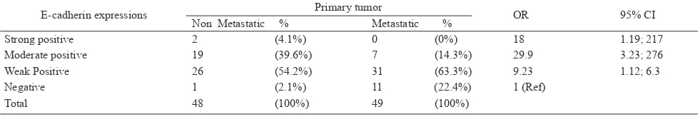
Table 1 shows that the chance of weak and moderate
positive E-cadherin expression to prevent metastasis was nine and thirty times of that with negative E-cadherin expression, respectively.
Further, the chance of positive expression of NM23H1 to prevent metastasis is 11.3 times of negative expression
of NM23H1, 95% CI= 4.29;30, p < 0.001.
Comparison of NM23H1 expression grade shows that
the chance of weak positive NM23H1 expression to
prevent metastasis is eight times as much as that of
negative expression (95% CI= 2.94; 21.4, p < 0.0001).
The chance of moderate positive NM23H1 expression
to prevent metastasis is 50 times as much as that of negative expression (95% CI= 5.82; 439; p < 0.0001).
Strong positive E-cadherin and NM23H1 expression cannot be valued due to the minimum number of samples and the presence of zero in both E-cadherin and NM23H1 expressions. Expression gradation comparison
shows an increase in Odds ratio from weak to moderate positive that is eight to ifty times (Table 2).
The number of E-cadherin positive expression in lymph nodes is high in E-cadherin positive primary tumors
with a 0.111-time decrease in positive expression in
Lymph nodes compared to E-cadherin negative primary
tumors (95% CI = 0.0025; 0.802, p< 0.001).
A high number of positive NM23H1 expressions occured
in lymph nodes of NM23H1 negative primary tumors
with a 0.034-time decrease in positive expression in
lymph nodes compared to NM23H1 positive primary
tumors (95% CI= 0.00079; 0.208, p < 0.001). The disagreement is due to a “0” (zero) in one cell.
Table3 shows positive and negative results of E-cadherin and NM23H1 expressions in non metastasized compared to metastasized primary tumors. In both positive and combination of positive and negative expression the
chance of invasion and metastasis prevention is 29 and 9 times of that in both negative expressions.
Table 1. Chi square test of gradation of E-cadherin expressions in non-metastasic compared to those in metastatic primary tumor
E-cadherin expressions Primary tumor OR 95% CI
Non Metastatic % Metastatic %
Strong positive 2 (4.1%) 0 (0%) 18 1.19; 217
Moderate positive 19 (39.6%) 7 (14.3%) 29.9 3.23; 276
Weak Positive 26 (54.2%) 31 (63.3%) 9.23 1.12; 6.3
Negative 1 (2.1%) 11 (22.4%) 1 (Ref)
Total 48 (100%) 49 (100%)
Table 2. Chi square test results of gradation of NM23H1 expressions in non metastatic primary tumor compared metastatic primary tumors
NM23H1 Expression Non Metastatic % Metastatic %Primary tumor OR 95% CI
Strong positive 0 (0%) 0 (0%) 3.89 0.221; 68.4
Moderate positive 12 (25%) 0 (0%) 50.6 5.82; 439
Weak positive 28 (58.3%) 15 (30.6) 7.93 2.94; 21.4
Negative 8 (16.7%) 34 (69.4) 1 (Ref)
Total 48 (100%) 49 (100%)
x2 =26.74 p < 0.0001
Table 3. Chi square test of E+/N+; E+/N-, E-/N+, E-/N- expressions in non metastatic and metastatic primary tumor
E/N expressions Primary Tumors
Total % OR 95% CI p
Non-metastatic % Metastatic %
E+/N+ 40 (72.7%) 15 (27.3%) 55 (100%) 29.333 3. 481; 247.179 0.002 E+/N- 7 (23.3%) 23 (76.7%) 30 (100%) 8.762 3.117; 24.626 0.000 E-/N- 1 (8.3%) 11 (91.7%) 12 (100%) REFERENCE
Total 48 (100%) 49 (100%) 97 (100%)
E+ = Positive e-cadherin, E - = Negative e-cadherin, N+ = Positive NM23H1, N - = Negative NM23H1
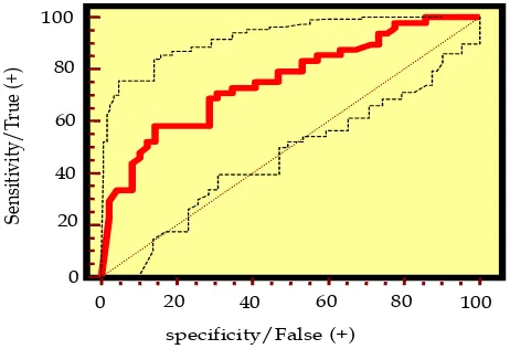
ROC curve is to illustrate the sensitivity and speciicity of each marker – E-cadherin and NM23H1 – in the
prediction of metastasis and histological grade of malignancy. The ROC curve can predict the best
marker. The sensitivity and the speciicity of E-cadherin
expression to predict the occurrence of metastasis was
59.2% and 87.5%, respectively, with a cutoff point of ≤ 40, and an area under the curve of 0.755.
The sensitivity and the speciicity of NM23H1 expression to predict the occurrence of metastasis is 69.4% and 83.3% respectively, with a cutoff point of <0, and an area under the curve of 0.827.
The two curves show that as a metastasis marker, E-cadherin expression is speciic, while NM23H1 expression is both speciic and sensitive. Comparison
of the curve of E-cadherin and NM23H1 expressions in metastasis prediction shows an area of difference
of 0.048. The ability of E-cadherin and NM23H1 to predict metastasis does not differ signiicantly when the difference limit is 0.05 (Figure 1).
Figure 1. The curve of relationship between E-Cadherin expres-sions and metastasis
the Results of Immunohistochemistry staining
The results of immunohistochemistry staining of E-Cadherin and NM23H1 can be seen in Figure 2-3.
Two pathologists’ calculations of immunohistochemical
readings gave likelihood ratios of 11.2 and 4.31.
E-cadherin prediction of the occurrence of metastasis is eleven, while NM23H1 prediction is four. In lymph
nodes, likelihood calculation is not performed because
the purpose is different. Metastasis prediction before and after immunohistochemistry with a priori compared to a posteriori probability according to Veneis method
showed an increase of 49.4% for E-cadherin and
49.27% for NM23H1.9
Figure 4a shows that all primary tumors with moderate
positive E-cadherin expressions have moderate positive expression in lymph nodes. Some primary tumors with
weak positive E-cadherin expressions (approximately twenty-ive percent) have moderate positive E-cadherin expressions in lymph nodes and the rest (approximately seventy-ive percent) have weak positive E-cadherin
expressions in lymph nodes. In primary tumors with
negative E-cadherin expressions, there are weak
positive, strong positive and also negative E-cadherin expressions in the lymph nodes.
Figure 4b shows that weak positive NM23H1 expressions
in primary tumors have positive NM23H1 expressions in lymph nodes, which is roughly one-third that have
moderate positive, while the rest (approximately two-thirds) have weak positive expressions in the lymph
nodes. Negative NM23H1 expressions in primary
tumors have negative, weak positive, and strong
positive NM23H1 expressions.
0 20 40 60 80 100
100
80
60
40
20
0
specificity/False (+)
Se
ns
it
iv
it
y/
Tr
ue
(+
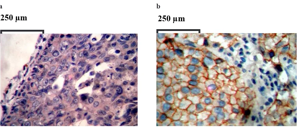
Figure 2. Negative and strong positive E-cadherin expression in invasive ductal breast carcinoma Magniication 400x a= negative expression, b= strong positive expression
Figure 3. Negative and moderate positive NM23H1 expression in invasive ductal breast carcinoma. Magniication 100x a= negative expression, b= moderate positive expression
= 250 µm
= 250 µm
a
b
1mm
a
b
1mm
prim
metastasis
Agreement of E-Cadherin expressions of Primary Tumor and
its Metastasis in lymph Nodes
++ + Negative Primary tumor
Lymph node +++ ++ + Negative
Figure 4. E-cadherin, and NM23HI expression in primary tumor and its metastasis in lymph nodes a= E-cadherin, b= NM23HI
L
ymph node
30
20
10
0
Ekspresi NM23H1 dari Tumor Primer dan Metastasisnya di Kelenjar Getah Bening
NM23H1 expression of the primary tumor and its metastases in lymph nodes
Figure 9.
prim
NM23H1 expressions of primary tumors and their
metastases in lymph nodes
Lymph node +++ ++ + Negative
Weak positive Negative Primary tumor
L
ymph node
40
30
20
10
0
dIsCusIoN
Among carcinoma sub types, such as lobular, papillary
and medullary, invasive ductal breast carcinoma
incidence has the highest rate. As a top referral hospital,
the breast carcinoma patients who come to Cipto
Mangunkusumo central national hospital may represent breast carcinoma patients in Indonesia. During the search
of the cases, various problems appeared so that the
expected minimum samples required were not fulilled.
Therefore, some samples were obtained from some
other hospitals in Jakarta and Bandung, as mentioned in
the methods, and it is expected that the samples will represent breast cancer patients in Indonesia.
Reliable parafin blocks that met the inclusion criteria
from ninety-seven cases were obtained. The selection and the reading of immunohistochemical slides were carried out by two experienced anatomic pathologists so that the internal validity is expected to be good.
The cases used are mastectomy cases of invasive ductal breast carcinoma with grade malignancy one, two and three. In situ ductal breast carcinoma was not included due to the scarcity of cases diagnosed. In situ ductal carcinoma gives strong positive expressions, especially E-cadherin expression, because the cell-cell adhesion in in situ ductal carcinoma is very strong, and micro-invasion has not occurred yet. Therefore, in this study, in situ ductal carcinoma was used as positive controls of E-cadherin and NM23H1 immunohistochemistry staining.
It is known that metastasis occurs not only to axillary
lymph nodes, as metastasis may occur in internal mammary lymph node, areas of clavicula, or other organs. However, all invasive ductal carcinoma cases in this study came from mastectomy with metastasis only to axillary lymph node, as far metastasis is contra indication to mastectomy.
Table 2 shows an increase in Odds Ratio from weak
positive and moderate positive to eight and ifty times
compared to that of negative expression. These results
agree with one of NM23H1 working mechanisms,
which is as metastasis suppressor that reduces cell motility and lowers cell differentiation to malignancy that may reduce or prevent the occurrence of invasion and metastasis.10
Various studies have tried to relate the decrease in E-cadherin expression to tumor growth and metastasis in breast and in other carcinomas. E-cadherin is a transmembrane glycoprotein that mediates intercellular adhesion that depends on calcium, and is speciically involved in epithelial cell-cell adhesion. E-cadherin gene is located at chromosome 16q22.1, and works as a very important morphogenetic regulator. In carcinoma, decrease
in E-cadherin expression is one of the changes that lead to invasive phenotype characters. Moreover, data from several researchers support E-cadherin’s role as tumor suppressor gene and as suppressor of invasion and metastasis.11
Decreased E-cadherin expression is related to high
invasion and advance stage of prostate, colon, colorectal, and breast carcinomas. Especially in breast carcinoma, decreased E-cadherin expression is related to negative estrogen receptor and high metastasis.11 However, our
study showed that positive expression is a good marker
for invasion and metastasis in lymph nodes.
Metastasis is a serious problem for doctors in treating breast carcinoma. The problems will be more complicated due to different therapy protocols that should be administered to breast carcinoma patients with or without metastasis.
Despite the so many studies in this ield, metastasis
processes, either locoregionally to lymph node or systematically to far organs, are still confusing.
Among so many identiied groups of gene suppressors, cadherin should get speciic attention.12 It agrees with
the results of the studies stating that loss or disturbance of E-cadherin expression can increase motility, local invasion, and metastasis.13-16 Graff et al. (cited by
Madhavan, et al. 2001) found that decreased E-cadherin
expression is caused by CpG island hypermethylation of E-cadherin gene promoter region.12
Table 2 shows an increase in Odds ratio of NM23H1
expression from weak positive to moderate positive.
This result agrees with one of NM23H1 roles, which is as a metastasis suppressor that reduces cell motility, lowers cell differentiation, thus reducing or preventing
invasion and metastasis. Decreased NM23H1 will activate RAF-MOS. Activated RAF-MOS will
phosphorilize MEK 1-2, and activated MEK 1-2 will activate ERK, which promotes metastasis.17
E-cadherin and NM23H1 expression in most of the lymph nodes in our study can be explained by the
results of Kowalczyk et al.1994, who found decreased
E-cadherin expression in invasive ductal carcinoma that caused the carcinoma to grow and metastasized to both lymph nodes and far sites. Cancer cells can re-express their E-cadherin as soon as they reach the far sites. Re-expression of E-cadherin by cancer cells can be found in all metastasis of ductal carcinoma and their expression level can be the as high as or even higher than the expression in the primary tumors.10 By learning E-cadherin expression in metastasis of breast carcinoma
in lymph nodes, Bukholm et.al (cited by Kowalczyk et al. 1994) showed that in nineteen out of twenty metastasis
in lymph nodes strongly re-express the E-cadherin.10
Graff et al. (cited by Madhavan, et al. 2001) found that
re-express in its metastasis deposit.12 E-cadherin odds ratio
analysis in our study shows that down regulation can function
as metastasis predictive marker in breast carcinoma. This
fact shows the important role of E-cadherin in predicting the occurrence of metastasis in cases with negative lymph nodes, but eventually have micro metastasis that are
undetected/ undiagnosed histologically so that doctors
may under diagnose their patients. 10, 12
ROC curve to evaluate the relationship between markers,
such as E-cadherin or NM23H1 and metastasis shows
signiicant relationship of both markers and metastasis. The two ROC curves show that as metastasis markers, E-cadherin expression is speciic, and NM23H1 expression is both sensitive and speciic. Comparison
of the curve of E-cadherin and NM23H1 expressions in metastasis prediction shows an area of difference of
0.048. Therefore, the ability of E-cadherin and NM23H1
to predict metastasis does not differ signiicantly. This fact agrees with molecular biology theory stating that NM23H1 is a metastasis suppressor, while E-cadherin
is a tumor and/or invasion suppressor, so that either individually or collectively the two markers are signiicant
in preventing tumor growth, invasion, and metastasis.
The chance of both positive E-cadherin and NM23H1 expressions to prevent invasion and metastasis is twenty-nine times the chance of negative E-cadherin and NM23H1 expressions. Further, the chance of positive E-cadherin and negative NM23H1 expressions to prevent metastasis and invasion is nine times the chance of both negative E-cadherin and NM23H1 expression. Therefore, it can be concluded that E-cadherin and NM23H1 expressions can be used as invasion and metastasis markers, both individually and collectively.
The result of gradation analysis, ROC curves, and combined tables of E-cadherin and NM23H1 support
the signiicant association between either E-cadherin or NM23H1 and both markers with metastasis, which is speciic and sensitive. Taking all the analyses into
account, it can be concluded that E-cadherin and
NM23H1 are good invasion and metastasis markers. A priori and a posteriori probability comparison shows
the percentage of metastasis prediction before and after immunohistochemistry staining. In this study, there is an increase of E-cadherin metastasis prevention
from a priori of 50.5% to a posteriori of 99.9%, which shows an increase of 49.4%. The signiicant increase
in a posteriori probabilities in this study shows the effectiveness of immunohistochemistry staining to predict the occurrence of metastasis. Our results agree
with the application of immunohistochemistry markers by Veneis (1997) in his research on cancer. The aim is to
provide a gold standard of immunohistochemical staining to predict the occurrence of metastasis for clinical use.9
The most valuable result of this study is that the chance of positive E-cadherin expression to prevent metastasis and invasion is 13.6 times the chance of negative E-cadherin
expression. The chance of weak positive NM23H1
expression to prevent metastasis is eleven times the chance of negative NM23H1 expression. Further, gradation
analysis of combined expressions of weak positive E-cadherin and NM23H1 shows a signiicant increase in Odds ratio that is sensitive and speciic for metastasis.
Impairments in the expression of certain genes may be due to epigenetic disorder or mutation. Epigenetic
disorder occurring in E-cadherin is only about 25-30% and the rest is due to mutation, while most
disorders occurring in NM23H1 are epigenetic. Epigenetic disorders are expected to response to demethylation therapy, which recently widely used.18 The hypermethylated genes are expected to become normal after demethylation therapy. Further studies using RT-PCR to distinguish cases with mutation from
epigenetic/hypermethylation are urgent, so that doctors
can give optimum treatments to their patients.
In conclusion, E-cadherin and NM23HI expressions
can be used as invasion and metastasis markers, but cannot be used as markers for the degree of histological
malignancy in invasive ductal carcinoma.
REfERENCEs
Department of Health and Human Services, Centers for
1.
disease Control and Prevention. National Program of cancer
statistics (USCS). 2007 Top Ten Cancer’s [Internet]. [Cited
2011 April 1]. Available from: http://www.cdc.gov/uscs.
Tjindarbumi D, Mangunkusumo R. Cancer in Indonesia,
2.
present and future. J Clin Oncol. 2002;32:17-21.
Ramli M. Kanker payudara, deteksi dini dan penatalaksanaan
3.
masa kini. Seminar sehari deteksi dini kanker. Muktamar V/PIT XII:Jogjakarta;2000. [Indonesian].
Leong AS. Short course of molecular immunohistology. 4.
The sixth annual scientiic meeting Asia-Paciic society for molecular immunohistology (APSM). The Sunway Medical Center of Malaysia; 2011.
Sastroasmoro S, Ismael S. Dasar-dasar metodologi 5.
penelitian klinis. 2nd edition. Jakarta ; Sagung Seto: 2002. [Indonesian].
Departemen Ilmu Kedokteran Komunitas FKUI. Kumpulan
6.
bahan kuliah epidemiologi biostatistik. Jakarta: Departemen Ilmu Kedokteran Komunitas; 2004. [Indonesian].
Nichols GE, Frierson HF, Boyd JC, Hanigan MH. 7.
Automated immunohistochemical assay for estrogen status in breast cancer using monoclonal antibody CC4-5 on the Ventana ES. Am J Clin Pathol. 1996;106:332-8.
MedCalc version 9.6.4.0. for Windows. Statistics for biome-8.
dical research software manual. Mariakerke: Microsoft; 2003. Vineis P. Sources of variation in biomarkers. In:Toniolo P, 9.
Kowalczyk AP, Palka HL, Luu HH, Nilles LA, Anderson 10.
JE, Wheelock MJ, et al. Posttranslational regulation of
plakoglobin expression. Inluence of the desmosomal cadherins on plakoglobin metabolic stability. J Biol Chem. 1994;269:31214-23.
Mastracci TL, Tjan S, Bane AL, Malley FPO, Andrulis
11.
IL. E-cadherin alterations in atypical lobular hyperplasia and lobular carcinoma in situ of the breast. Mod Path.
2005;18:741-51.
Madhavan M, Srinivas P, Abraham E, Ahmed I, Mathew
12.
A, Vijayalekshmi NR, et al. Cadherins as predictive markers of nodal metastasis in breast cancer. Mod Pathol. 2001;14:423-7.
Berx G, Roy V. The E-cadherin/Catenin complex: an
13.
important gatekeeper in breast cancer tumorigenesis and malignant progression. Breast Cancer Res. 2001;3:289-93.
Knudsen KA, Wheelock M J. Cadherin and the mammary 14.
gland. J Cell Biol. 2005;95:488-96.
Dickson RB, Lippman ME. Molecular basis of breast cancer. 15.
In: Mendelsohn J, Howley PM, Israel MA, Liotta LA,
editors. The molecular basis of cancer. 2 ed. Philadelphia;
WB Saunders; 2001. p. 326-9.
Steeg PS. Metastasis suppressors alter the signal transduction 16.
of cancer cells. Nature Reviews Cancer. 2003; 3: 55-63. Steeg PS, Palmieri D, Ouatas T, Salerno M. Histidine kinases 17.
and Histidine phosphorylated proteins in mammalian cell biology, signal transduction and cancer. Cancer Letters.
2003; 1-12. [cited 2006 Dec 15]. Available from: http://
www.sciencedirect.com.
Roymans D, Willems R, Van Blockstaele DR, Slegers H. 18.