accredited by DGHE (DIKTI), Decree No: 51/Dikti/Kep/2010 199
An Age Estimation Method to Panoramic Radiographs
from Indonesian Individuals
Anny Yuniarti, Agus Zainal Arifin, Arya Yudhi Wijaya, Wijayanti Nurul Khotimah Laboratory of Vision, Image Processing and Graphics, Department of Informatics
Faculty of Information Technology, Institut Teknologi Sepuluh Nopember Kampus ITS Sukolilo, Surabaya 60111, Indonesia, Telp/fax 031-5939214/031-5913804
e-mail: [email protected], [email protected], [email protected], [email protected]
Abstrak
Fitur-fitur gigi dapat dipertimbangkan sebagai kandidat terbaik untuk pengidentifikasian PM. Jika data AM tidak tersedia, maka bantuan ahli forensik gigi diperlukan untuk membatasi ruang populasi data dan meningkatkan ruang pencarian data dengan pembuatan profil gigi pasca kematian atau postmortem dental profiling. Usia adalah salah satu faktor yang penting dalam membangun atau menentukan identitas seseorang. Inspeksi manual pada radiografi gigi memiliki dua kelemahan, yaitu intraobserver error dan interobserver error. Pada makalah ini diusulkan sebuah sistem semi-otomatis untuk estimasi usia. Terdapat dua fase dalam pengembangan sistem yang diusulkan, yaitu: fase pemodelan dan fase estimasi. Fase pemodelan adalah tahap untuk menurunkan rumus estimasi berdasarkan data yang sudah diketahui. Pada penelitian ini, digunakan data dari suku Jawa. Fase estimasi meliputi proses penentuan region of interest (ROI), penghitungan panjang gigi secara otomatis, dan estimasi usia berdasarkan rumus pemodelan yang telah dibentuk sebelumnya. Percobaan pada penelitian ini menunjukkan hasil yang menjanjikan, yaitu nilai rata-rata absolute error sebesar 5,2 tahun, jika dibandingkan dengan aplikasi metode Kvaal pada orang-orang Turki yang menghasilkan selisih lebih dari 12 tahun.
Kata kunci: estimasi usia, radiografi panoramik, regresi, identifikasi manusia, pengolahan citra.
Abstract
Dental features can be considered as the best candidate feature for post-mortem identification. If ante-mortem data is unavailable, then forensic experts are needed for reducing the search space by creating post-mortem dental profiling. Age is one of important factors in dental profiling. Manual inspection of dental radiographs suffers from two drawbaks, i.e., intraobserver error and interobserver error. This paper proposed a semi-automatic system for age estimation. There are two phases in developing the proposed system, i.e., the modeling phase and the estimation phase. The modeling phase is the stage for deriving an estimation formula based on known data. In this paper, we use data taken from Javanese people. The estimation phase include the process of defining a Region of Interest (ROI), automatic length computation, and age estimation based on the derived modeling formula. Our experiments showed a promising result, i.e., an average absolute error of 5.2 years, compared to application of the Kvaal method to panoramic radiographs from Turkish individuals that yields a difference of more than 12 years.
Keywords: age estimation, panoramic radiographs, regression, human identification, image processing.
1. Introduction
caused by fire or collision [1]. Dental features can be considered as the best candidate for postmortem identification. This is due to the strengthness and variation of dental features available.
In traditional methods, dental based identification depends on information such as missing teeth or teeth performance. With the enhancement of dental medicine studies and dental treatment methods by dentists, those traditional methods are unreliable now. Therefore, it is very important to develop new methods using dental features for identification [2], [3].
If antemortem data is not available, then an aid from dental forensic experts is required to reduce the data population space and increase the antemortem search space. This process is generally called as postmortem dental profiling. Information from this process can result in more focused antemortem data searching.
Age is one of many important factors used to develop or define an identity. An age estimation is a procedure commonly performed by anthropologists, archaeologists, and forensic experts. Dental radiographs are inspected and compared with radiograph images in order to produce scores that helps defining subject’s age. The inspection process is performed manually.
There are two disadvantages of manual inspection, especially because the characteristics of dental radiograph images that have low contrast. Firstly, the manual inspection may produce an intra observer error, that is, a different inspection analysis result by an expert in two different observation times. Secondly, the manual inspection can result in an error inter observer, that is, a different inspection analysis result by two different experts.
In case of age estimation, some researchers have been using computer aided application for determining parts of dental radiographs (pulp, tooth boundary, root). Examples of the application are AutoCAD2000 [4], Adobe Photoshop [5], etc. However, those applications still need expert decisions such as how to determine pulp area or tooth boundary. Thus, they still suffer from intra observer and inter observer error.
This paper proposes an automatic approach to age estimation using panoramic radiographs. We firstly develop a model for estimation by analyzing dental features on Indonesian individuals. Secondly, using the model from the previous step, we analyze the performance of the model. Lastly, we develop an age estimation system that receives panoramic images and estimate the age of the subject requested.
2. Research Method
Panoramic radiographs used in this paper were obtained from 31 individuals by the same dentist. The population were from West Java, Indonesia, consisted of 30 women and only 1 man, with ages ranging from 50 to 73 years.
Initially, we developed a model for estimation by analyzing dental features. We measured the length of selected teeth then analyze the measurements using MS Excel 2007 software. We selected teeth from both sides of the jaw and only those teeth presented in [6] were selected.
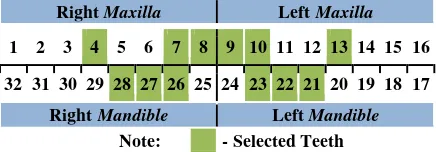
Figure 1 shows the international dental numbering system which we used as our numbering system in this paper. There are 32 teeth in adult people, sixteen teeth on each jaw. There are two jaws, maxilla and mandible. Each jaw is divided into two groups, left and right. Thus, each group consists of eight teeth comprised of two bicuspid, one cuspid, two premolar teeth, and three molar teeth. In this research we only selected maxillary central and lateral incisors, maxillary second premolar, mandibular lateral incisor, mandibular canine, mandibular first premolar (see Figure 1).
Right Maxilla Left Maxilla
1 2 3 4 5 6 7 8 9 10 11 12 13 14 15 16
32 31 30 29 28 27 26 25 24 23 22 21 20 19 18 17
Right Mandible Left Mandible Note: - Selected Teeth
After measuring manually the lengths of selected teeth, we analyze the correlation value each tooth’s length and the age. Next, we derived linear regression models using tooth lengths that were significantly correlated with age.
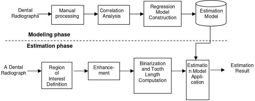
In addition, we developed an automatic system for computing tooth lengths. Given a digital panoramic radiograph, users can determine the top left and the right bottom boundary of a particular tooth. Next, the system will process this Region of Interest (RoI) into a binary image using our previous methods in [7]. Using the binary image, we can compute the tooth length using a vertical projection based method [8, 9]. Using the derived regression model, we estimate the age based on the tooth length. The design of our proposed age estimation system is as shown in Figure 2.
Figure 2. Design of the proposed age estimation system
3. Results and Analysis
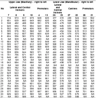
The measurements from manual observations of selected tooth length are shown in Table 1. Some images does not contain some teeth, therefore we put NA (not available) and do not take into account those teeth in our next stages.
After we measured the selected tooth length, next we calculate the correlation value between the lengths of each numbered tooth with the chronological age provided in our dataset as in Equation (1).
The correlation between selected tooth length and person’s age is as shown in Table 2. We can see that the higher correlation score is achieved by canines in mandibular (left and right) followed by premolars also in mandibular (lower jaw).
Using the highest correlation value as in Table 2, we develop a regression model to estimate age based on mandibular canine (tooth number 27) lengths. The estimation formula derived was as in Equation (2).
1
Next, we computed the estimation age using formula in Equation (2). Our results showed that the average absolute difference between the predicted and the chronological age was relatively small, i.e. 4.2 years.
Based on the derived estimation formula, we developed an automatic system able to estimate age based on a dental panoramic radiography. Firstly, the user provides top-left and bottom-right corners of a canine. Our system will automatically crop the panoramic radiographt
into a region of interest (ROI) based on the corners provided. Next, the system enhances the ROI and transforms it into a binary one. After that, the tooth length can be computed based on it’s vertical integral projection. Finally, using a particular age estimation formula, a predicted age can be computed.
Table 1. Manual measurements
No
Upper Jaw (Maxillary) - right to left Lower Jaw (Mandibular) - right to left
Lateral and Central
Table 2. Correlation Analysis Results
Tooth number Position Tooth type Number of individuals Correlation value
27 Mandibular Canine 29 -0.463376336 22 Mandibular Canine 28 -0.266467997 28 Mandibular Premolar 25 -0.193957433 21 Mandibular Premolar 23 -0.191435637 26 Mandibular Lateral incisor 28 -0.179354086 7 Maxillary Lateral incisor 25 -0.29823082 23 Mandibular Lateral incisor 30 0.12305
Figure 2. A dental panoramic radiograph
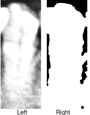
Left Right
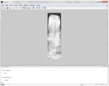
Figure 3. Left: A sample region of interest (ROI). The user chose the top-left and right-bottom corners of a canine. Right: A binary version of an ROI.
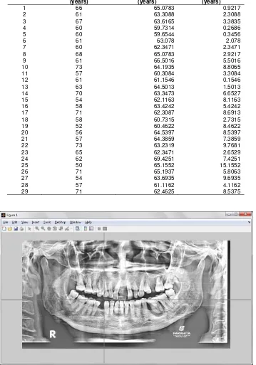
As an illustration, Fig. 2 shows a sample radiography with a known chronological age = 66. Fig. 3 (left) shows a cropped ROI, which is a mandibular canine and it’s binary version is as shown in Fig. 3 (right). The computed length was 516 pixels, resulting in an estimated age = 64.6936. Table 3 shows the results of our estimation system on our dataset. The average absolute error of our estimation system was 5.2 years.
4. Conclusion
This research uses a limited number and limited race of Indonesian individuals, i.e. Javanese people only. Therefore, in the future, the database should be added by more individuals from different races.
However, our experiments showed a promising estimation result, i.e. an average absolute error of 5.2 years, compared to application of the Kvaal method to panoramic radiographs from Turkish individuals that yields a difference of more that 12 years [6] .
Table 3. Estimation Results
No Chronological Age (years)
Estimated Age (years)
Absolute Error (years)
1 66 65.0783 0.9217
2 61 63.3088 2.3088
3 67 63.6165 3.3835
4 60 59.7314 0.2686
5 60 59.6544 0.3456
6 61 63.078 2.078
7 60 62.3471 2.3471
8 68 65.0783 2.9217
9 61 66.5016 5.5016
10 73 64.1935 8.8065
11 57 60.3084 3.3084
12 61 61.1546 0.1546
13 63 64.5013 1.5013
14 70 63.3473 6.6527
15 54 62.1163 8.1163
16 58 63.4242 5.4242
17 71 62.3087 8.6913
18 58 60.7315 2.7315
19 52 60.4622 8.4622
20 56 64.5397 8.5397
21 57 64.3859 7.3859
22 73 63.2319 9.7681
23 65 62.3471 2.6529
24 62 69.4251 7.4251
25 50 65.1552 15.1552
26 71 65.1937 5.8063
27 54 63.6935 9.6935
28 57 61.1162 4.1162
29 71 62.4625 8.5375
Figure 5. A process of defining the bottom-right corner of an ROI.
Figure 6. The output of the proposed system.
Acknowledgment
References
[1] P. Lin, Y. Lai and P. Huang. An Effective Classification and Numbering System for Dental Bitewing Radiographs using Teeth Region and Contour Information. Pattern Recognition Journal. 2010; 1380-1392.
[2] J. Zhou and M. Abdel-Mottaleb. Automatic Human Identification Based on Dental X-ray Images. The SPIE Conference on Defense and Security – Biometric Technology for Human Identification. 2004. [3] M. Abdel-Mottaleb, O. Nomir, D. Nasser, G. Fahmy and H. Ammar. Challenges of Developing an
Automated Dental Identification System. The 64th IEEE Midwest Symposium on Circuits and Systems. Cairo, Egypt. 2004.
[4] S. Singaraju, P. Sharada. Age Estimation using Pulp/Tooth Area Ratio: A Digital Image Analysis. J Forensic Dent Sci [serial online]. 2009; 37-41.
[5] M. Babshet, A. B. Acharya and V. G. Naikmasur. Age Estimation from Pulp/Tooth Area Ratio (PTR) in an Indian Sample: A Preliminary Comparison of Three Mandibular Teeth used Alone and in Combination. Journal of Forensic and Legal Medicine.2011; 350-354.
[6] H. O. Erbudak, M. Ozbek, S. Uysal and E. Karabulut. Application of Kvaal et al.’s Age Estimation Method to Panoramic Radiographs from Turkish Individuals.Forensic Sci. Int.2012.
[7] A. Yuniarti, A. Z. Arifin, B. Amaliah and A. S. Nugroho. Teeth Separation and Molar Premolar Classification on Dental Radiographs. Proceeding of the Information Systems International Conference (ISICO). Surabaya, Indonesia. 2011.
[8] A. Yuniarti, A. S. Nugroho, B. Amaliah and A. Z. Arifin. Classification and Numbering of Dental Radiographs for An Automated Human Identification System. Telkomnika Indonesian Journal of Electrical Engineering. 2012; 10(1): 137-146.
[9] Hermawati FA, Koesdijarto R. A Real-Time License Plate Detection System for Parking Access.
