Chronic thromboembolic pulmonary hypertension in young
woman with history of caesarian section
Abstrak
Hipertensi pulmonal tromboemboli kronik merupakan salah satu subgrup dari hipertensi pulmonal. Penyakit ini merupakan kondisi medis yang sangat serius dan seringkali tidak terdiagnosis. Hipertensi pulmonal tromboemboli kronik umumnya tidak terdiagnosis karena
gejala yang tidak spesiik dan kurangnya alat diagnostik.
Tujuan presentasi ini adalah untuk menjelaskan mengenai etiologi, faktor resiko, diagnosis dan manajemen hipertensi pulmonal tromboemboli kronik. Wanita berusia 36 tahun datang dengan keluhan utama mudah lelah dan sesak
bila beraktiitas sejak dua tahun yang lalu. Gejala ini
timbul sekitar tiga bulan setelah melahirkan dengan seksio caesaria atas indikasi preeklampsi. Anamnesa lebih
lanjut, pemeriksaan isik, EKG dan ekokardiograi sangat mendukung ke arah diagnosis hipertensi pulmonal. Dari
pemeriksaan sonograi vaskuler femoral tidak didapatkan adanya trombosis vena dalam (DVT). Scan perfusi paru
menunjukkan bahwa pasien ini mengalami hipertensi pulmonal tromboemboli kronik. CT pulmonary angiography
(CTPA) tidak mendapatkan adanya trombus sehingga
pasien ini tidak layak dioperasi endarterektomi pulmonal.
Pasien ini kemudian diterapi dengan vasodilator pulmonal,
ACE inhibitor, diuretik dan mendapat antikoagulan oral untuk seumur hidup.
Abstract
Chronic thromboembolic pulmonary hypertension (CTEPH) is one of subgroups of pulmonary hypertension. This is a serious medical condition that severely under diagnosed. CTEPH is commonly underdiagnosed due to
non speciic symptoms and lack of diagnostic tools. The aim of this presentation is to discuss the etiology, risk
factors, diagnosis and management of CTEPH. A 36-year-old woman presented with easily fatigue and dyspneu on effort since two years ago. The symptom occured about three months after she gave birth with caesarian
section due to preeclampsia. Further history taking,
physical examination, electrocardiography (ECG) and echocardiography were highly suggestive of pulmonary hypertension. No deep vein thrombosis (DVT) was found on vascular femoral sonography. It was found after the lung perfusion scintigraphy performed that she actually had CTEPH. This patient was categorized as inoperable because CT pulmonary angiography showed no thrombus. The patient got pulmonary vasodilator and oral anticoagulant for lifelong.
Keywords: CTEPH, CTPA, lung perfusion scan, oral anticoagulant, pulmonary hypertension
pISSN: 0853-1773 • eISSN: 2252-8083 • http://dx.doi.org/10.13181/mji.v23i4.1067 • Med J Indones. 2014;23:232-8
Correspondence author: Nitia A. Asbarinsyah, [email protected] C a s e R e p o r t
Copyright @ 2014 Authors. This is an open access article distributed under the terms of the Creative Commons Attribution-NonCommercial-ShareAlike 4.0
International License (http://creativecommons.org/licenses/by-nc-sa/4.0/), which permits unrestricted non-commercial use, distribution, and reproduction in any medium, provided the original author and source are properly cited.
Chronic thromboembolic pulmonary hypertension (CTEPH) is one of subset of pulmonary hypertension (PH). It develops when thromboembolic material obstructs pulmonary artery branches and mean
pulmonary arterial pressure (mPAP) rises to ≥ 25 mmHg. CTEPH is responsible for signiicant levels
of morbidity and mortality, especially when left untreated. Historically, 3-year mortality is reported as high as 90% in patients with a mean pulmonary artery pressure (mPAP) of > 50 mmHg.1,2
The exact prevalence of CTEPH is still unknown.
In the United States, registry data suggest that the incidence of CTEPH is between 3 and 30 per 1 million in the general population. It is estimated that an acute PE occurs in 0.5 to 0.6 million individuals each year, and CTEPH occurs in 3.8% of patients with a history of acute pulmonary embolism (PE).3,4
This igure may underestimate the true frequency of
CTEPH, as the disease is often misdiagnosed because
of nonspeciic symptoms and a variable disease
course. This is hindered by the observation that up
to 40-60% of the patients have no known history or
clinically apparent of acute pulmonary embolism episodes.3,5 In this case report, we present CTEPH
case with no history of acute pulmonary embolism episode. The aim of this presentation is to discuss
the etiology, risk factors, diagnosis and management
of CTEPH.
CASE ILLUSTRATION
A 36-year old woman presented to the outpatient clinic of National Cardiovascular Center Harapan Kita (NCCHK) with chief complaint of dypsneu on effort which occurred since almost two years ago. The symptoms started at around three months after she had caesarian section due to preeclampsia. The limitation of daily activity progressed slowly and even asymptomatic in some period, but was worsening in two months before she came to the clinic. Since the symptoms occured at the degree of her ordinary activities, she was in functional
class III of New York Heart Association (NYHA) classiication. There was no orthopneu, nor
paroxysmal nocturnal dyspneu. She had recurrent history of edema in lower feet, and non productive cough since two months ago. There was no history of syncope, chest pain or hemoptysis. She denied any routine drugs usage or history of contraceptive pill consumption. She had no history of asthma, allergy or history of previous lung infection. No history of congenital heart disease in the family.
She had history of hypertension grade I which occured since she was pregnant two years ago. No dislipidemia, diabetes mellitus, or family history were found in this patient. 3 CmH2O. Heart auscultation revealed normal irst heart sound and accentuated pulmonary component of second heart sound. No murmur nor gallop were heard. The breath sound was vesicular in both lungs, without rhales nor wheezing. Her abdomen was soft with palpable liver approximately 2 cm below costal border, and peristaltic sound was normal. Extremities were warm and there were minimal pretibial edema in both feet. No peripheral cyanosis were noted.
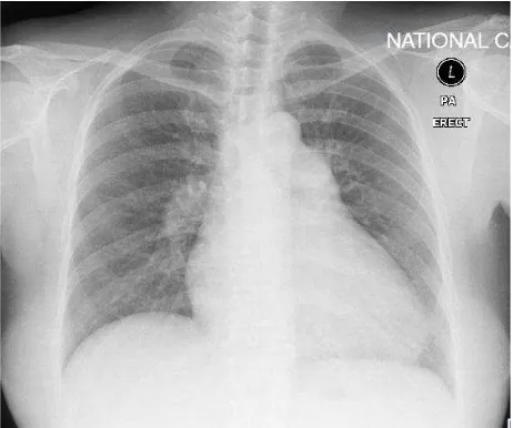
Electrocardiography (ECG) showed sinus rhythm with QRS rate of 94 beats/minute, QRS axis was 105º, with pulmonal P wave morphology. PR interval was 0.16s, QRS duration 0.08s, R wave was dominant over S wave with R/S > 1 in right precordial lead V1, and T wave inversion on the V1 – V6, I, II, III, avF, suggesting right ventricular strain (Figure 1). The conclusion of the ECG recording was sinus rhythm with right axis deviation with right ventricular hypertrophy and right atrial enlargement. Chest X-ray showed 60% of cardiothoracic ratio with normal aortic segment. The pulmonic segment was prominent with the cardiac apex pointing upward. Cardiac waist was normal, without sign of
iniltration nor congestion (Figure 2).
Figure 1. The ECG show sinus rhythm with right axis deviation, Pulmonal P wave, with R/S > 1 in lead V1 suggesting right ventricular hypertrophy, and T wave inversion on the V1 – V6, I, II, III, avF leads, suggesting right ventricular strain
Figure 2. Chest X-ray showed 60% of cardiothoracic ratio with normal aortic segment. The pulmonic segment was prominent with the cardiac apex pointing upward. Cardiac waist was nor-mal, without sign of iniltration nor congestion
Vascular femoral sonography examination revealed
normal low on arteries and veins. There were no sign of chronic venous insuficiency nor deep vein
thrombosis at both lower extremities.
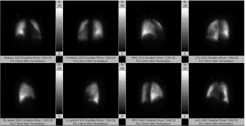
Lung perfusion scan was then performed. On
the right lung radioisotope uptake activity on the
upper and lower lobus was homogen, no segmental perfusion was detected. On the left lung, segmental perfusion defects were detected in anteromediobasal
and superior segment of lower lobus (Figure 3). It was concluded from the lung perfusion scan that there were chronic pulmonary embolism at the left lung. Chest CT scan and pulmonary angiography showed a cardiomegaly with dilated right atrial and right ventricle with enlargement of main pulmonary artery. But no thrombus on pulmonary arteries were found.
To conirm the diagnosis of pulmonary hypertension,
she underwent right heart catheterization (RHC), which showed mean pulmonary arterial pressure of 54 mmHg and there was no structural abnormality or defect of the heart.
This patient was then given sildenail 25 mg (t.i.d),
furosemide 40 mg (as needed) and Warfarin 2 mg (o.d). Two months after the treatment, her functional
capacity were improved. The result of 6 minute walk
test was 360 metres with aerobic capacity of 6.9 METS.
DISCUSSION
Etiology and risk factors of CTEPH
CTEPH occurs in about 0.5% to 4% of patients with a history of PE.1 International registry of
Figure 3. Lung perfusion scan showed segmental perfusion defects were detected in anteromediobasal and superior segment of lower lobus on the left lung
vein thrombosis.6 This is in contrasts with previous
retrospective reports indicating no history of venous thromboembolism in 40% to 60% of the patients.5 It
is possible that PE is simply undiagnosed in some patients. In the majority of patients, months or even years (the so-called “honeymoon period”) may pass
after PE before clinically signiicant pulmonary
hypertension manifests. This presumed gradual progression of pulmonary hypertension occurs in the absence of documented recurrent pulmonary
embolic events and is thought to relect progressive
remodeling of the unobstructed pulmonary
vasculature, stimulated by increased blood low
through these vessels. When there is no history of PE, the etiology is uncertain.7
In our patient, no docummented event of acute PE episode. But the symptoms started three months after she had caesarian section due to preeclampsia. Hypercoagulability during pregnancy
is associated with a 5-fold increased risk in
venous thromboembolism. Pulmonary embolism complicating pregnancy has incidence rates ranging from 0.11 to 0.73 per 1000 deliveries.8
Furthermore, women with pre-eclampsia have a
small but signiicantly higher risk of subsequent
thromboembolic disease.9 It is very likely that this
patient actually had ‘silent pulmonary embolism’ after her caesarian section and had ‘honeymoon
period’ before the irst symptom developed.
Certain medical conditions are associated with
an increased risk of CTEPH, including previous
splenectomy, the presence of a ventriculo-atrial shunt,
infected pacemaker, previous and recurrent VTE,
chronic venous ulcers, thyroid hormone replacement therapy and malignancy.10 In this patient, vascular
femoral sonography found no deep vein thrombosis (DVT) or other medical conditions associated with CTEPH. Thus, the diagnosis of CTEPH in this patient was nearly missed.
Clinical manifestations and initial non invasive assessment of CTEPH
Signs and symptoms
There are no speciic signs or symptoms for CTEPH.
The symptoms of CTEPH are indistinguishable
from other subgroups of PH, making the diagnosis
more challenging.1 Patients generally present with
progressive dyspnoea on exertion, oedema and/or signs of right heart dysfunction including fatigue, chest pain and syncope. Cardiac auscultation may reveal an accentuated pulmonic component to the second heart sound and delayed closure of the pulmonic valve (split P2). Other signs may include a palpable left parasternal heave, a right ventricular S3 or S4 gallop, prominence of the jugular A wave
or V wave, hepatojugular relux, and lower extremity
In our patient, the symptoms started to occur two years ago and progressing in two months, with worsening functional capacity and recurrent pretibial edema. There was no episode of syncope or exertional angina. At physical examination, there was an accentuated pulmonic component of second heart sound. There were an increased of jugular venous pressure and hepatomegaly in the patient. The symptoms and signs of these patient were highly suggestive of pulmonary hypertension and right heart dysfunction.
Electrocardiography
Common electrocardiogram indings on PH include
right atrial enlargement, right axis deviation, and right ventricular enlargement, often with a strain pattern.12 T wave inversion, representing the
repolarization abnormalities associ ated with right ventricular hypertrophy, is usually seen in the
anterior precordial leads and may be mistaken for
anteroseptal ischemia.11,13 ECG of our patient showed
normal sinus rhythm, right axis deviation (RAD), right atrial enlargement (RAE), and right ventricular hypertrophy (RVH) which were compatible with
ECG indings on PH. When ECG indings are
present, they may be helpful prognostically because the presence of right atrial enlargement has been
associated with a 2.8-fold greater risk of death over
a 6-year period of observation, and RVH carried a
4.3-fold greater risk of death.11 And in one study,
increased P wave amplitude was found to correlate with worse survival.12 In our case, the presences
of RAE and RVH were associated with a worse prognosis for this patient.
Chest X-ray examination
The chest X-ray can help determine the presence and
cause of pulmonary hypertension, but the indings are often nonspeciic.12 Findings on the chest radiograph
in the early stages of CTEPH may be normal, but as disease progresses, signs that suggest the presence of PH include an enlarged main and hilar pulmonary artery shadows with “pruning” or attenuation of
peripheral pulmonary vascular markings and right
ventricle (RV) enlargement would be occured. Central pulmonary artery enlargement often occurs
with more advanced disease. The lung ields may be clear, have areas of hypoperfusion (Westermark
sign), or have evidence of previous infarction (Hampton hump).14 The chest radiograph also
allows to show indings consistent with underlying
disease processes, such as hyperinlation (COPD), kyphosis (restrictive lung disease), or pulmonary
venous congestion (pulmonary venous hypertension, pulmonary veno-occlusive disease, or pulmonary capillary hemangiomatosis).12 Our patient’s chest
X-ray were supportive for the evidence of PH in advanced stage, which showed cardiomegaly with upright apex and prominence of the right heart border (sign of RV enlargement), and prominent pulmonal segment with “pruning” of pulmonary
artery. Westermark sign and Hampton hump weren’t
present. No sign of lung disease or pulmonary venous
congestion from the X-ray make the diagnosis of PH
group 3 (due to lung disease) and PH group 2 (due to left heart disease) could be excluded.
Echocardiography
Echocardiography has the advantage of detecting underlying causes or comorbidities, such as left-sided cardiac disease, valvular abnormality, chamber mass, or intracardiac shunt.15 Findings
on echocardiogram that are suggestive of elevated pulmonary pressure include enlarged right atrium and ventricle, RV hypertrophy, globally reduced RV systolic function, pulmonary artery dilatation, interatrial septal bowing to the left, and LV to appear D-shaped. Value of pulmonary systolic pressure and tricuspid regugirtation velocity could be used to determine the possibility of PH diagnosis.2,12,16
Echocardiography of the patient showed enlarged RA and RV, reduced RV contractility (TAPSE 1.6 cm), IVS paradox and LV D-shaped. PA systolic pressure > 50 mmHg (83 ± 5 mmHg) with features
suggesting of elevated pulmonary pressure make the diagnosis of PH in these patients are likely (Class I,
level of evidence (LOE) B). And in the patient, there was no any sign of left ventricular dysfunction or hypertrophy, left atrium was within normal limit and no other abnormality found in valves beside tricuspid
valve make the cause of pulmonary hypertension
due to left heart disease in this patient was excluded
inely.
Diagnostic modalities of CTEPH
algorithms have consistently recommended the use of a radionuclide ventilation/perfusion (V/Q) scan to screen for CTEPH.17 V/Q scans are particularly
useful in determining CTEPH and differentiating CTEPH from other causes of PH. V/Q scanning demonstrated a sensitivity of 90%-100% and
speciicity of 94%-100% for differentiation between
idiopathic pulmonary arterial hypertension (IPAH) and CTEPH.18 Normal V/Q scan rules out CTEPH
and perfusion scans (rather than ventilation) are abnormal in virtually all CTEPH patients.2 Recent
data showed that 98.7% of patients had abnormal perfusion scans and 19.0% had abnormal ventilation scans.1,6 Underutilization of V/Q scans in screening
PH invites potential misdiagnosis of PAH. In the recent report from the Pulmonary Arterial Hypertension Quality Enhancement Research Initiative registry in USA, 43% of PAH patients never had a V/Q scan leading up to their diagnosis.19
CT pulmonary angiography (CTPA) has the advantage of being a non-invasive, cross sectional
technique with a spatial resolution close to
conventional pulmonary angiography.20 CTPA
may reveal organised thrombi lining the proximal pulmonary vessels, abrupt tapering or amputation
of vessels or subtle intraluminal ibrous webs may be seen in CTEPH operable patients. Other indings
include dilatation of proximal pulmonary arteries and right heart chambers, scarring and a mosaic perfusion pattern.21 Contrast-enhanced CTPA may
be used as a complementary tool in CTEPH but does not replace the ventilation/perfusion scan.2
Eventhough multidetector CTPA had speciicity
of 99%, it only had sensitivity of 51% in detecting CTEPH.17 Screening PH with only CT angiogram
potentially misses CTEPH.1
Management of CTEPH
Surgical pulmonary endarterectomy (PEA) has become the gold standard for curative treatment of
proximal CTEPH, resulting in a signiicant reduction
of PA pressure and improvement in right ventricular
function, quality of life and survival.5 For our
patients, the PEA has not become a considerable option management since the location of their thrombus was not accessible from CTPA which means the thrombus are either microthrombus or located in the distal arteries.
For medical management of CTEPH, the patient should receive life-long anticoagulation (usually
with oral vitamin K antagonists adjusted to a target INR between 2.0 and 3.0) to prevent in situ pulmonary artery thrombosis and recurrent venous thromboembolism.1,2 Speciic PAH drug therapy
may still play a role in CTEPH patients, especially when the surgical therapy is not an option (Class IIb, LOE C). The reason is from the histopathological examination of distal arteries in CTEPH patients reveals vascular changes similar to those in patients with idiopathic PAH and as in PAH.17 There are
severals studies and small trials suggesting beneits of
endothelin receptor antagonists, Phosphodiesterase type 5 (PDE-5) inhibitors and prostanoid to CTEPH
patient, but the data is still limited and requires further studies. At present, no speciic medical therapy has
been widely approved for CTEPH.5,18 Our patients
got Warfarin for the oral anticoagulant, diuretics and renin angiotensin aldosterone system (RAAS)
blockers for the heart failure and PDE-5 inhibitors as
a pulmonary vasodilator. Two months after she was given the medical therapy, her functional capacity was improved from NYHA functional class III to class II. Her congestive symptoms were still occured
infrequently, and they were could be managed by
oral diuretic (Furosemide). She was planned to have re-evaluation by echocardiography six months after the medical therapy were started.
Acknowledgments
We thank to dr. Indriwanto Sakidjan, SpJP (K) as
the academic supervisor in Cardiology and Vascular Department, Faculty of Medicine, Universitas Indonesia.
REFERENCES
1. Jenkins D, Mayer E, Screaton N, Madani M. State-of-the-art chronic thromboembolic pulmonary hypertension diagnosis and management. Eur Respir Rev. 2012;21(123):32-9.
2. Galiè N, Hoeper MM, Humbert M, Torbicki A, Vachiery J-L, Barbera JA, et al. Guidelines for the diagnosis and treatment of pulmonary hypertension: the Task Force for the diagnosis and treatment of pulmonary hypertension of the European Society of Cardiology (ESC) and the European Respiratory Society (ERS), endorsed by the International Society of Heart and Lung Transplantation (ISHLT). Eur Heart J. 2009;30(20):2493-537.
3. Tanabe N, Sugiura T, Tatsumi K. Recent progress in the diagnosis and management of chronic thromboembolic pulmonary hypertension. Respir Investig. 2013;51(3):134-46.
5. Moraca RJ, Kanwar M. Chronic thromboembolic pulmonary hypertension. Heart Fail Clin. 2012;8(3):475-83.
6. Pepke-Zaba J, Delcroix M, Lang I, Mayer E, Jansa P, Ambroz D, et al. Chronic thromboembolic pulmonary hypertension (CTEPH): results from an international prospective registry. Circulation. 2011;124(18):1973-81. 7. McNeil K, Dunning J. Chronic thromboembolic pulmonary
hypertension (CTEPH). Heart. 2007;93(9):1152-8. 8. Ho YK, Wang CP, Wu YL, Lee TH, Ying TH, Lee MS.
Pulmonary embolism after cesarean section and successful treatment with early application of extracorporeal membrane oxygenation system and anticoagulant agents. Taiwan J Obstet Gynecol. 2014;53(2):273-5.
9. Van Walraven C, Mamdani M, Cohn A, Katib Y, Walker M, Rodger MA. Risk of subsequent thromboembolism for patients with pre-eclampsia. BMJ. 2003;326(7393):791-2. 10. Bonderman D, Wilkens H, Wakounig S, Schäfers H-J, Jansa
P, Lindner J, et al. Risk factors for chronic thromboembolic pulmonary hypertension. Eur Respir J. 2009;33(2):325-31. 11. Foria PR, Trow TK. Diagnosis of pulmonary arterial
hypertension. Clin Chest Med. 2013;34(4):665-81. 12. McLaughlin VV, Davis M, Cornwell W. Pulmonary arterial
hypertension. Curr Probl Cardiol. 2011;36(12):461-517. 13. Rich S. Pulmonary Hypertension. In: Bonow RO, Mann
DL, et al., editors. Braunwald’s heart disease: A textbook of cardiovascular medicine. 9th ed. Philadephia: Elsevier Saunders; 2012. p. 1696-717.
14. Marshall PS, Kerr KM, Auger WR. Chronic thromboembolic pulmonary hypertension. Clin Chest Med. 2013;34(4):779-97.
15. Frazier AA, Burke AP. The imaging of pulmonary hypertension. Semin Ultrasound CT MR. 2012;33(6):535-51.
16. Foria PR, Vachiéry JL. Echocardiography in pulmonary arterial hypertension. Am J Cardiol. 2012;110(6 Suppl):16S-24S.
17. Kim NH, Delcroix M, Jenkins DP, Channick R, Dartevelle P, Jansa P, et al. Chronic thromboembolic pulmonary hypertension. J Am Coll Cardiol. 2013;62(25Suppl): D92-9. 18. Auger WR, Kim NH, Trow TK. Chronic thromboembolic pulmonary hypertension. Clin Chest Med. 2010;31(4):741-58. 19. McLaughlin VV, Langer A, Dragomir A, Cassanova A, Tan M, Clements PJ, et al. Contemporary trends in the diagnosis and management of PAH: an initiative to close the care gap. Chest. 2010;138(4_MeetingAbstracts):351A.
20. Mehta S, Helmersen D, Provencher S, Hirani N, Rubens FD, De Perrot M, et al. Diagnostic evaluation and management of chronic thromboembolic pulmonary hypertension : a clinical practice guideline. Can Respir J. 2010;17(6):301-34.