Vol6, No I, January - March 1997 Prognostic Factor on Nasopharyngeal Cancer 45
Flow-Cytometric
DNA
Content
and
S-Phase
Fraction
Values
as a
Prognostic
Factor
on
Nasopharyngeal
cancer
-
a
preliminary
Report
S. Gondhowiardjo*, R. Susworo*, S.B. Kresno**, S.
Hartini**,
A. Roezin***, A.N. Kurniav/an****
Abstrak
Kandungan DNA inti sel dan aktivitas proliferasi sel telah diketahui berperan sebagaifaktor prognostik beberapa kanker, seperti kanker payudara, prostat, dan sentil<s utei; tetapi untuk karsinoma nasofaring (KNp), belum ai*"àn i. penelitian pendahuluan ini menggunakan pemeriksaanflow-cytometry yang relatif sederhana, cepat, Iebih akurat dan lebih objektif dibandingkan metode lainnya. Pemeriksaan terhadap 24 KNP tak terdiferensiasi dai 25 spesimen biopsi KNP memperlihatkan adanya distribusi luas nilainilai SpF (4'2% - 44'8Vo), pola ploidi (26Vo dan 74Vo untuk diploid dan aneuploid), serta indeks DNA (0.75 - 2.72). Penelitian lebih lanjut mengenai hubungan Parameter-parameter ini dengan respons radiasi, Iocal and distant control, serta angka kelangsungan-hidup Âasih berlangsung.
Abstract
It is known that the DNA content of the nuclei and proliferation activity can sewe as useful prognostic factors in a variety of neoplasms, such as cancer of the breast, prostate, and cervical uterine. However, there is still n-o ciear data aiout this co*elation in the nasopharyngeal cancer (NPC). Flow-cytometric analysis provides an accurate, more objective, rapid and relatively simple examination,comparedwithothertechniques,Ourpreliminaryresults of 24undifferentiatedcarcinomaoutof25NpCbiopsyspecimens demonstrated that there were wide distributions of SPF value (4.2% - 44.8 Vo), ploidy pattern (26Vo vs 74Vo for diploid an-d aneuploid respectively), and DNA index value (0.75-2.72). Further study analyzing the responsibility aU of those parameters to the radiation response, local and distant control, and sumival rate
is
still underway.Keyworils : DNA content, proliferation activity, DNA index, ploidy panern and S-phase fraction, pCNA, KI-67.
The prognostic indicators
of
head and neck tumors arelargely
determinedby
the combination
of
mor-phologic-diagnosis and clinical stage of the tumor.2'4In
fact,
despite the improvementin
surgical and radiotherapy techniques combinedwith
chemothe-raphy, these tumors continue to have a fatal outcomeTransformation of normal cells into neoplastic growth subsequently
is
associated with changesin
cellbe-havior.'
Cell kinetics, as one aspectof
the cellularbehavior, can be represented by several proliferation parameters such as thymidine labeling index (TLI), cytometric or flow-cytometric DNA content and S-phase fraction (SPF) values, T potential doubling time
(T pot) by
the bromo-deoxyuridine (thymidine
analog), proliferatingcell
nuclear anrigen (PCNA), and also Ki 67 fractions.r-) \Vhereby, cancer treatment modalities such as chemo and radio-therapies are potentially.only effective in a certain cellular prolifera-tion phase.oIn Indonesia, nasopharyngeal cancer (NpC) is the most frequently found malignancy in head and neck region. Moreover, it was the third highest from all malignant diseases treated by radiation. Most of the NpC patients are males within the productive ages, who seek medi-cal help at the locally advanced stage, which is well-known to have a high incidence of local failure after treatment, which subsequently causes mortality.T'8
Surgical intervention of the NpC is hampered by the difficulties of the tumor localization.n-, I
ùor"ou".,
up D e p a rt m enl s of Radi o t he rapy*, C l i ni c al p at ho lo p y* *Ea r - N o s e and Th roa t*' ", Anato mic al p a I ho lo gyr* * *, Faculty of Medicine, University of Indonesia./
46
Gondhowiardio et aLto now, chemotherapy does not show a good reslrlt-' Thus, radiation remains the treatment of choice'g'I1
However, in order to increase the radiation result, a lot
of efforts are still needed, such as changes in fractiona-tion method of irradiation which should
be
based on cellular proliferation activity'9Since there is no clarification concerning the role of cell kinetic and DNA content as predictor of radiation
response in the NPC, we conducted an investigation
to
ixp\ore
the po\enilù rD\Èbl
$ese ptrtrse\ersrs
relation to the existing malignancy indicators'
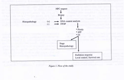
The first part
of
this studywill
be dealing with the relationship between proliferation activity and DNAcontent values yield through flow-cytometric analysis,
i.e., the S phase fraction (SPF) value, DNA index and ploidy pattern of the NPC cells as compared to WHO histo-pathologic grading. The second part
will
high-light the relationship of these values as compared tothe clinical radiation response, local control and sur-vival of the patients in order to establish the role of this parameters as a prognostic indicator (Figure 1)'
METHODS
Biopsy specimens obtained
from
suspected NPCpatients were directly placed in Ep-pendorf tube
con-iisting TOVo alcohol and stored in 4oC until processed'
Med J Indones
Specimens were
fragm
or
en-zymatically bY using
1
ma, St. Louis, USA) to forma
After centrifuging the suspension at 1.500 rpm for 5 minutes at 40 C and removal of the supernatant, the suspension was washed twice with 1 ml phosphat buffered salinesolution. Then, it was incubated with 1
ml
propidium iodide (PI, Sigma, St. Louis, USA) working solutionfor 30 minutes. The PI working solution was prepared
earlier, by mixing 10 ml PBS solution with 300
pl PI
stsckss\utiq\
t
stgRl(&ase (Sigma. St. Louis ', USA)and25
pl
Triton X-100.The cell suspension was filtered with 40 micron nylon mesh prior to flow cytometric analysis. The automatic
measurement of the SPF value, DNA index and ploidy
pattem were done by using the "cell fit program" from the Beckton-Dickinson flow-cyto-meter facs scan.
SPF value indicated the number
of
cellular fractions during the S phase within the population of tumor cells.The ploidy pattern consisted of an-euploid, diploid, and tetra-ploid. An-euploid pattern was judged when the abnormal DNA content was distributed beyond the
l\Vo
deviation-rangeof
the DNA content within thenormal cells. Abnormal DNA content deviation-range which was located two times beyond the normal value was defined as tetra-ploid. The ratio between the
ab-normal DNA content of the tumor as compared to the
normal cell was named DNA index.
Histopathology (+) C)
NPC suspect
t
Biopsy +
+
DNA content analYsis+
STOPI
* Ploidy
* SPF
*DI
Stage Histopathology
[image:2.595.60.563.462.785.2]Radiation response Local control, Survival rate'
Vol 6, No l, January - March 1997
Histopathologic data \Mere obtained from the medical records and were done by the Department
of
Pathol-ogy, Faculty of Medicine, University of Indonesia. No effort was done to re-evaluate the histopathologicdiag-nosis by the respective pathologist.
Statistical description and analysis
The frequency distribution and other descriptive
statis-tics (mean, median, range and SD) were calculated
with the SPSS program, after all the SPF, DNA index
values, ploidy pattern and the histologic pattern of the
NPC were compiled into a computerized data file.
Later on all those variables means and the end point values
of
this study such as the radiation response,local and distant control, and 2 years survival rate were tested using the two tailed student-t or chi-square test.
The linear correlation between bi-parameter variables
was estimated by Pearson product limit test.
RESULTS
DNA content
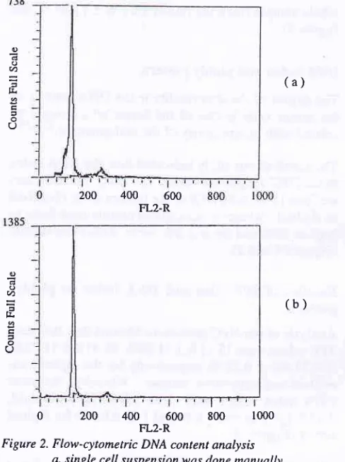
Figure 2 shows the FCM DNA content analysis
his-togram from the alcohol fixed specimen biopsy of NPC patients, which is separated according to method of
making the single cell suspension, manually
or
en-zimatically. [t seems that no different histograms rilere presented using these two kinds of technique.
Histopathologic diagnosis
There were 25 biopsy NPC specimens available for
this study.
The
average age of the patients was 41.6years (ranged between 12-65 years), 687o of them were
male. Twenty three out
of
the 25 NPC patients wereclassified as stage
fV (AJC^ICC
592), and the resrof them were recurrent cases.
One out
of
the25
specimens was morphologicallyjudged as squamous cell carcinoma (SCC) type, while the rest were classified as undifferentiated cancer
(WHO histopathological classification).
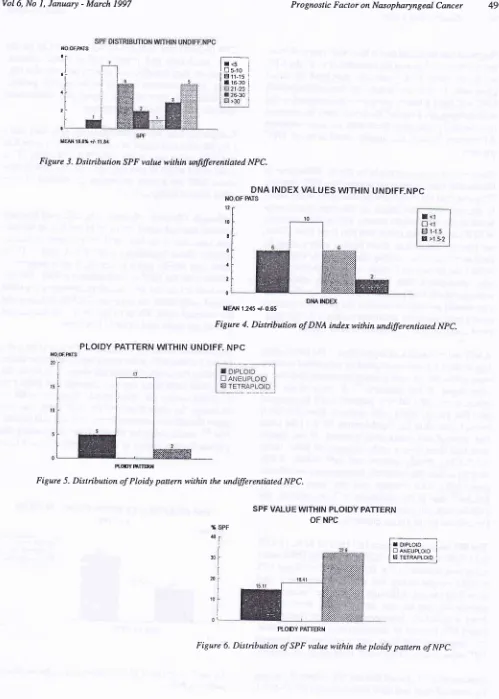
SPF values
Since clinically undifferentiated NPC has shown a
wide variation in treatment out-come, we examined
the SPF value as the simplest objective proliferation
parameter. Our study demonstrated that the SPF values
were
distributed
between 4.2Vo-44.8Vo from thePrognostic Factor on Nasopharyngeal
Cancer
47(a)
400
600FL2.R
(b)
[image:3.595.313.554.80.745.2]o
2oo
ooo.rr-f, 8oo
loooFigure 2. Flow-cytometric DNA content analysis
a. single cell suspensionwas done manually
b. single cell suspension was done enzimatically
Table I
.
Result of flow-cytometric DNA content analysis on 25 ofNPC patients
No Stage Histopathology SPF (7o) DI PLOIDI
o d o (t) a h a o (J 200 o o v) h c
,
o OI
Residive2
tln3m0 (lV)3
tln3mO (IV)4
t4n3m0 (lV)5
t2n3m0 (IV)6
t4n3m0 (IV)7
t3n3m0 (IV)8
t4n3m0 (IV)9
t3n3m0 (IV)l0
tln2mo (lv) II
t2n2m0 (IV)12
tln3m0 (lV)l3
t4n3m0 (lV)14
rln2m0 (lV)l5
t4nlm0 (IV)l6
r4n tm0 (IV)l7
t4n2m0 (lV)l8
tln2m0 (IV)l9
t2n2m0 (lV)20
rln3mo (lv)2l
Residive22
tln2m0 (lV)23
t2n2m0 (lV)24
rln3m0 (lV)25
t2n2m0 (lV)undiff.ca undiff.ca undiff.ca undiff.ca undiff.ca undiff.ca undiff.ca undiff.ca undiff.ca undiff.ca undiff.ca undiff.ca
undiff ca
undiff ca
undiff ca
undiff ca
undiff ca
undiff ca
undiff ca
undiff ca
undiff ca
scc undiff ca
undiff ca
undiff ca
1.72
al.l2
al.a2
d0.75
at.05
d1.08
dl.l7
a1.30
^0.82
at.06
d1.,10
al.l0
a1.70
a1.96
r1.86
a2.22
a2-72
a2.m
t0.75
a1.03
d1.65
a0.70
a0.70
aO.75
r
1.33
a25.4 04.2 05.4 26.1 06.5 17.7 t4.3 19.2 05.3
r r.5
08.8 08.6 25.4 32.8 09.0 l0
r 3.8
[image:3.595.314.558.84.411.2]48
Gondhowinrdio et al.whole sample fractions (mean 18.7 Vo + 11.84 Vo, sae
Figure 3).
DNA index and PloiilY Pattern
The degree of the abnormality in the DNA content of
the tumor cells is one of the factor which might be
related with progressivity of the malignan"i",'2I2'13
The result of our study indicated that the DNA index of theNPC rangedbetween
0.75'2.72'
Furthermore, we found that only 247o of thetumors were classified as diploid, whereby an-euploid tumors contribute as highàs 68Vo and the rest 8Vo werc tetra-ploid tumors(Figures 4 and 5).
Relation of SPF vatue and
DNA
indexto
ploidypattern
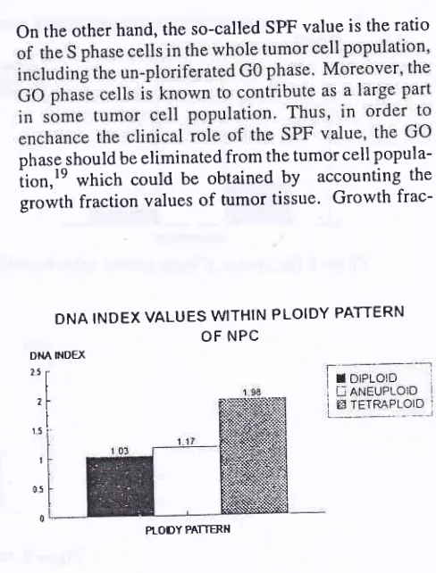
Analysis of our NPC specimens showed that the mean SPF values were 15.1 l%o + ll.86Vo,l8.4l7o + ll'727o, and 32.67o + 0.28 Eo respectively for the diploid, an-euploid and tetra-ploid tumors. Whereby, the mean
DNA index values
were
1.98 + 0.02 for tetra-ploid,l.l7 +
0.75 for aneuploid and 1.03t
0.049 for diploid tumor (Figure 6).Although
it
is not yet clear, evidence indicated thatdiploid pattern and a lower SPF value were related to a better prognosis of the tumor'2'12'13
DISCUSSION
The methods of cell proliferation kinetic assessment
are usually carried out by the following procedures: auto-radiographic determination
of
thethymidine-labelled cells, estimation of the DNA contents
distribu-tion through cytometric or flow-cytometric machine and detection
of
bromo-deoxyuridine-labelled cellsusing monoclonal antibody through immuno-histo-cherii stry method or fl o*-rytometrlc analysi s. 3-5
Flow-cytometric DNA contents analysis provides the
simplest technique with more quantitative, objective,
and
rapid
measurementsof
the cyto-kinetic
or proliferation ratesof
the tumor as compared to theother cellular proliferation examination measure-ments. This machine-dependent, automatic cellular
assessment involving a large number of cells, thus,
it
will
subsequently raise the accuracy of themeasure-ment and the objectivity of the analysis' Moreover,
it
also serves double purpose, namely to detect thenon-ploid tumor stem-lines and to assess their proliferation Ly *"asuring the SPF uul l"'3'4'l 3 This information has
Med
I
Indonesbeen proven to be useful in supplementing the clinical and pathologic classification
'
of several malignantdis-13-14
-eases.Recently, a technique to obtain a single cell suspen-sions from paraffin embedded tissue, formaldehyde or
alcohol fixed specimens was described and proved to
be successfully effective in examining the DNA cgnl tents value thiough flow-cytometric âalysis.13'15-18
This method certainly open a chance for an
examina-tion
of
various typesof malignancies
in
eitherretrospective or in prospective ways.
Accumulating evidence supports the claim that
detec-tion
of
Gl DNA content in tumor cells, in most cases,is
a reliable markerof
malignancy. Moreover, the proportionof DNA
abnormality (DNA Index) and, ôr DNA proliferation activities (SPF) might reflectthe degree
of
malignancy and its biological be-havior. l;5' I 3' I 5In this present study, we used the prospective study to
analyze DNA content value and proliferative activity of NPC in relation to the treatment outcome.
The NPC single cell suspension was made from aq
alcohol fixed biopsy
,p""i'*"nr,
while Johnson et al.l3used paraffin-block specimens for various epidermoid head and neck tumor. However, our results were
rela-tively similar in view
of
mean and distribution ranges (18.7V", 4.2-44.8Vo versus l9%o, 4-45Vo and 1.2,0'75'2.72 versus 1.42,0.''t-3'5 respectively for SPF value
and DNA
index).
Moreover, this SPF values were higher than those observed in the other malignancies, such as breast, lung, uterine, cervical and colon(12-l57o). Thus, we were confined that our techniques were reliable.
SPF values indicate the proportion
of
within the tissue which is actively synthesizing its DNA, and could bethought to be the proliferation status of the tumor.
Although microscopic-based morphology detection
and classification of malignant diseases have
under-gone progressive development in order to achieve ac-curate diasnosis and sub-classification
of
human n"oplusrn,1: and cellular differentiation orhisto-mor-phology
of
the tumors have long been regarded as animportant factor related to the natural history and treat-,nÀt ."rponse,l8 there has been uncertainty toward its reliability. This is particularly true when it is used to
predict some biological behaviors such as growth or
r{o-oF.mTs
ti
I
I i-I I .i
I r I
2l
Vol6, No l, January - March 1997 Prognostic Factor on Nasopharyngeal Cancer 49
l-..r
I!trro
II gii-15 !
[image:5.595.54.553.44.743.2]l!razo i
Figure 3. Dsitribution SPF value within unfiferentiated NPC.
DNA INDEX VALUES WTHIN UNDIFF.NPC
12 t0
6
,t
2
0
IEAN 12,15 .,r- 0.65
Figure 4. Distibution of DNA indexwithin undifferentiated NpC.
PLOIDY PATTERN WITHIN UNDIFF. NPC
R(X)YffTEruI
Figure 5. Distribution of Ploidy pattern within the undifferentiated NPC.
SPF VALUE WITHIN PLOIDY PATTERN
E;'t-rl
I D ereuproro I
ljJgrilggi
PLODY PATTERN
Figure 6. Distibution of SPF value within the ploidy pattern of NpC.
NO-OF mrs
Dt{AINDE(
OF NPC
,10
30
20
10
0
50 Gondhowiardjo et al.
diasnosis can be associated with a wide range of
treat-r"it
out"orn"r.4 A unique characteristicof
the NPCbehavior,
whichclinicians,
is
thatNPC
will
have a b well differentiatedis not entirely true, since there were radiation response differences among the similar conditions
of
NPCpatients.
This latter observation might be due
to
differences inthe cellular kinetic behavior amongst the NPC patients' This was confirmed with the present study which
clear-reflect the behavior
of
tumor cells' Moreover' it wasalso
anticipatedthat
these v indicator in selected Patients wh tion should be used or whethernee d a more energetic trsatment such as adjuvant
treat-ments, and the use
of
radio-sensitizer agents'9'16'17A SPF value which was higher than20Vo, DNA index higher than 1.5 and an-euploid or tetra-ploid patterns
(0.15-2.72), Ploidy pattern and SPF values (4'2Vo-44.87o) within the relatively homogenous undifferen-tiated NPC. This concept has also been noted by
Dischel8 that in the squamous cell carcinomas, the
cellular differentiation observed is quite unrelated to
the cellular proliferation characteristics'
The SPF value means were I 5. I I Vo
!
| |'86Vo, l8'417o+ ll.72Vo
and32.67o + 0.28Vo' while the DNA index value means were 1 .03+
0.049,l.l7 +
0'75 and l '98+
0.02 respectively
for
diploid,
aneuploid and tetraploid tumors.A
not Yet statisticallyproven,
derivedfrom a reiativelY
1't
ifferen-tiated NPC) seems
to
demonstrate a possibletenden-cy,
suggested that there was a relationship between SPF vaiue and the DNA index with the ploidy pattern'Johnson et all3 reported that the SPF values
of
variousepidermoid head and neck tumors were 797o (5-40Vo)'
Med J Indones
20lo (4-38Vo) and26Vo (I3-45Vo) respectively for the
well,
moderate andpoorly diffrentiated
tumors'Moreover, they found no differences in view of malig-nancy grade between diploid or un-diploid pattern' Unfortunately, they did not report.any correlation analysis with the clinical outcomes.''
Comparing these two studies, particularly in the poorly
1un;-àlffrentiated tumor group' it was clearly seen that
àur SPF value was lower (18.7Vo,4'2-443Vo)' This
differences might in part explain
why
theundifferen-tiated NPC has a better prognosis as compared to the other type of malignancies.
Although Chauvell showed that
cell
proliferationkinetics-showed significant role in predicting the
dis-ease outcome in the head and neck cancer, however' whether those hypothesis (higher SPF value, DNA index and ploidy pattern) are valid as a prognostic indicator foi the NPC are still unknown' Furthermore'
it is also not
Yet
is the mostrelated to the
loc
iseases andthe survival
rate
the secondpart of our study could clarify this issue'
On the other hand, the so-called SPF value is the ratio
ohase should be eliminated from the tumor cell
popula-iion,te which could be obtained
by
accounting thegrowth fraction values of tumor tissue' Growth
frac-DNA INDEX VALUES WITHIN PLOIDY PATTERN OF NPC
Dt{Â FIDEX 25 r
t
zl
,rl
I 05
[image:6.595.315.559.376.697.2]0
Figure 7. Distîributin
of
DNA index value within the ploidypattent og NPC.
Vol6, No I, January - March 1997
tion value which can be measured by Ki67 and PCNA
techniques recognizes
all
phasesof
the cell cycles, except the GO p-ha5e.3'a'20'ltWe propose the name
of
thisnew
value
as theproliferating SPF (p-SPF) value, which measures only
the SPF value of the proliferating cell phase (
Gl,
S,G2 and M). Further investigation to prove whether this p-SPF value has a greater correlation to the radiation
response in a clinical setting is underway.
REFERENCES
l. Brooks DJ, Garewal HS. Measures of tumor proliferative activity. Int J Clin Lab Res 1992;22:196-200.
2. Naus GJ, Zimmerman RL. Prognostic value of flow cytometric DNA content analysis in single treatment stage
IB-IIB squamous cell carcinoma of the cervix. Gynecol Oncol 1991;43:149-53.
3. Chauvel P, Courdi A, Gioanni J, Vallicioni J, Santini, Demard F. The labelling index: a prognostic factor in head and neck carcinoma. Radiother Oncol 1989;.14.,231-7.
4. Larsen JK. General review cell proliferation: Analysis by flow-cytometry. Nouv Rev Fr Hematol 1992;34:317-35. 5. Tubiana M, Courdi A. Cell Proliferation kinetics in human
solid tumors: relation to probability of metastatic dissemina-tion and long-term survival. Radiother 1989;15:l-8. 6. Withers HR. Biologic basis of radiation therapy. In: Perez
AC, Brady LW, eds. Principles and practice of radiation oncology. Phi ladelphi a: JB Li ppincott Co, 1992; 64-9 6. 7. Susworo. Kombinasi radiasi ekstema dan intrakaviter:
alnatif pengobatan karsinoma nasofaring yang responsif ter-hadap radiasi. Disertasi. Jakarta: Universitas Indonesia,
1990.
8. Dir-Jen Pelayanan Medik-Departemen Kesehatan R.I., Badan Registrasi Kanker Ikatan Ahli Patologi Indonesia, Yayasan Kanker Indonesia. Data histopatologik kanker di Indonesia tahun 1989.
PrognosticFactoronNasopharyngealCancer 5l
9.Perez CA. Nasopharynx. In: Perez CA, Brady LW, eds. Principles and practice of radiation oncology. philadelphia: JB Lippincott co, 1992:617 43.
10. Rahima M, Rakoowsky E,Bara;ilay J, Sidi J. Carsinoma of
the nasopharynx. Cancer 1986;58:843-9.
11. Pee ZE, Gun LP, Long CK. Radiation therapy of
nasopharyngeal cancer: prognostic factors based a lO-year follow-up of 1302 patients. Int I Radiat Oncol Biol physiol
I 989; I 6:301-5.
I 2. Dressler LG, Larry MA, Seamer MT, Owens M A, Clark GM, Mc Gure WL. DNA flow cytometry and prognostic factors
in
1331 frozen breast cancer specimens. Cancer1988;61:420-7
-13. Johnson TS, Williamson KD, Cramer MM, Peters LL Flow cytometric analysis of head and neck carcinoma DNA index
and S-fraction from parafin-embedded sections; comparison with mali gnancy grading. Cytometry I 985 ;6:46 I -70.
14. Rew DA. Clinical applications of low cytometry. BrJ Hosp
Md
1992;48:171-5.15. Hedley DW. Flow cytometry using parafin-embedded
tis-sue: five years on cytometry 19891'10:229-41.
16. Begg AC, Hofland I, Moonen L, et al. The predictive value of cell kinetic measurement in European trial of accelerated
fractionation in advanced head and neck carcinoma: an
in-terim report. Int J Radiat Oncol Biol Physiol. 1990;19:1449-53.
17. Begg AC. Critical apraisal of insitu cell kinetic measurement as response predictors in human tumors. In: Duran RE, ed.
Cell kinetic-aplications to cancer therâpy. Semin Radiat Oncol 1993;3:144-51.
18. Dische S, Saunders MI, Bennet MH, Wilson GD, McNally NJ. Cell proliferation and differentiation in squamous cell
cancer. Radiother Oncol 1989;5: l9-23.
19. Nakano T, Oka K. Differential values of Ki-67 index and
mitotic index of proliferating cell population. Cancer 1993;72:2401-8.
20. Allegranza A, Girlando S, Arrigoni GL, et al. Proliferating
cell nuclear antigen expression in central nervous systems neoplasms. Virchows Arch (A) 199 I :419 :417 -23.