Vol. 17 No. 1, p 31-37 EISSN: 2086-4094
Inhibition of Urokinase-Type Plasminogen Activator Expression by
Macelignan in
Porphyromonas gingivalis
Supernatant-Induced
Human Oral Epithelial Cells
YANTI
School of Biotechnology, Atma Jaya Catholic University, Jalan Jenderal Sudirman 51, Jakarta 12930, Indonesia
Phone: +62-21-5703306 ext 335, Fax: +62-21-5719060, E-mail: [email protected]
Received December 1, 2009/Accepted Februari 22, 2010
This study was to investigate the effect of macelignan on Porphyromonas gingivalis supernatant-induced uPA expression via regulating mitogen-activated protein kinase (MAPK) and activating protein-1 (AP-1) signaling pathways in human oral epithelial KB cells using casein zymography, Western blotting, reverse transcription-PCR and reporter gene assays. Zymographic analysis of secreted enzymes identified the main caseinolytic band at 54 kDa. Macelignan inhibited the expression of uPA protein and mRNA, as well uPA secretion, in KB cells exposed to P. gingivalis supernatant. Consistent with these findings, macelignan suppressed phosphorylation of p38 and c-Jun N terminal kinase (JNK) in P. gingivalis supernatant-induced KB cells. The levels of c-Fos and phosphorylated c-Jun, which together form AP-1, the transcription factor that is involved in uPA gene expression, were partially reduced by macelignan. Macelignan also blocked P. gingivalis supernatant-induced AP-1 activity in these cells. These results suggest that macelignan decreased P. gingivalis supernatant-induced uPA expression by blocking AP-1 activity, which may be mediated by inhibition of phosphorylation of p38 and JNK in KB cells. Macelignan may potently use for the modulation of periodontal inflammation.
Key words: macelignan, Myristica fragrans, urokinase plasminogen activator, Porphyromonas gingivalis, human epithelial cells
___________________________________________________________________________
http://journal.ipb.ac.id/index.php/hayati DOI: 10.4308/hjb.17.1.31
INTRODUCTION
Nutmeg (Myristica fragrans Houtt.), a perennial herb native to Indonesia and to the Molucca islands, in particular, has traditionally been used for culinary and medicinal purposes (Janssens et al. 1990; Olajide et al. 1999; Sonavane et al. 2002; Narasimhan & Dhake 2006; Cho et al. 2007). Macelignan is a bioactive compound with molecular weight of 328 present in nutmeg and has been reported to possess antioxidant, anti-inflammatory, anti-diabetic, hepatoprotective and skin-whitening properties (Jin et al. 2005; Cho et al. 2008; Han et al. 2008; Sohn et al. 2008). Our previous studies demonstrated that macelignan possesses anticariogenic and anti-biofilm activities that are effective against oral pathogens such as Streptococcus mutans, S. sanguinis, and Actinomyces viscosus (Chung et al. 2006; Yanti et al. 2008), suggesting potential application of macelignan in treatment of dental plaque. During progression of periodontal disease, plaque accumulation in response to bacterial products triggers the host cells to release inflammatory mediators and leads to the breakdown of extracellular matrix. The process of connective tissue breakdown is facilitated by the action of serine proteinases, such as matrix metalloproteinases (MMPs) and plasminogen activators (PAs).
The plasminogen/plasmin system plays a central role in extracellular proteolysis by facilitating connective tissue breakdown, which leads to inflammation. Two PAs, i.e. urokinase-type plasminogen activator (uPA) and
tissue-type plasminogen activator (tPA), convert plasminogen into plasmin; plasmin is another serine protease that degrades fibrin and activates MMPs (Andreasen et al. 1997). uPA is a 54 kDa serine protease and is highly expressed in inflamed and healing cells in response to cytokines and bacterial products. Several studies have demonstrated that regulation of uPA expression in gingival fibroblasts is tightly controlled by the growth factors epidermal growth factor (EGF) and transforming growth factor (TGF)-â1, the cytokine interleukin-1â and the bacterial product Porphyromonas gingivalis
lipopolysaccharide (LPS) (Ogura et al. 1999, 2001; Smith
et al. 2004; Smith & Martinez 2006).
Identification of a potent natural inhibitor of uPA has been the subject of a great deal of research interest because of the potential benefits for periodontal therapy. To date, only a few bioactive compounds that exhibit an inhibitory effect on uPA expression and low toxicity to normal cells have been identified. Genistein and curcumin suppress EGF-induced uPA expression in human gingival fibroblasts (Smith et al. 2004), strongly suggesting that these compounds will be useful in the control of periodontal inflammation. Also, epigallocatechin-3-gallate (EGCG) from green tea has been shown to have an effect on uPA expression in human oral cancers and fibrosarcomas (Kim
expression in human oral epithelial KB cells exposed to
P. gingivalis supernatant in vitro. The molecular mechanism by which macelignan modulates the relevant signaling molecules, i.e., mitogen-activated protein kinase (MAPK) pathway and activating protein-1 (AP-1), involved in uPA expression in P. gingivalis supernatant-induced KB cells was also investigated.
MATERIALS AND METHODS
Isolation of Macelignan. The dried seed kernels of
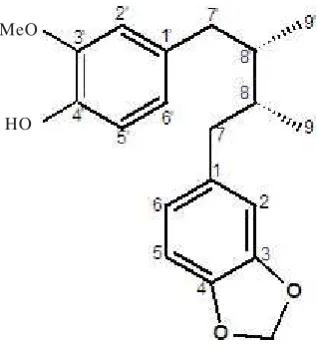
M. fragrans Houtt. (Myristicaceae) were collected in Jakarta, Indonesia, and identified at Department of Oriental Medicinal Materials and Processing, Kyunghee University (Yongin, Korea). A voucher specimen (H010) is deposited in the Department of Biotechnology, Yonsei University (Seoul, Korea). The dried seed kernel of M. fragrans Houtt. (100 g) was extracted with 100% ethanol (400 ml), and the extract (7 g) was further fractionated with ethyl acetate. The ethyl acetate fraction (4.2 g) was applied to a silica gel column (60, 70-230 mesh, Merck) and eluted with n-hexane and ethyl acetate solution (10:1, v/v) to give six fractions (FI to FVI). FIII was further separated with n-hexane and ethyl acetate solution (20:1, v/v), yielding FIII-B (0.52 g). FIII-B was eluted with 80% methanol using Rp-18 column chromatography (LiChropep, 25-40 ìm, Merck), yielding compound III-B-2 (0.5 g). Comparison of several spectral data of compound III-B-2 including 13C-NMR, 1H-NMR, 13C-DEPT, 1H-1H COSY, 1H-13C HSQC, 1H-13C HMBC, and FAB-MS with that in the literature (Woo et al. 1987) suggested the chemical structure to be macelignan 99% (Figure 1) or (8R,8S )7( 3 , 4 m e t h y l e n e d i o x y p h e n y l ) 7 )7( 4 h y d r o x y 3 -methoxyphenyl)-8,8-dimethylbutane.
Instrumentation. NMR spectra were recorded on a Bruker Avance-500 spectrometer at 600 MHz for 1H- and 13C-NMR in CDCl
3 with TMS as an internal standard. Complete proton and carbon assignments were based on 1D (1H-, 13C-, 13C-DEPT) and 2D (1H-1H COSY, 1H-13C HSQC, 1H-13C HMBC) NMR experiments. Mass spectra (FAB-MS) were measured using a JMS-700. All instrumental spectra are available upon request.
Cell Culture and Cell Viability. KB cells were cultured in a 5% CO2 atmosphere at 37 oC in Dulbecco’s Modified Eagle’s Medium (DMEM; Gibco) supplemented with 100 U/ml of penicillin A, 100 U/ml of streptomycin and 10% heat-inactivated fetal bovine serum (FBS). The cells were seeded at a concentration of 2 x 10⁵ cells/ml per 75 cm2 flask and cultured for 24 hours. Confluent cells were detached by trypsinizing for 2 minutes and aliquots of separated cells were subcultured. The effects of
P. gingivalis supernatant and macelignan on cell viability were evaluated with an MTT (3-(4,5-dimethyl-2-thiazolyl)-2,5-diphenyl-2H-tetrazolium bromide; Sigma-Aldrich) colorimetric assay according to the method of Mosmann (1983).
Bacterial Supernatant and Sample Treatment. P.
gingivalis supernatant was prepared as described by Chang et al. (2004) with a slight modification. Cells were seeded at a concentration of 2 × 10⁵ cells/ml in 6-well plates and cultured for 24 hours in DMEM-FBS. After washing twice with Dulbecco’s phosphate-buffered saline (DPBS), the cells were incubated in serum free-DMEM without P. gingivalis supernatant (negative control group), with 10% P. gingivalis supernatant (positive control group) or with 10% P. gingivalis supernatant plus various concentrations of macelignan or MAPK inhibitors (U0126, SB203580, and SP600125) or uPA inhibitor (amiloride) or MMP inhibitor (GM6001). The cellular lysates were also collected for this experiment.
Casein Zymography. Secretion of uPA in the conditioned medium was measured by casein zymography (Bodet et al. 2007). Briefly, the conditioned media from the negative control, positive control, and treatment group (macelignan) were collected and subjected to electrophoresis with 10% SDS polyacrylamide gels containing 0.5% skim milk and human plasminogen 2 mg/ml. uPA was detected at 54 kDa as clear zones against the dark background. Effects of MAPK inhibitors (U0126, SB203580, and SP600125), uPA inhibitor (amiloride) and MMP inhibitor (GM6001) on uPA secretion in the conditioned media were also determined by casein zymography.
Reverse Transcription-PCR. uPA mRNA in the cellular lysates was determined by RT-PCR. Total RNAs from cellular lysates of negative control, positive control and treatment group (macelignan) were extracted as previously described (Yanti et al. 2009). The human oligonucleotide primers for uPA and glyceraldehyde 3-phosphate dehydrogenase (GAPDH) were designed according to a PCR primer selection program at the website of the Virtual Genomic Center from the GenBank database. uPA primers were designed as 5’CTGCCTGCCCTGGAACTCTG3’ for forward and 5’CCTTGCGTGTTGGAGTTAAG3’ for reserve. GAPDH primers were 5’ATTGTTGCCATCAATG ACCC3’ for forward and 5’AGTAGAGGCAGGGATGAT3’ for reverse. PCR consisted of 35 amplification cycles and each cycle was carried out for 30 seconds at 94 oC, 30 seconds at annealing temperature (56 oC for uPA and 48 oC for GAPDH) and 1 minute at 72 oC in a thermal cycler (Gene Amp PCR System 2700). The human GAPDH MeO
HO
housekeeping gene was used as an internal control to standardize the relative expression levels for uPA. PCR products were separated electrophoretically in a 2% agarose DNA gel and stained with ethidium bromide.
Western Blotting. To determine uPA protein expression, the conditioned media from the negative control, positive control, and treatment group (macelignan) were concentrated with Fast-Con Protein Concentration kit and subjected to Western blotting. To determine the expression of MAPK phosphorylation, c-Jun phosphorylation and c-Fos expression, cellular lysates from the negative control, positive control and treatment group (macelignan) were prepared and assayed by Western blot analysis. Proteins (50 µg) were resolved by 10% SDS-PAGE and transferred to nitrocellulose transfer membranes. The membranes were blocked with 5% skim milk for 1 hour at room temperature and then probed with the following primary antibodies: anti-rabbit polyclonal uPA, anti-mouse monoclonal p-ERK1/2, anti-mouse monoclonal p-p38, mouse monoclonal p-JNK, anti-mouse monoclonal c-Jun or anti-rabbit polyclonal c-Fos at a 1:1000 dilution overnight at 4 oC. After three washes, the blots were subsequently incubated with the secondary antibody peroxidase-conjugated mouse IgG or anti-rabbit IgG at a 1:4000 dilution for 2 hours at room temperature. The blots were stained with SuperSignal West Femto Maximum sensitivity substrate (Thermo Scientific) and visualized using a LAS 3000 Bio Imaging Analysis System. Equal loading of blots was demonstrated by stripping blots and reprobing with antibodies for anti-rabbit polyclonal ERK1/2, anti-rabbit polyclonal p38, anti-rabbit polyclonal JNK, anti-rabbit polyclonal c-Jun or anti-mouse monoclonal α-tubulin.
Reporter Gene Assay. Cells were seeded at a concentration of 1 × 10⁶ cells/ml in 6-well plates and cultured in DMEM-FBS for 6 hours at 37 oC in a 5% CO
2 atmosphere prior to the experiments. For transient transfection, a 2.0 µg AP-1 luciferase reporter plasmid was mixed with Lipofectamine reagent in 100 µl of serum-free DMEM. A 0.25 µg of â-galactosidase plasmid was used as the internal control. Cells were incubated for 5 hours. For luciferase assays, cells were washed twice with DPBS after 5 hours of transfection, and treated with various doses of macelignan (5, 10, and 25 µM), amiloride (25 µM), and GM6001 (25 µM) for 48 hours. Cell lysates were collected and luciferase activity was tested using a luciferase assay system according to the manufacturer’s instructions. Firefly luciferase activities were standarized for â -galactosidase activity.
Statistical Analysis. Triplicate experiments were performed throughout this study. All data are presented as the mean + standard deviation (SD). The significance of differences between control and treated groups were statistically analyzed by the paired Student’s t-test.
RESULTS
Cytotoxicity in KB Cells Treated with P. gingivalis
Supernatant and Macelignan. KB cells were found to
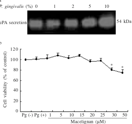
produce primarily uPA and secretion of uPA was upregulated by exposure to P. gingivalis supernatant for 48 hours in culture. At a concentration of 10%, P. gingivalis supernatant optimally increased the level of uPA expression compared to expression in unstimulated cells (Figure 2a) but had no effect on cell viability. Macelignan treatment up to a concentration of 25 ìM had no effect on the viability of either untreated or P. gingivalis supernatant-treated KB cells, suggesting that the inhibitory effect of macelignan on uPA expression was not attributable to cytotoxicity (Figure 2b).
Macelignan Inhibited the Expression of uPA Secretion, Protein, and mRNA Induced by P. gingivalis Supernatant. The primary band detected by casein zymography in conditioned medium of untreated and P. gingivalis
supernatant-treated KB cell cultures had a molecular weight of 54 kDa and was identified as uPA (Figure 3). Addition of 10% P. gingivalis supernatant to the culture medium optimally upregulated uPA secretion compared to secretion in control cells. As determined by casein zymography and Western blotting, treatment with 2-25 ì M macelignan inhibited uPA secretion and protein expression in P. gingivalis supernatant-treated KB cells in a dose-dependent manner (Figure 3a). At the level of the gene, 10% P. gingivalis supernatant also induced the expression of uPA mRNA in KB cells. The level of uPA mRNA was effectively decreased after treatment with macelignan (Figure 3a). We also investigated the effects of amiloride and GM6001, inhibitors of uPA, and MMP, respectively. The degree of suppression of uPA secretion by amiloride (2-25 ìM) was equivalent to that of macelignan in P. gingivalis supernatant-induced KB cells (Figure 3b). In contrast, GM6001 had no effect on uPA secretion in P. gingivalis supernatant-treated KB cells (Figure 3c).
P. gingivalis (%)
uPA secretion 54 kDa
0 1 2 5 10 a
b 1 2 0
1 0 0
80
60
40
20
0
Pg (-) Pg (+) 1 5 10 15 20 25 30 50
Cell viability (% of control)
* *
Macelignan (µM)
Figure 2. (a) Dose-dependent effect of P. gingivalis supernatant
on uPA secretion in KB cells assayed by casein
zymography. (b) Effect of P. gingivalis and macelignan
on KB cell viability. Values represent the mean + SD of
triplicate experiments. * indicates P < 0.05 against P.
Macelignan Decreased MAPK Phosphorylation Induced by P. gingivalis Supernatant. Inhibition of uPA expression via MAPK signaling pathways in P. gingivalis
supernatant-treated KB cells was first determined by casein zymography of conditioned medium from cells that had been cultured in the presence of the specific MAPK inhibitors U0126, SB203580, and SP600125 at a concentration of 10 ìM. Figure 4a shows that the p38 inhibitor SB203580 and the JNK inhibitor SP600125 significantly reduced uPA secretion in P. gingivalis
supernatant-treated KB cells. We then carried out Western analysis to determine whether macelignan interfered with MAPK signaling pathway-mediated uPA expression in P. gingivalis supernatant-induced KB cells (Figure 4b). Exposure to P. gingivalis supernatant increased the activation of MAPKs ERK1/2, p38 and JNK by phosphorylation. Macelignan treatment decreased the levels of phosphorylated p38 and JNK in P. gingivalis
supernatant-induced KB cells in a dose-dependent manner comparable to that seen following treatment with the specific MAPK inhibitors U0126, SB203580, and SP600125. In contrast, macelignan exhibited a lesser inhibitory effect on ERK1/2 phosphorylation than on phosphorylation of p38 and JNK.
Macelignan Suppressed c-Jun Phosphorylation and c-Fos Expression Induced by P. gingivalis Supernatant. Because the JNK and p38 signaling pathways have been associated with the transcription factors c-Jun and c-Fos, the effect of P. gingivalis supernatant on c-Jun phosphorylation and c-Fos expression in KB cells was evaluated. Phosphorylation of c-Jun and expression of c-Fos were upregulated by P. gingivalis supernatant treatment
in KB cells compared to control cells. Macelignan treatment resulted in a partial reduction in c-Jun phosphorylation and c-Fos expression in these cells (Figure 4c).
Macelignan Blocked Transcription Factor AP-1 Activity Induced by P. gingivalis Supernatant. AP-1, a transcription factor involved in uPA gene expression, is composed of homo- and heterodimers of c-Jun and c-Fos. The effect of P. gingivalis supernatant on activation of AP-1 in KB cells was further tested using a luciferase assay. Figure 5 shows that exposure of KB cells to P. gingivalis supernatant increased AP-1 activation by P. gingivalis (10%)
54 kDa
Macelignan (µM)
uPA secretion
uPA protein
uPA mRNA
GAPDH mRNA
0 0 2 5 10 25 - + + + + +
54 kDa
480 bp
565 bp a
0 0 2 5 10 25 - + + + + + b
P. gingivalis (10%)
Amiloride (µM)
uPA secretion 54 kDa
0 0 2 5 10 25 - + + + + + c
P. gingivalis (10%)
GM6001 (µM)
uPA secretion 54 kDa
Figure 3. (a) Effect of macelignan on the expression of uPA
secretion, protein, and mRNA in P. gingivalis
supernatant-induced KB cells assayed by casein zymography, Western blotting and RT-PCR. (b-c) Effects of amiloride and GM6001 on uPA secretion in P. gingivalis supernatant-induced KB cells assayed by casein zymography.
P. gingivalis (10%) - + + + +
54 kDa
U0126 (10 µM) +
-SB203580 (10 µM) +
-SP600125 (10 µM) - - - - +
uPA secretion a
P. gingivalis (10%) - + + + + +
Macelignan (µM) 0 0 0 5 10 25
U0126 (10 µM) +
-p-ERK1/2
ERK1/2 b
SB203580 (10 µM) +
-p-p38
p 3 8
SP600125 (10 µM) +
-p-JNK
JNK
P. gingivalis (10%) - + + + + +
Macelignan (µM) 0 0 5 10 25
p-c-Jun
c-Jun
c-Fos
α-Tubulin
c
Figure 4. (a) Effect of MAPK inhibitors on uPA secretion in P.
gingivalis supernatant-induced KB cells assayed by casein
zymography. (b) Effect of macelignan on P. gingivalis
supernatant-induced activation of MAPK signaling pathways in KB cells assayed by Western blotting. (c)
Effect of macelignan on P. gingivalis
approximately 3.2-fold over untreated cells. Macelignan blocked AP-1 activity in a dose-dependent manner in treated KB cells, and at the highest concentration (25 ìM), the inhibitory activity of macelignan was greater than that of the uPA inhibitor amiloride and the MMP inhibitor GM6001.
DISCUSSION
Periodontal disease is marked by alternating periods of extracellular matrix tissue breakdown and specifically indicated by an increase in the expression of PAs, including uPA. The uPA system also includes its endogenous inhibitors, plasminogen activator inhibitor (PAI) types 1 and 2. Imbalance in the activities of uPA and PAIs has been shown to contribute to the destruction of periodontal connective tissue (Xiao et al. 2001). Smith and Martinez (2006) reported the differential expression of uPA in healthy and diseased human gingival tissues, suggesting that uPA might play a key role in the process of periodontal inflammation. Regulation of uPA expression is an important strategy in prevention and treatment of periodontal inflammation; however, the molecular mechanisms controlling this process are not fully understood. An important area of periodontal research involves the quest for potent agents that will control inflammation with little or no toxicity to normal cells and investigation of the mechanisms by which these agents regulate uPA expression.
We previously reported that macelignan exerts strong antibacterial and antibiofilm activities that are effective against periodontopathogens, suggesting the potential use of macelignan as an antiplaque agent (Chung et al. 2006; Yanti et al. 2008) and in periodontal therapy. Here,
the inhibitory effect of macelignan on uPA expression and its associated signaling pathways in P. gingivalis
supernatant-induced KB cells was clearly defined. Our results showed that uPA production was upregulated by exposure of KB cells to P. gingivalis supernatant for 48 hours in culture (Figure 2a). P. gingivalis, a gram-negative black-pigmented anaerobic bacterium, is recognized as the main oral pathogen participating in dental plaque accumulation. In periodontal disease, bacterial products trigger the host cells to secrete inflammatory mediators such as cytokines, prostaglandin and MMPs, and lead to extracellular tissue breakdown (Schwartz et al. 1997). P. gingivalis culture supernatant contains virulence factors such as gingipain, LPS and fimbriae, and induces the expression of MMPs (MMP-1, MMP-2, and MMP-9) and PAs (uPA and tPA) in various periodontal cell types (Chang et al. 2002a,b, 2004, 2006; Yang et al. 2003; Smith
et al. 2004; Smith & Martinez 2006). Our results demonstrated that P. gingivalis supernatant treatment resulted in upregulation of uPA secretion, protein and mRNA in KB cells; treatment with macelignan resulted in a dose-dependent decrease in these parameters (Figure 3a). The effect of macelignan on uPA inhibition is very similar to that of the uPA inhibitor amiloride (Figure 3b). One group has reported that P. gingivalis LPS stimulates uPA expression in human gingival fibroblasts (Ogura et al. 1999); however, it is not known which components of
P. gingivalis supernatant share this activity with LPS and how the effect is mediated in other periodontal cell types, including KB cells.
The MAPK signaling pathways, ERK1/2, p38 and JNK, have been associated with regulation of uPA gene expression, although the profiles of MAPK activation appear to vary in a cell type-dependent manner (Ward et al. 2001; Adeyinka et al. 2002). Considering that MAPK regulation of uPA expression may play a role in periodontal inflammation, we investigated the ability of macelignan to interrupt MAPK signaling pathways involved in uPA expression in P. gingivalis supernatant-treated KB cells. Casein zymographic analysis demonstrated that the p38 inhibitor SB203580 and the JNK inhibitor SP600125 significantly reduced uPA secretion induced by P. gingivalis supernatant in KB cells (Figure 4a); this data suggests that the p38 and JNK signaling pathways are involved in uPA expression. Smith et al. (2004) reported that inhibitors of ERK1/2 and JNK are involved in EGF-induced uPA expression in human gingival fibroblasts. Consistent with the results of casein zymography, macelignan was found to reduce the levels of p38 and JNK phosphorylation in KB cells exposed to P. gingivalis
supernatant (Figure 4b). This finding suggests that macelignan downregulates P. gingivalis supernatant-induced uPA gene expression by blocking the activation of p38 and JNK phosphorylation in KB cells. Bioactive compounds such as curcumin and genistein have been reported to modulate MAPK signaling pathway-mediated uPA expression in human gingival fibroblasts in response to EGF (Smith et al. 2004). In addition, EGCG derived from green tea also alters the production of uPA in human P. gingivalis (10%) - + + + + + +
Figure 5.Effect of macelignan on AP-1 activity induced by P.
gingivalis supernatant in KB cells determined by luciferase assay. Values represent the mean + SD of triplicate experiments. # indicates P < 0.01 against
untreated cells. *and ** indicate P < 0.05 and P < 0.01
fibrosarcoma cells through ERK1/2 and JNK phosphorylation (Kim et al. 2004). These studies show that the higher levels of uPA expression were associated with an imbalance in the levels of uPA and its endogenous inhibitors (PAIs), causing extracellular matrix breakdown, marked periodontal inflammation and cancer progression. A major role of MAPKs is regulation of transcription factor activity and transmission of extracellular signals to the nucleus, where target gene expression is induced. The JNK and p38 signaling pathways have been associated with induction of transcription factors, including phosphorylation of c-Jun and increasing the expression level of c-Fos; these events lead to activation of the AP-1 complex, the transcription factor responsible for induction of uPA expression (Rao 2003). Because the JNK and p38 signaling pathways involved in uPA expression were significantly inhibited by macelignan, we investigated the effect of macelignan on the phosphorylation of c-Jun and expression of c-Fos induced by P. gingivalis supernatant treatment of KB cells.
P. gingivalis supernatant upregulated MAPK activation, followed by an increase in the phosphorylation of c-Jun and expression of c-Fos, which comprise the AP-1 complex (Figures 4b,c). These results imply that P. gingivalis
supernatant-induced uPA expression in KB cells is tightly regulated at the transcriptional level and that MAPK signaling pathways are critically involved in uPA expression in these cells. These results are in agreement with those of previous studies (Miralles et al. 1998; Parra
et al. 2000; Shin et al. 2003).
It has been clearly shown that macelignan decreased the levels of c-Jun phosphorylation and c-Fos expression (Figure 4c) and blocked AP-1 activity (Figure 5). Because the AP-1 complex is comprised of c-Jun and c-Fos, the significant effect of macelignan was linearly correlated with a reduction in AP-1 activity. AP-1-regulated uPA transcription is dependent not only on the proportion of c-Jun and c-Fos in the complex, but also on the levels of c-Jun phosphorylation and c-Fos expression (Waskiewicz & Cooper 1995). Therefore, macelignan alters uPA gene expression in P. gingivalis supernatant-induced KB cells by partially decreasing c-Jun phosphorylation, c-Fos expression and AP-1 activity. Several studies aimed at investigating the molecular mechanisms underlying regulation of uPA expression by natural agents in relation to periodontal inflammation are currently underway. However, simple comparisons are difficult to make due to differences in the cellular sources of uPA, differences in culture conditions, the nature of the compounds under investigation and the concentrations used, and the signaling pathways targeted by the agents. Although studies using natural agents such as curcumin, genistein and EGCG have been reported (Kim et al. 2004; Smith et al. 2004), these studies focused primarily on the role of signal transduction in uPA protein expression and not on events at the transcriptional level.
In summary, our results strongly suggest that macelignan suppresses expression of uPA induced by P. gingivalis supernatant by blocking c-Jun phosphorylation,
c-Fos expression and AP-1 activity which may be facilitated by the decreased levels of p38 and JNK phosphorylation in KB cells. Macelignan may be considered a potent candidate for use as a preventive agent in the control of periodontal inflammation in oral care functional foods.
ACKNOWLEDGEMENT
This work was supported in part by Yonsei Biomolecule Research Initiative of the two-step Brain Korea 21 Project. I thank Jae-Kwan Hwang at Laboratory of Natural Products and Biomaterials, Department of Biotechnology, Yonsei University, Seoul (Korea) for his kind assistance.
REFERENCES
Adeyinka A, Nui Y, Cherlet T, Snell L, Watson PH, Murphy LC. 2002. Activated mitogen-activated protein kinase expression during human breast tumorigenesis and breast cancer
progression. Clin Cancer Res 8:1747-1753.
Andreasen PA, Kjoller L, Christensen L, Duffy MJ. 1997. The urokinase-type plasminogen activator system in cancer
metastasis: A review. Int J Cancer 72:1-22.
Bodet C, Andrian E, Tanabe S, Grenier D. 2007. Actinobacillus
actinomycetemcomitans lipopolysaccharide regulates matrix metalloproteinase, and plasminogen activator production by human gingival fibroblasts: A potential role in connective
tissue destruction. J Cell Physiol 212:189-194.
Chang YC, Chu SC, Yang SF, Hsieh YS, Yang LC, Huang FM. 2004. Examination of the signal transduction pathways leading to activation of gelatinolytic activity by interleukin-1á and Porphyromonas gingivalis in human osteosarcoma cells. J Periodont Res 39:168-174.
Chang YC, Ho YC, Chou LS, Huang FM. 2006. Signal transduction pathways involved in the stimulation of tissue type plasminogen activator by interleukin-1alpha and Porphyromonas gingivalis in human osteosarcoma cells. J Periodont Res 41:374-380.
Chang YC, Lai CC, Yang SF, Chan Y, Hsieh YS. 2002b. Stimulation
of matrix metalloproteinases by black-pigmented Bacteriocides
in human pulp and periodontal ligament cell cultures. J Endod
28:90-93.
Chang YC, Yang SF, Lai CC, Liu JY, Hsieh YS. 2002a. Regulation of matrix metalloproteinase production by cytokines, pharmacological agents and periodontal pathogens in human
periodontal ligament fibroblast cultures. J Periodont Res
37:196-203.
Cho JY, Choi GJ, Son SW, Jang KS, Lim HK, Lee SO, Sung ND, Cho KY, Kim JC. 2007. Isolation and antifungal activity of
lignans from Myristica fragrans against various plant
pathogenic fungi. Pest Manag Sci 63:935-940.
Cho Y, Kim KH, Shim JS, Hwang JK. 2008. Inhibitory effects of
macelignan isolated from Myristica fragrans Houtt. on melanin
biosynthesis. Biol Pharm Bull 31:986-989.
Chung JY, Choo JH, Lee MH, Hwang JK. 2006. Anticariogenic
activity of macelignan isolated from Myristica fragrans
(nutmeg) against Streptococcus mutans. Phytomedicine
13:261-266.
Han KL, Choi JS, Lee JY, Song J, Joe MK, Jung MH, Hwang JK. 2008. Therapeutic potential of peroxisome proliferators-activated receptor-alpha/gamma dual agonist with alleviation of endoplasmic reticulum stress for the treatment of diabetes. Diabetes 57:737-745.
Ho YC, Yang SF, Peng CY, Chou MY, Chang YC. 2007. Epigallocatechin-3-gallate inhibits the invasion of human oral cancer cells and decreases the productions of matrix
metalloproteinases and urokinase-plasminogen activator. J
Janssens J, Laekeman GM, Pieters LA, Totte J, Herman AG, Vlietinck AJ. 1990. Nutmeg oil: identification and quantification of its most active constituents as inhibitors of
platelet aggregation. J Ethnopharmacol 29:179-188.
Jin DQ, Lim CS, Hwang JK, Ha I, Han JS. 2005. Anti-oxidant and anti-inflammatory activities of macelignan in murine hippocampal cell line and primary culture of rat microglial
cells. Biochem Biophys Res Commun 331:1264-1269.
Kim MH, Jung MA, Hwang YS, Jeong M, Kim SM, Ahn SJ, Shin BA, Ahn BW, Jung YD. 2004. Regulation of urokinase plasminogen activator by epigallocatechin-3-gallate in human
fibrosarcoma cells. Eur J Pharmacol 487:1-6.
Miralles F, Parra M, Caelles C, Nagamine Y, Felez J, Munoz-Canoves P. 1998. UV irradiation induces the murine urokinase-type plasminogen activator gene via the c-Jun N-terminal kinase signaling pathway: Requirement of an AP-1 enhancer
element. Mol Cell Biol 18:4537-4547.
Mosmann T. 1983. Rapid colorimetric assay for cellular growth and survival: Application to proliferation and cytotoxicity
assays. J Immunol Methods 65:55-63.
Narasimhan B, Dhake AS. 2006. Antibacterial principle from Myristica fragrans seeds. J Med Food 9:395-399.
Ogura N, Nagura H, Abiko Y. 1999. Increase of urokinase-type plasminogen activator receptor expression in human gingival
fibroblasts by Porphyromonas gingivalis lipopolysaccharide.
J Periodontol 70:402-408.
Ogura N, Tobe M, Tamaki H, Nagura H, Abiko Y. 2001. IL-1beta increases uPA and uPA receptor expression in human gingival
fibroblasts. IUBMB Life 51:381-385.
Olajide OA, Ajayi FF, Ekhelar AI, Awe SO, Makinde JM, Alada AR.
1999. Biological effects of Myristica fragrans (nutmeg)
extract. Phytother Res 13:344-345.
Parra M, Lluis F, Miralles F, Caelles C, Munoz-Canovez P. 2000. The c-Jun N-terminal kinase (JNK) signaling pathway mediates induction of urokinase-type plasminogen activator
(uPA) by the alkylating agent MNNG. Blood 96:1415-1424.
Rao JS. 2003. Molecular mechanisms of glioma invasiveness: The
role of proteases. Nat Rev Cancer 3:489-501.
Schwartz Z, Goultschin J, Dean DD, Boyan BD. 1997. Mechanisms
of alveolar bone destruction in peridontitis. Periodontol
2000?? 14:158-172.
Shin BA, Yoo HG, Kim HS, Kim MH, Hwang YS, Chay KO, Lee KY, Ahn BW, Jung YD. 2003. p38 MAPK pathway is involved in the urokinase plasminogen activator expression in human
gastric SNU-638 cells. Oncol Rep 10:1467-1471.
Smith PC, Martinez J. 2006. Differential uPA expression by
TGF-beta1 in gingival fibroblasts. J Dent Res 85:150-155.
Smith PC, Santibanez JF, Morales JP, Martinez J. 2004. Epidermal growth factor stimulates urokinase-type plasminogen activator expression in human gingival fibroblasts. Possible modulation
by genistein and curcumin. J Periodont Res 39:380-387.
Sohn JH, Han KL, Kim JH, Rukayadi Y. Hwang JK. 2008. Protective effects of macelignan on cisplatin-induced
hepatotoxicity is associated with JNK activation. Biol Pharm
Bull 31:273-277.
Sonavane GS, Sarveiya VP, Kasture VS, Kasture SB. 2002.
Anxiogenic activity of Myristica fragrans seeds. Pharmacol
Biochem Behav 71:239-244.
Ward Y, Wang W, Woodhouse E, Linnoila I, Liotta L, Kelly K. 2001. Signal pathways which promote invasion and metastasis: Critical and distinct contributions of extracellular signal-regulated kinase and Ral-specific guanine exchange factor
pathways. Mol Cell Biol 21:5958-5969.
Waskiewicz AJ, Cooper JA. 1995. Mitogen and stress response pathways: MAP kinase cascades and phosphatase regulation
in mammals and yeast. Curr Opini Cell Biol 7:798-805.
Woo WS, Shin KH, Wagner H, Lotter H. 1987. The structure of
macelignan from Myristica fragrans. Phytochemistry
26:1542-1543.
Xiao Y, Bunn CL, Bartold PM. 2001. Effect of lipopolysaccharide from periodontal pathogens on the production of tissue plasminogen activator and plasminogen activator inhibitor 2
by human gingival fibroblasts. J Periodont Res 36:25-31.
Yang SF, Hsieh YS, Huang FM, Yang LC, Chang YC. 2003. Effect of black-pigmented bacteria on the plasminogen-plasmin
system in human pulp and osteoblastic cells. Oral Surg Oral
Med Oral Pathol Oral Radiol Endod 95:621-625.
Yanti, Oh HI, Anggakusuma, Hwang JK. 2009. Effects of
panduratin A isolated from Kaempferia pandurata Roxb. on
the expression of matrix metalloproteinase-9 by Porphyromonas
gingivalis supernatant-induced KB cells. Biol Pharm Bull 32:110-115.
Yanti, Rukayadi Y, Kim KH, Hwang JK. 2008. In vitro anti-biofilm
activity of macelignan isolated from Myristica fragrans Houtt.
against oral primary colonizer bacteria. Phytother Res