Natalia, et al.
Pediatric myocarditis and cardiac magnetic resonance
174 Med J Indones, Vol. 23, No. 3,
August 2014
Clinical presentation of myocarditis in pediatric: the role of
cardiac magnetic resonance in diagnosis
Abstrak
Miokarditis merupakan suatu penyakit inlamasi miokard dengan presentasi klinis dan onset yang bervariasi. Kesulitan dalam diagnosis seringkali menyebabkan pasien yang datang dalam kondisi akut tidak terdiagnosis sebagai miokarditis, lalu mendapatkan penanganan yang tidak tepat. Hal ini akan membahayakan karena menyebabkan cedera miokard lebih parah, bahkan dapat menyebabkan kematian mendadak. Di lain sisi, pasien dapat pula datang setelah lanjut, dalam kondisi kardiomiopati dilatasi (DCM) atau kardiomiopati aritmogenik (ARVD). Oleh karena itu diagnosis miokarditis ini menjadi hal yang sangat penting. Kasus ini menunjukkan bahwa cardiac magnetic resonance
(CMR) merupakan alat diagnostik non-invasif yang penting dalam mendiagnosis miokarditis dengan keakuratan yang tinggi pada seorang anak laki-laki dengan keluhan nyeri dada. Pemeriksaan CMR ini menunjukkan morfologi jantung yang normal, fungsi ejeksi ventrikel kanan dan kiri yang normal, gambaran edema miokard dan jaringan ibrosis yang mengkonirmasi diagnosis miokarditis sesuai dengan kriteria konsensus Lake Louise. CMR merupakan alat diagnostik non-invasif yang penting dalam mendiagnosis miokarditis dengan keakuratan yang tinggi.
Abstract
Myocarditis is an inlammatory disease of myocard with varied onset and clinical presentation which lead to diagnosis dificulties. These dificulties often cause incoming patients with acute condition are not diagnosed as acute myocarditis that subsequently may lead to improper therapy. This circumstance will probably cause more severe myocardial injury and even sudden death. On another circumstance, patients may also come late in conditions of dilated cardiomyopathy (DCM) or aritmogenik cardiomyopathy (ARVD) due to complexities of myocarditis early detection. Therefore, myocarditis diagnosis is extremely important. This case shows how CMR is important and highly accurate in diagnosing myocarditis through a case whereby a boy had complaints of chest pain and his CMR examination showed normal cardiac morphology with normal function of right and left ventricular, but with myocardial edema and tissue ibrosis, which conirmed the diagnosis of myocarditis according to the Lake Louise Consensus criteria. CMR is an important and high accuracy non-invasive diagnostic tool in myocarditis diagnosis.
Keywords: cardiac magnetic resonance, myocarditis, myocardial oedema
pISSN: 0853-1773 • eISSN: 2252-8083 • http://dx.doi.org/10.13181/mji.v23i3.654 • Med J Indones. 2014;23:174-81 Correspondence author: Sisca Natalia, [email protected]
C a s e R e p o r t
Copyright @ 2014 Authors. This is an open access article distributed under the terms of the Creative Commons Attribution-NonCommercial-ShareAlike 4.0 International License (http://creativecommons.org/licenses/by-nc-sa/4.0/), which permits unrestricted non-commercial use, distribution, and reproduction in
any medium, provided the original author and source are properly cited.
Sisca Natalia, Octavia Lilyasari, Sony H. Wicaksono, Manoefris Kasim
Department of Cardiology and Vascular Medicine, Faculty of Medicine, Universitas Indonesia/National Cardiovascular Center Harapan Kita, Jakarta, Indonesia
Myocarditis is an inlammation of the myocardium caused by nonspeciic response to various triggers such as viral or bacterial infections, cardiotoxic agents, infarction, or mechanical injury. Most of these etiologic factors may already be evident from
the patient’s history.1 However, diagnosing this
disease appears to be dificult due to heterogeneity of presentations ranging from asymptomatic to severely symptomatic. These symptoms include clinical heart failure and ventricular dilatation, fulminant heart failure and severe left ventricular dysfunction, with
or without cardiac dilatation, and a recent lulike syndrome accompanied by fever, arthralgias, and
malaise.2
There are various ways to conirm those diagnoses such as clinical examination, pathological examination, or by using diagnostic criteria which are presented in the forthcoming parts. Furthermore, this paper elaborates and focuses on cardiac magnetic resonance (CMR) examination
Case illustration
A 10 year-old boy, came to the National Cardiac Centre Harapan Kita (NCCHK) Hospital and had already had complaints of repeated chest pain since one day before admission. The pain was severe and located in the middle of chest which caused dificulty in breathing. He initially went to another hospital, pursued electrocardiogram (ECG) test and
eventually was diagnosed with heart attack. Later on, he was given 2 tablets salicylic acid and 2 tablets
of clopidogrel, and referred to the NCCHK Hospital. The patient also complained of fever, without any
cough, cold, or diarrhea. Additionally, he had
maternal history of normal childbirth and growth as well. At the emergency room of NCCHK, the physical examination was within normal limits, no abnormality was found.
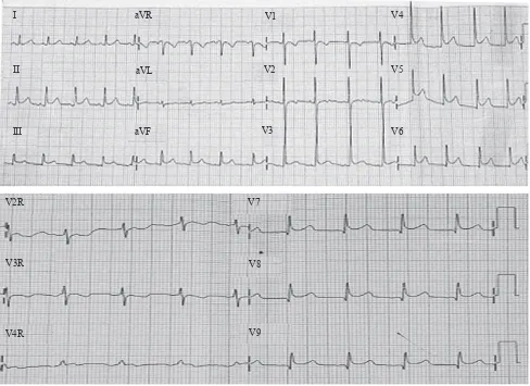
ECG showed following characteristics: sinus rhythm (SR), QRS rate 101x/m, 30°QRS axis, normal P wave, PR interval of 0.08”, QRS duration of 0.04’’, ST segment elevation V3-V9, II, III, aVF, R/S in
Figure 1. ECG at emergency room NCCHK hospital
V1 < 1, R/S in V6 > 1. Some additional laboratory results were normal, while AST (79 U/L), ALT (26 U/L), CKMB (134 u/L), and hsTrop T (1029 ng/L) were increasing. Furthermore, chest X-rays showed normal heart and lungs. Echocardiographic examination showed normal cardiac morphology, with normal left ventricle global contractility (EF 65%), and normal RV contractility (TAPSE 2.3 cm) as well. There was no atrial septal defect (ASD), no ventricular septal defect (VSD) and no persistent ductus arteriosus (PDA). MSCT examination
showed normal coronary artery, no stenosis and no coronary anomaly.
According to CMR, the morphology were: situs soli
-tus, atrioventricular (AV) concordance, ventriculoarte
-rial (VA) concordance, all pulmonary veins (PV) to left atrial (LA), no ASD, no VSD, no PDA, all valves are normal, normal left aortic arch, normal coronary artery; ventricle volume, function and low measurement: left ventricle ESV/EDV/EF: 19.4/71.7/72.9%, right ven
-tricle ESV/EDV/EF : 33.7/65.8/48.7%, stroke volume aorta 67 mL, pulmonal artery 65.1 mL; T2WSTIR:
V3R
V4R
V2R V7
V8
V9 I
II
III
aVR
aVL
aVF
V1
V2
V3
V4
V5
Natalia, et al.
Pediatric myocarditis and cardiac magnetic resonance
176 Med J Indones, Vol. 23, No. 3,
August 2014
Figure 2. (A, B, C, D) normal cardiac morphology from different window. Situs solitus with concordance atrio-ventricle and ventri-culo-arterial. No shunt ASD, VSD nor PDA. All valves are normal; (E) myocardial edema at septal and inferolateral wall; (F, G) ibrotic tissue at mid wall in septal and inferolateral, basal anterior wall; (H) normal coronary artery with left aortic arch; (I) normal thoracic aorta, without aneurysm, coarctation or dissection; (J) normal cali -ber of pulmonary artery
myocardial edema at septal and inferolateral wall; late enhancement: ibrotic tissue at mid wall in septal and inferolateral, basal anterior wall; MRA: the thoracic
aorta is normal, without aneurysm, coarctation or
disec-tion. The pulmonary artery is normal in caliber. There are four pulmonary veins that enter the left atrium. LVOT: left ventricle outlow tract (Figure 2).
On the second day of treatment, CMR was performed,
that showed normal cardiac morphology, with tissue
edema and ibrosis in the mid area of the myocardial wall, as sign of myocarditis.
DISCUSSION
Myocarditis
Myocarditis is clinically and pathologically
deined as myocardial inlammation, which is an important cause of myocardial diseases such as
dilated cardiomyopathy and arrhythmogenic right
ventricular cardiomyopathy. Until recently, the classiication, diagnosis and therapy of myocarditis
were still debatable.3 Incidence of myocarditis
in young adults with sudden death reached 12%. Non-fatal incidence is likely higher than actually diagnosed due to dificulty of establishing the
diagnosis in standard clinical settings. 4
There are 3 phases in the development of enteroviral myocarditis in experimental animals .5 Virus entry into
myocytes mediated by a speciic receptor coxsackie virus group B and a common transmembrane receptor (coxsackie virus and adenovirus receptor [CAR]) on some adenovirus genome for viral internalization
into myocytes.6 Coxsackie virus using delecting
decayaccelerating factor (DAF) and adenoviruses special integrins (αvβ3 and αvβ5) as co-receptors. If CAR is absent in the cardiac myocytes, virus infection and inlammation will not occur.7 Acute
injury to heart muscle cells occurs after viral entry,
which is subsequently induced by virus replication.8
Acute phase of myocarditis occurred for just a few days, followed by a subacute phase characterized by an immune reaction. This phase can occur several weeks to several months, characterized by activation of virus-speciic T lymphocytes, which can attack the organs of the patient because of the similarity of molecules. Activation of cytokines (tumor necrosis factor - alpha [TNF-α]), interleukin [IL] -1 and -6), viral protein of the heart can aggravate heart damage and result in impaired contractility function.8
Several diagnostic modalities: challenges in the diagnosis of myocarditis
One major challenge in diagnosing myocarditis
is requirement for various data derived from anamnesis, physical examination, and non-invasive examination which potentially have different
A B
C D
H
I J
E F
Natalia, et al. Pediatric myocarditis and cardiac magnetic resonance 177 Med J Indones, Vol. 23, No. 3,
August 2014
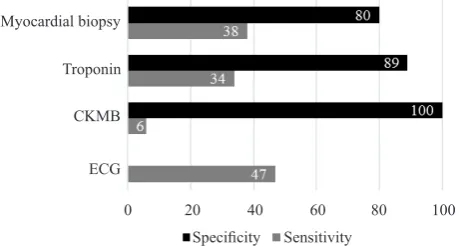
Figure 3. Proportional eficacy of some examinations for myo -carditis with various range of sensitivity and speciicity. The speciicity of ECG itself is unknown, according to this report. Modiied from Liu PP9
sensitivity and speciicity. As shown by Liu PP, et al9
there are none perfect examinations with high both sensitivity and speciicity that really good enough to
diagnose myocarditis, but one each other is needed in establishing a correct diagnosis.
History and physical examination
Patient presentation may be virtually normal, with non-speciic symptoms, or with clinical acute signs of myocardial infarction or heart failure. Physical examination is often without any abnormalities.10
Certain literature stated that major cardiac symptoms in the acute phase were chest pain (71%), and 61% of myocarditis patients reported a history of cardiac infection before symptoms appear, such as respiratory tract infections, gastroenteritis, skin infections.11 In
the case reported here, the patient complained about
repeated chest pain and no abnormality was found in physical examination.
Electrocardiogram
ECG indings of myocarditis may include ST segment changes, T-wave, Q-waves, atrioventricular block
and bundle branch block. Additionally, arrhythmias such as ventricular tachycardia and ventricular
ibrillation may also occur. ECG in myocarditis has a low diagnostic value. ST segment elevation or T inversion is the most sensitive criterion in 50% of patients. Morgera, et al examined the clinical and prognostic value of the ECG performed consecutively in 45 histologically proven myocarditis cases (29 men and 16 women). In patients with onset of less than one month, ECG at the time of admission was AV block and repolarization abnormalities, whereas the pattern obtained from fulminant myocarditis was
pseudoinfarction (Q-wave coupled with ST-segment elevation). Left atrial enlargement, atrial ibrillation, right ventricular enlargement and blocks were found
in patients with chronic symptoms.12 In this case, the
ECG abnormalities at the time of admission were ST-segment elevation in leads V3-V9, II, III, aVF. In children, ST-segment elevation more than 0.2 mV in more than one lead found in some cases of myocarditis
and pericarditis.13,14 According to Gazit, et al15
ST-segment changes, diagnostic of transmural myocardial
infarction in adults may be seen in pediatric patients
without coronary artery occlusion.
Biomarkers
Serum biomarkers of myocardial injury such as CK, CKMB, and troponin can increase depending on the severity and time of testing during the course of disease.16 In this case, the cardiac enzymes were
increased (CKMB 134u/L, hs Troponin T 1029ng)
due to injury.
Echocardiography
Cardiac wall motion abnormalities will often be found in chronic cases, both regionally and globally. Ventricular dysfunction is not speciic to inlammation and its sensitivity is low as well. Biventricular dysfunction in myocarditis has been reported to be a major predictor of death and transplantation.17 In this
case there is no ventricular dysfunction.
Biopsy
The Dallas criteria 1987 for myocarditis is the inding of iniltrating lymphocytes induced by myocyte injury in the absence of ischemia. Endomyocardial biopsy (EMB) is the gold standard examination to diagnose myocarditis, which viral genomes identiied.18 This
criterion is very speciic but only has a sensitivity of 10-22%, the lack of precision being caused by sampling errors.19 Severe complications due to EMB
(perforation, tamponade) occurred in 0.1 -0.5% and the overall complication occurred for about 6%.20
The biopsy was not performed to this patient due to
those considerations.
CMR on myocarditis
The advance in medicine has helped us to understand the progression from acute to chronic
myocarditis and even to dilated cardiomyopathy
accompanied by life threatening symptoms.9 The
Troponin 89
ECG CKMB Troponin
Myocardial biopsy
0 20 40 60 80
Speciicity Sensitivity
Natalia, et al.
Pediatric myocarditis and cardiac magnetic resonance
178 Med J Indones, Vol. 23, No. 3,
August 2014
Friederich Laissy Mahrholdt Abdel-Aty
Sensitiity (%) 84 100 88 76
Speciicity (%) 100 100 91 95
Tabel 1. Proportion of some studies on the accuracy of CMR in detecting acute myocarditis
diverse onset and clinical presentations would mean
that some patients will come after serum markers and electrocardiographic changes of acute injury
have already subsided. Furthermore, the clinical
presentations of acute and chronic myocarditis frequently overlap. The differentiation between acute and chronic myocarditis affects the clinical management of the patients, such as physical exercise within the acute phase of myocarditis is known to have harmful effects on the severity of myocardial
injury and may even lead to sudden cardiac death.11
Therefore diagnosis of myocarditis is very important. This can be done by non-invasive diagnostic tool with high accuracy with cine CMR, early and late contrast enhancement CMR, and T2WSTIR.9 Gagrialdi, et al20 showed the use of
CMR as a non-invasive diagnostic tool in acute myocarditis of 11 children. Compared to biopsy, T2-weighted spin echo CMR had 100% speciicity and 100% sensitivity.20 This small study was followed
by a second report by the same researchers in 75 pediatric patients with acute symptomatic heart
failure. From biopsy, they identiied 51 patients with
acute myocarditis and 24 patients with idiopathic
dilated cardiomyopathy. By using biopsy as the diagnostic standard, T2 weighted CMR showed 100% sensitivity and 90% speciicity. Then all of these patients were followed for 2 years and CMR was performed every 6 months. The sensitivity and speciicity remained high during the evolution when
the disease continues.21 Combination of several
techniques will be beneicial, especially in cases that are not clear. Indeed, CMR is currently the
most accurate diagnostic method both, in guiding
biopsy or in seeing the development of disease.1
Indication
CMR examination is indicated in patients with signs and symptoms that lead to myocarditis, signiicant evidence of myocardial injury and suspicion of viral etiology. Potential CMR examination performed
in patients with chest pain, increased troponin and
normal coronary arteries. Another indication is in
patients with myocarditis due to the ECG indings that lead to signs of myocarditis.22 In this case CMR
examination was indicated in the presence of chest pain, ECG abnormalities, increased of troponin, and age of 10 years as well.
Procedure
CMR techniques used to assess myocarditis are SSFP (steady-state free precession) to assess left ventricular function and volume, T2 weighted (T2W) to assess myocardial edema as well. The use of T2W imaging (a sensitive pulse sequence) is to
see an increase in myocardial water both regionally
and globally, which is the substantial inding as an inlammatory response in myocarditis. Additionally, early global relative enhancement (GRE) or dynamic irst-pass perfusion myocardial imaging during entry of 0.05-0.1 mmol/kg of gadolinium chelate injection shows the increased of capillary leakage, and late T1 weighted after gadolinium injection (late gadolinium enhancement) relects irreversible injury.11,23
CMR indings
CMR indings in myocarditis may vary depending on time elapsed from onset of symptoms to time when CMR is done. Focal form of acute myocarditis was found in the irst 5 days and will develop into more spacious. CMR can detect ongoing inlammation, the extent, severity, and can distinguish an acute or chronic myocarditis. In the irst days of the disease, myocardial edema will be found in about 30% of cases and is seen as hypersignal on T2 weighted. Edema predominantly in the inferolateral wall with or without thickening of the walls of the heart, as it is also found in autopsy.13 Pericardial effusion presents
in approximately 20% of cases and is usually moderate. Cine CMR may demonstrate wall motion abnormalities of the heart that frequently diffuse global hypokinetic. In severe cases (fulminant) a decreased ejection fraction can be found without LV
dilatation.
Friederich, et al24 stated acute myocarditis
characterized by focal contrast enhancement on day
2, will spread on the 7th day become diffuse and
then settle down to 2-4 weeks. On the 84th day, the
contrast signal will subside as the group control.
Roditi, et al25 examined 12 patients with suspected
acute myocarditis. They found focal enhancement of myocardial in 10 out of 12 patients, with regional
wall motion abnormalities in hipokinetic, akinetic,
or dyskinetic forms.Accumulation of gadolinium in these affected areas is due to membrane rupture, causing an increase in extracellular gap, inlammatory edema, permeability of blood vessels
that lead to an increase in volume distribution, and
lower distribution of gadolinium clearance as well.9,13
Myocarditis usually has a nodular or diffuse patchy distribution that does not follow the segmental vascular distribution. Patchy contrast enhancement on epicardial and lateral walls was found in 88% cases of myocarditis. Enhancement patterns in myocarditis generally do not involve subendocardium except in eosinophilic myocarditis. Furthermore, the type of virus and the pattern of myocardial damage suggests a relationship. Contrast enhancement in the lateral wall was found in the majority of myocarditis caused by parvovirus B19 (PVB19), whereas in the midwall
septal interventricular midwall it was usually caused
by human herpes virus 6 (HHV6).26 Thus the contrast
enhancement is not only able to distinguish myocarditis
from infarction, but also can distinguish variations in viral cause. In this patient, whose ejection fraction were normal both on the right and left ventricles, myocardial edemas were found in the septal and inferolateral wall on T2 Weighted, and tissue ibrosis was found in the anterior and inferolateral wall of the mid and basal septum at LGE as well. The exposed walls were epicardial area and patchy form.
CMR criteria for myocarditis (“The Lake Louise criteria”)
There are criteria for the diagnosis of myocarditis by The Lake Louise consensus criteria.10 In this case,
the diagnosis of myocarditis is established based on the indings of myocardial edema and tissue ibrosis on CMR examination, in addition to the results of normal coronary arteries on MSCT.
Myocarditis versus myocardial infarction
Acute myocarditis is often diagnosed as myocardial infarction, especially in patients who present with
chest pain and ST segment elevation on the ECG. If patients have low risk proile or have a history of inluenza, fever and cough, coronary angiography is an option to prevent the thrombolytics in cases of myocarditis. CMR was an important tools to conirm the diagnosis of myocarditis in the case of no stenosis on coronary angiography/CT angio examination.13
There are several studies that distinguish ischemic cardiomyopathy from non-ischemic. McCrohon, et al revealed LGE in 90 patients with heart failure and LV systolic dysfunction. All patients with
ischemic cardiomyopathy have subendocardial or transmural enhancement. In contrast, the
non-ischemic cardiomyopathy group had 3 different patterns which is no enhancement (59%), patchy
or longitudinal striae enhancement at midwall
consistent with ibrosis (28%), and the remained can
not be distinguished.27 While Hunold, et al stated
non-ischemic patients showed no involvement of subendocardial layer, GLE found only localized
or epicardial or midmyocardial wall.28 Myocardial
infarction in these patients excluded by the indings of CMR picture, and the normal characteristic on coronary MSCT.
Follow-up
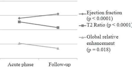
Several case reports have demonstrated the beneits of CMR in the evaluation of therapy in patients with myocarditis. LGE will decrease signiicantly in the acute phase toward healing phase. If T2W and GRE are done together, the results will 100% negative predictive value to differentiate acute myocarditis with convalescent myocarditis. Uchida et al reported that the changes of T2W is rare in
the chronic conditions.29 Improvement on T2W and
GRE are associated with clinical improvement and parameters of left ventricular function as shown in igure 4. Thus, has been also reported by Gutberlet et al that CMR can be used to evaluate disease
progression or regression in response to therapy.30
Supportive therapy is the irst treatment of choice in patients with myocarditis. Heart failure caused by
myocarditis must be given standard therapies such
as diuretics to lower ventricular illing pressure, ACE-i to lower vascular resistance, and beta-blockers after clinically stable. Arrhythmias should be monitored and treated. Patients who received imunogluobulin improved ejection fraction in 17
units. Another therapy is prednisone.31 Steroid
therapy decreased tissue inlammation.32 CMR can
Natalia, et al.
Pediatric myocarditis and cardiac magnetic resonance
180 Med J Indones, Vol. 23, No. 3,
August 2014
Figure 4. Accordance of improvement of global left ventricu -lar function and CMR parameters of acute myocardial injury in myocarditis. The lines showed changes of left ventricular ejec -tion frac-tion, T2 ratio, and GRE in myocarditis evalua-tion. Im -provement in ejection fraction is obviously in parallel with con -current normalization of T2 and GRE. Modiied from Zagrosek A et al.11 acute injury have not been found anymore. Recent guidelines recommend restriction of activities in
patients with acute myocarditis.11 Some researches
were conducted to follow myocarditis patients up to 3 months. The prognosis will be good if contractile function improve in sequence with signiicantly decreased of delayed enhanced of myocardial tissue
damage.11,23 This patient came with complaints
of chest pain, no symptoms of heart failure, a good pump function, and without arrhythmia as
well. Furthermore, the patient was given
anti-inlammatory drug (steroids). Throughout treatment
the patient’s condition was good, without chest
pain, then patient discharged. CMR to conduct a re-evaluation was planned after 1 month therapy. CMR results after 1 month of therapy showed no myocardial edema, with less ibrotic tissue. These
results will lead to good prognosis as shown in Figure 4.
In conclusion, CMR is a non-invasive diagnostic tool, which is essential for diagnosing myocarditis in
pediatric patients who came with clinical presentation
of chest pain. CMR revealed tissue edema on T2W and ibrosis on LGE, which conirmed the diagnosis of myocarditis.
REFERENCES
1. Skouri HN, Dec GW, Friedrich MG, Cooper LT. Noninvasive imaging in myocarditis. J Am Coll Cardiol. 2006;48(10):2085-93.
2. Feldman AM, McNamara D. Myocarditis. N Engl J Med. 2000;343(19):1388-98.
3. Magnani JW, Dec GW. Myocarditis: current trends in diagnosis and treatment. Circulation 2006;113(6):876-90.
4. Fabre A, Sheppard MN. Sudden adult death syndrome and other non-ischaemic causes of sudden cardiac death. Heart. 2006;92(3):316-20.
5. Kawai C. From myocarditis to cardiomyopathy: mechanisms of inlammation and cell death: learning from the past for the future. Circulation. 1999;99(8):1091-100. 6. Freimuth P, Philipson L, Carson SD. The coxsackievirus
and adenovirus receptor. Curr Top Microbiol Immunol. 2008;323:67-87.
7. Shi Y, Chen C, Lisewski U, Wrackmeyer U, Radke M, Westermann D, et al. Cardiac deletion of the Coxsackievirus-adenovirus receptor abolishes Coxsackievirus B3 infection and prevents myocarditis in vivo. J Am Coll Cardiol. detecting myocardial inlammation. J Am Coll Cardiol. 2005;45(11):1823-5.
10. Friedrich MG, Sechtem U, Schulz-Menger J, Holmvang G, Alakija P, Cooper LT, et al. Cardiovascular magnetic resonance in myocarditis: A JACC White Paper. J Am Coll Cardiol. 2009;53(17):1475-87.
11. Zagrosek A, Abdel-Aty H, Boyé P, Wassmuth R, Messroghli D, Utz W, et al. Cardiac magnetic resonance monitors reversible and irreversible myocardial injury in myocarditis. JACC Cardiovasc Imaging. 2009;2(2):131-8. 12. Morgera T, Di Lenarda A, Dreas L, Pinamonti B, Humar F,
Bussani R, et al. Electrocardiography of myocarditis revisited: clinical and prognostic signiicance of electrocardiographic changes. Am Heart J. 1992;124(2):455-67.
13. Towbin JA, Bricker JT, Garson A Jr. Electrocardiographic criteria for diagnosis of acute myocardial infarction in childhood. Am J Cardiol. 1992;69(19):1545-8.
14. Spodick DH. Electrocardiogram in acute pericarditis. Distributions of morphologic and axial changes by stages. Am J Cardiol. 1974;33(4):470-4.
15. Gazit AZ, Avari JN, Balzer DT, Rhee EK. Electrocardiographic diagnosis of myocardial ischemia in children: is a diagnostic electrocardiogram always diagnostic? Pediatrics. 2007;120(2);440-4.
16. Lauer B, Niederau C, Kühl U, Schannwell M, Pauschinger M, Strauer BE, et al. Cardiac troponin T in patients with clinically suspected myocarditis. J Am Coll Cardiol. 1997;30(5):1354-9.
17. Caforio AL, Calabrese F, Angelini A, Tona F, Vinci A, Bottaro S, et al. A prospective study of biopsy-proven myocarditis: prognostic relevance of clinical and aetiopathogenetic features at diagnosis. Eur Heart J. 2007;28(11):1326-33.
19. Shanes JG, Ghali J, Billingham ME, Ferrans VJ, Fenoglio JJ, Edwards WD, et al. Interobserver variability in the pathologic interpretation of endomyocardial biopsy results. Circulation. 1987;75(2):401-5.
20. Gagliardi MG, Bevilacqua M, DiRenzi P, Picardo S, Passariello R, Marcelletti C. Usefulness of magnetic resonance imaging for diagnosis of acute myocarditis in infants and children, and comparison with endomyocardial biopsy. Am J Cardiol. 1991;68(10):1089-91.
21. Gagliardi MG, Polletta B, Di Renzi P. MRI for the diagnosis and follow-up of myocarditis. Circulation. 1999;99(3):458-9. 22. Assomull RG, Lyne JC, Keenan N, Gulati A, Bunce NH,
Davies SW, et al. The role of cardiovascular magnetic resonance in patients presenting with chest pain, raised troponin, and unobstructed coronary arteries. Eur Heart J. 2007;28(10):1242-9.
23. Laissy JP, Garot J. Non-invasive imaging of myocardial infarction and myocarditis by cardiac magnetic resonance and multi-slice computed tomography. European Cardiology Review. 2006;2(2):40-4.
24. Friedrich MG, Strohm O, Schulz-Menger J, Marciniak H, Luft FC, Dietz R. Contrast media-enhanced magnetic resonance imaging visualizes myocardial changes in the course of viral myocarditis. Circulation. 1998;97(18):1802-9.
25. Roditi GH, Hartnell GG, Cohen MC. MRI changes in myocarditis - evaluation with spin echo, cine MR angiography and contrast enhanced spin echo imaging. Clin Radiol. 2000;55(10):752-8.
26. Olimulder MA, van Es J, Galjee MA. The importance of cardiac MRI as a diagnostic tool in viral myocarditis-induced cardiomyopathy. Neth Heart J. 2009;17(12):481-6. 27. McCrohon JA1, Moon JC, Prasad SK, McKenna WJ,
Lorenz CH, Coats AJ, et al. Differentiation of heart failure related to dilated cardiomyopathy and coronary artery disease using gadolinium-enhanced cardiovascular magnetic resonance. Circulation. 2003;108(1):54-9. 28. Hunold P, Schlosser T, Vogt FM, Eggebrecht H, Schmermund
A, Bruder O, et al. Myocardial late enhancement in contrast-enhanced cardiac MRI: Distinction between infarction scar and non-infarction-related disease. AJR Am J Roentgenol. 2005;184(5):1420-6.
29. Uchida Y, Nakamura F, Hirose J, Oshima T, MoritaT, Morizuki S, et al. Cardioscopic spectrum of the left ventricular endocardial surface and its relation to histologic changes in idiopathic myocarditis. Am Heart J. 1996;131(1):107-14.
30. Gutberlet M, Spors B, Thoma T, Bertram H, Denecke T, Felix R, et al. Suspected chronic myocarditis at cardiac MR: diagnostic accuracy and association with immunohistologically detected inlammation and viral persistence. Radiology. 2008;246(2):401-9.
31. Dennert R, Crijns HJ, Heymans S. Acute viral myocarditis. Eur Heart J. 2008;29(17):2073-82.