Effects of
Hibiscus sabdariffa
Linn. on insulin-like growth factor
binding protein 3 (IGFBP-3) to prevent overtraining syndrome
Abstrak
Latar belakang: Latihan isik berat (overtraining) dapat meningkatkan produksi reactive oxygen species
(ROS). Salah satu indikator sindrom overtraining adalah penurunan kadar insulin-like growth factor binding protein 3 (IGFBP-3) dalam darah. Hibiscus sabdariffa Linn., suatu antioksidan kuat, diharapkan mampu mencegah sindrom overtraining. Penelitian ini bertujuan mengetahui pengaruh pemberian H. sabdariffa pada tikus yang diberi “latihan isik overtraining”.
Metode: Penelitian eksperimental ini menggunakan 30
tikus Rattus norvegicus galur Wistar jantan (200-250
gram), dibagi menjadi 5 kelompok: 1) tikus kontrol (C); 2) tikus kontrol diberi H. sabdariffa (C-Hib); 3) tikus dengan latihan isik aerobik ringan (A-Ex), 4) tikus dengan latihan isik overtraining (OT); 5) tikus dengan latihan
isik overtraining dan diberi H. sabdariffa (OT-Hib). H.
sabdariffa diberikan secara oral dengan dosis 400 mg/ kgBB/hari selama 11 minggu. IGFBP-3 diukur dengan teknik ELISA dan dianalisis dengan uji ANOVA. Hasil: Kadar IGFBP-3 pada kelompok C adalah 17,4 ±
10 mIU/L, paling rendah pada kelompok OT (10,7 ± 9,9 mIU/L) dan paling tinggi pada kelompok OT-Hib (31,5 ± 6,2 mIU/L), p < 0,05. Terdapat perbedaan bermakna kadar IGFBP-3 pada kelompok OT dengan A-Ex (10,7 ± 9,9 vs 23,5 ± 9,7 mIU/L; p < 0,05. Perbedaan kadar IGFBP 3 pada kelompok C dan OT-Hib adalah (17,4 ± 10 vs 31,5 ± 6,2; p < 0,05).
Kesimpulan: Pemberian H. sabdariffa dapat mencegah penurunan kadar IGFBP-3 pada tikus dengan overtraining, yang menunjukkan H. sabdariffa dapat mencegah terjadinya sindrom overtraining.
Abstract
Background: Excessive physical exercises (overtraining) can increase the production of reactive oxygen species (ROS). One of the indicators of overtraining syndrome is a decrease in insulin-like growth factor binding protein 3 (IGFBP-3). Administration of Hibiscus sabdariffa Linn., a powerful antioxidant, is expected to boost endogenous antioxidants, and thus prevents overtraining. The aim of this study is to determine the effect of H. sabdariffa on IGFBP-3 levels in rats under ”overtraining physical excersice”.
Methods: This experimental study was conducted on 30 male rats (Rattus norvegicus 200-250 grams), randomly allocated into 5 groups: 1) control group (C); 2) control with H. sabdariffa (C-Hib); 3) mild aerobic exercise (A-Ex); 4) overtraining exercise (OT); 5) overtraining exercise with H. Sabdariffa (OT-Hib). H. sabdariffa (400 mg/kg/d, 11 weeks) were administered orally via syringe cannula. IGFBP-3 was measured by using ELISA (Cusa bio kit) and data were analyzed with ANOVA test.
Results: Plasma level of IGFBP-3 in the C and OT groups were 17.4 ± 10 mIU/L, the lowest in OT groups (10.7 ± 9.9 mIU/L) and the OT-Hib group had the highest level (31.5 ± 6.2 mIU/L). There was signiicant difference of the level IGFBP-3 in OT groups with A-Ex groups (10.7 ± 9.9 vs 23.5 ± 9.7 mIU/L; p < 0,05). The signiicant difference was also observed in the level of IGFBP 3 between C groups and the OT-Hib groups (17.4 ± 10 vs 31.5 ± 6.2; p < 0.05).
Conclusion: Administration of H. sabdariffa can prevent the decrease of IGFBP-3 levels in overtraining rats, indicating its role in preventing overtraining syndrome.
Keywords: Hibiscus sabdariffa Linn., IGFBP-3, overtraining
pISSN: 0853-1773 • eISSN: 2252-8083 • http://dx.doi.org/10.13181/mji.v23i4.991 • Med J Indones. 2014;23:187-91 Correspondence author: Ermita I.I. Ilyas, [email protected]
B a s i c M e d i c a l R e s e a r c h
Copyright @ 2014 Authors. This is an open access article distributed under the terms of the Creative Commons Attribution-NonCommercial-ShareAlike 4.0 International License (http://creativecommons.org/licenses/by-nc-sa/4.0/), which permits unrestricted non-commercial use, distribution, and reproduction in any medium, provided the original author and source are properly cited.
Ermita I.I. Ilyas,1 Neng T. Kartinah,1 Trinovita Andraini,1 Roman A. Goenarjo,1 Donna N. Kahandjak2
1 Department of Physiology, Faculty of Medicine, Universitas Indonesia, Jakarta, Indonesia
Overtraining syndrome is a result of excessive volume of exercise which, in the long run, will cause performance declining. This decrease in performance is particularly associated with physical, immunological and emotional disturbances.1-4
Overtraining syndrome frequently occurs due to long-term trainings before a competition with the exercise volume exceeding the capabilities of an athlete. It is also caused by imbalance between training and resting days, and between competitions and recovery.2,4 The incidence of overtraining
syndrome in elite athletes is 7-20% and occurs more commonly in predominantly aerobic sports such as swimming, running, and cycling.1,5,6 However, this
high incidence of overtraining syndrome is related to various and less known factors that precede or follow the overtraining syndrome.
Some theories try to explain the pathophysiology and indicators of overtraining syndrome. However, the existing theories are still unable to completely explain the pathophysiology of overtraining, because each theory has its advantages and disadvantages. One of the theories states that overtraining symptoms is associated with the disruption in the hypothalamic regulation of hormones. Other studies indicate a decrease in the hormone insulin-like growth factor binding protein 3 (IGFBP-3) in athletes who suffer overtraining.7,8 The secretion of IGFBP-3, a carrier
protein for insulin-like growth factor 1 (IGF-1), is stimulated by growth hormone (GH). The decrease in IGFBP-3 is proposed to be used as the indicator of overtraining in athletes.
Overtraining syndrome often occurs in athletes following aerobic type exercises with high oxygen consumption. Some studies suggest a link between physical exercise, increased oxygen consumption and reactive oxygen species (ROS) production. Therefore, the excessive ROS production during overtraining may result in an imbalance between the endogenous antioxidant system and pro-oxidants, which is referred to as oxidative stress.
Indonesia is rich of herbs which contain antioxidants such as Hibiscus sabdariffa Linn. This plant belongs to the family of Malvaceae and is often used as antihypertensive, antioxidant, anticancer,
antipyretic, antibacterial, anti-inlammatory, and
anti-hyperlipidemic phytotherapy.9,10 Calyx is the
major part of H. sabdariffa used in phytomedicine with anthocyanin being the main active component.11
Under certain conditions, anthocyanin shows higher
antioxidant activity compared to vitamin E, ascorbic
acid, and β-carotene.10,12
The aim of this study was to evaluate the effect of H. sabdariffa on overtraining by measuring the IGFBP-3.
METHODS
The protocol of this study has been approved by the Ethical Committee of the Faculty of Medicine,
Universitas Indonesia/Cipto Mangunkusumo
Hospital (No. 289/H2.F1/ETIK/2013).
Thirty male Wistar rats (Rattus norvegicus) weighing
200-250 g were divided randomly into ive groups:
1) control group (C), 2) control group given H. sabdariffa (C-Hib), 3) the mild aerobic exercise group (A-Ex), 4) the overtraining exercise group (OT), and 5) the overtraining exercise group given H. sabdariffa (OT-Hib). Before and during intervention, the rats were maintained properly according to the ethics of experimental animal usage. Rats were given standardized food and water ad libitum. The cages were arranged 12 hours of light (18:00 pm to 6:00 am) and 12 hours of dark (6:00 am to 18:00 pm), thus a treatment of overtraining exercise could be done during the day. Room temperature was set at 23°C ± 1°C. The extract of H. sabdariffa was administered orally via a syringe cannula to the rats in C-Hib and OT-Hib groups at a dose of 400 mg/
kgBW/day, ive days a week (except Saturdays and
Sundays) for 11 consecutive weeks.
H.sabdariffa was extracted by the following process: one kg of calyx H. sabdariffa was mixed with 5 liters of water for 3 hours. The mixture was kept for 24
hours at room temperature, then iltered and dried in
a Rotavapor (Büchi, Switzerland).
Blood sample were drawn on day-3 after treatment, to avoid the acute effects on overtraining. The rats were anesthetized with ether, the chest was opened and blood was collected by cardiac puncture. The blood was centrifuged at 1000 x g for 15 minutes to obtain plasma for the IGFBP-3 measurement. The level of free IGFBP-3 (i.e. not bound to IGF-1) was measured by ELISA (CusaBio kit).
Data analysis
All data in the experiments were analyzed using one way Anova. Multiple comparisons were done with post-hoc LSD method. The p < 0.05 was taken as the
limit of statistical signiicance.
RESULTS
Body weight
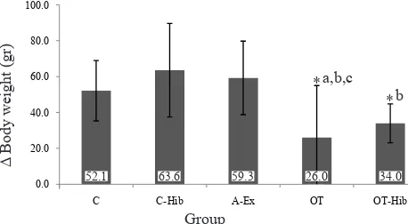
Figure 1 shows the differences in the average body weights between pre-test and post-test measurement of all groups. The rats in all groups gained weight in the course of the training programs; however, the OT group had the lowest post-test body weight compared
to other groups. There were signiicant differences in Δ
body weight between OT and C group, between groups OT and C-Hib, and between OT and A-Ex group (p
< 0.05). There was no signiicant difference in the Δ
body weight between OT and OT-Hib group (p > 0.05).
Nevertheless, there was a tendency to higher Δ body
weight in OT than in OT-Hib group.
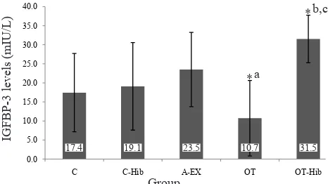
Serum levels of IGFBP-3
The levels of IGFBP-3 in the ive group are presented
in igure 2. The level in A-Ex group (23.5 ± 0.7
Time of
experiment Phase of training Speed (m/min) Duration (min) Frequency/day
Resting time between sessions (hours)
week-1 A1 15 20 1 24
week-2 A1 20 30 1 24
week-3 A1 22.5 45 1 24
week-4 A1 25 60 1 24
week-5-8 A2 25 60 1 24
week-9 T2x 25 60 2 4
week-10 T3x 25 60 3 3
week-11 T4x 25 60 4 2
Table 1. Program of aerobic exercise overtraining
A1: adaptation phase 1; A2: adaptation phase 2 with increased speed and duration; T2x: treadmill exercise twice/day; T3x: treadmill
exercise 3 times/day; T4x: treadmill exercise 4 times/day. Modiied from Margonis, et al13
Figure 1. Body weights before and after the intervention in the control (C), control-H. sabdariffa (C-Hib), mild aerobic cise (A-Ex), overtraining exercise (OT), and overtraining exer-cise-H. sabdariffa (OT-Hib).
*p < 0.05; acompared to C; bcompared to C-Hib; ccompared to A-Ex
mIU/L) was higher than in groups C (17.4 ± 10.2 mIU/L) and C-Hib (19.1 ± 11.4 mIU/L). The level of IGFBP-3 in C-Hib group was slightly higher than group C; the difference was statistically not
signiicant. The lowest IGFBP-3 level was obtained
in the OT group (10.7 ± 9.9 mIU/L), which differed
signiicantly from A-Ex and OT-Hib groups (31.4 ±
6.2 mIU/L) (p < 0.05) which was the highest level of all groups.
DISCUSSION
The results of this study showed that Δ body weight
in the OT group was lower than in controls. This difference is assumed to be due to the increased body metabolism and/or the skeletal muscle catabolism in OT compared to the control group. The OT group has a longer catabolic phase due to higher energy demand that is obtained from fat and protein. Xiao, et al14 reported that the body weight of rats in
0.0 20.0 40.0 60.0 80.0 100.0
C C-Hib A-Ex OT OT-Hib
Δ Body weight (gr)
Group
52.1 63.6 59.3 26.0 34.0
a,b,c *
overtraining group was 23.6% lower than control group. In addition, the decreased body weight in this study is also due to decreased appetite in OT rats, one of the markers of the overtraining syndrome. The loss of body weight in the OT group indicates that this group was experiencing the overtraining syndrome. Other supporting parameters of the overtraining condition in this study were signs of fatigue and decreased performance when the rats were put on treadmill training. According to Margonis, et al13 one of
the indicators of the overtraining syndrome is a decrease in the physical exercise capacity which is related to chronic fatigue of rats.
The OT-Hib group did not experience fatigue and decreased performance when running on a treadmill. The results showed that the performance in the OT-Hib group was better than in the OT group. In addition, the OT-Hib group had higher
Δ body weight than the OT group. This inding
suggests that the OT-Hib group did not experience the overtraining syndrome, since loss of body weight or decreased weight gain is one of the signs of overtraining syndrome.
The IGFBP-3 levels of the A-Ex group were higher than in group C and C-Hib. This suggests that physical exercise can increase IGFBP-3 levels. Exercise can increase the levels of GH, IGF-1 and IGFBP-3 because it is a powerful stimulus of growth hormone releasing hormone (GHRH) and GH release.15 The
increase of GH secretion from the pituitary gland will stimulate the liver and other tissues to produce IGF-1 and IGFBP-3.16-18 However, in the condition
of overtraining syndrome the level of IGFBP-3 will
decrease.7,8 Therefore, the decreased IGFBP-3 level
can be used as a speciic marker of the overtraining
syndrome, although the mechanism of such decrease is still unclear. The decrease of IGFBP-3 is allegedly due to disturbance in the GH /IGF-1 axis or disruption in IGFBP-3 synthesis in liver cells by oxidative stress. Heavy physical exercises can increase the ROS production resulting in oxidative stress.
It is well known that oxidative stress is an imbalance between free radicals production and antioxidant defends mechanism in the body.6 Production of
ROS and damage of lipids, proteins and genetic materials (DNA, RNA) through ROS are believed to be the main mechanisms of the biochemical and physiological changes that occur during physical exercise.19 Further studies are needed to disclose
the mechanisms of the decreased IGFBP-3 levels in the overtraining syndrome associated with oxidative stress.
In the present study, the IGFBP-3 level in the OT
group is lower than in the A-Ex group. This inding
suggests that exercise program given to the rats in this group leads to the overtraining syndrome. However, when the overtraining exercise program is accompanied by the provision of H. sabdariffa
extract the rats do not experience the overtraining syndrome. The IGFBP-3 levels in the OT-Hib group are even higher than in A-Ex and OT groups. It seems that H. sabdariffa works more effectively in overtraining animals compared to animals in normal condition. The higher level of IGFBP-3 in the OT-Hib group are likely caused by combination of physical exercise with the antioxidant role of H. sabdariffa.
Antioxidants are compounds that may prevent or inhibit the oxidation of proteins, DNA, and lipids in cell membranes.H. sabdariffa contains anthocyanins,
lavonoids, protocatechuic acid, ascorbic acid, and
other compounds.20 As anthocyanins are potent
antioxidants,H. sabdariffa can reduce free radicals by inhibiting the activity of xanthine oxidase and by increasing the activity of glutathione peroxidase (GPx).21 Therefore, we suggest H. sabdariffa may
prevent overtraining in heavy physical exercise. The capacity of H. sabdariffa to reduce ROS and increase GPx activity may alleviate ROS damage at the levels of molecules, cells, organs, and systems. This study found a tendency of H. sabdariffa to increase IGFBP-3 level in the C-Hib group, although
Figure 2. The IGFBP 3 level in the control (C), control-H. sabdariffa (C-Hib), mild aerobic exercise (A-Ex), overtraining exercise (OT), and overtraining exercise-H. sabdariffa groups *p < 0.05; acompared to A-Ex; bcompared to C; ccompared to OT
0.0 5.0 10.0 15.0 20.0 25.0 30.0 35.0 40.0
C C-Hib A-EX OT OT-Hib
IGFBP-3 levels (mIU/L)
Group
17.4 19.1 23.5 10.7 31.5
a *
the differences were not statistically signiicant
compared to control group.
It is suspected the high levels of IGFBP-3 in the OT-Hib group are caused by the combined effects of physical exercise and H. sabdariffa. Physical exercise can increase IGFBP-3 levels through the increase of GH secretion. There is a positive correlation between the intensity of the exercise with the magnitude of GH secretion.15 Increased GH
secretion will intensify liver stimulation to produce IGFBP-3. On the other hand, the greater exercise intensity will result in higher ROS production. Therefore, the highest level of IGFBP-3 found in OT-Hib may be caused by exercise in high intensity and the concomitant provision of H. sabdariffa.
From the above results, we conclude that H. sabdariffa can maintain the IGFBP-3 level and is expected to prevent overtraining syndrome in excessive physical exercise.
Acknowledgment
The authors are grateful to DRPM UI, which has funded this study through a grant from Universitas Indonesia 2013 (Hibah Riset Madya UI 2013).
Conlict of interest
The authors afirm no conlict of interest in this study.
REFERENCES
1. Bischler TK. The utility of resting levels of IGF-I and IGFBP-3 as markers of training status in elite athletes [thesis]. Canada: University of Lethbridge; 2007 [cited 2012 Jun 6]. Available from:https://www.uleth.ca/dspace/ handle/10133/651
2. Cunha GS, Ribeiro JL, Oliveira AR. Overtraining: theories, diagnosis and markers. Rev Bras Med Esporte. 2006;12(5):267e-71e.
3. Kreher JB, Schwartz JB. Overtraining syndrome: a practical guide. Sports Health. 2012;4(2):128-38.
4. Brenner JS. American Academy of Pediatrics Council on Sports Medicine and Fitness. Overuse injuries, overtraining, and burnout in child and adolescent athletes. Pediatrics. 2007;119(6):1242-5.
5. Fry AC, Schilling BK, Weiss LW, Chiu LZ. β2-Adrenergic
receptor downregulation and performance decrements during high-intensity resistance exercise overtraining. J Appl Physiol (1985). 2006;101(6):1664-72.
6. Marcello BM. Overtraining in sport: physiological, psychological and performance effect of participation in division I competitive softball [dissertation]. Texas: Baylor University; 2006 [cited 2012 jun 17]. Available from: https://beardocs.baylor.edu/xmlui/handle/2104/4959 7. Elloumi M, El Elj N, Zaouali M, Maso F, Filaire E, Tabka
Z, et al. IGFBP-3, a sensitive marker of physical training and overtraining. Br J Sports Med. 2005;39(9):604-10. 8. Capoluongo E, Pitocco D, Santonocito C, Concolino P,
Santini SA, Manto A, et al. Association between serum free IGF-I and IGFBP-3 levels in type-I diabetes patients affected with associated autoimmune disease or diabetic complications. Eur Cytokine Netw. 2006;17(3):167-74. 9. Wang SC, Lee SF, Wang CJ, Lee CH, Lee WC, Lee HJ.
Aqueous extract from Hibiscus sabdariffa Linnaeus ameliorate diabetic nephropathy via regulating oxidative
status and Akt/Bad/14-3-3γ in an experimental animal
model. Evid Based Complement Alternat Med. 2011;p.1-9. 10. Hohl R, Ferraresso RL, De Oliveira RB, Lucco R, Brenzikofer R, De Macedo DV. Development and characterization of an overtraining animal model. Med Sci Sports Exerc. 2009;41(5):1155-63.
11. Mahadevan N, Shivali, Kamboj P. Hibiscus sabdariffa
Linn.- An overview. NPR. 2008;8(1):77-83.
12. Kowalczyk E, Krzesiński P, Kura M, Szmigiel B, Błaszczyk J. Anthocyanins in medicine. Pol J Pharmacol.
2003;55(5):699-702.
13. Margonis K, Fatouros IG, Jamurtas AZ, Nikolaidis MG, Douroudos I, Chatzinikolaou A, et.al. Oxidative stress biomarkers responses to physical overtraining: implications for diagnosis. Free Radic Biol Med. 2007;43(6):901-10. 14. Xiao W, Chen P, Dong J. Effect of overtraining on sceletal
muscle growth and gene expression. Int J Sports Med. 2012;33(10):846-53.
15. Rosendal L, Langberg H, Flyvbjerg A, Frystyk J, Ørskov
H, Kjaer M. Physical capacity inluences the response
of insulin-like growth factor and its binding protein to training. J Appl Physiol (1985). 2002;93(5):1669-75. 16. Goodman HM. Basic Medical Endocrinology. 4th ed.
China: Elsevier; 2009.
17. Yadav S, Khrisnamurthy S. Insulin-like growth factors
and growth hormone deiciency. Indian Pediatr.
2007;44(5):349-53.
18. Taipale RS, Häkkinen K. Acute hormonal and force responses to combined strength and endurance loadings in men and woman: The “Order Effect”. PLoS ONE. 2013;8(2):e55051.
19. Banerjee AK, Mandal A, Chanda D, Chakraborti S. Oxidant, antioxidant and physical exercise. Mol Cell Biochem. 2003;253(1-2):307-12.
20. Essa MM, Subramanian P. Hibiscus sabdariffa affects ammonium chloride induced hyperammonemic rats. Evid Based Complement Alternat Med. 2007;4(3):321-8. 21. Daniel D, Aduwamai UH, Blessing V, Michael AA.