Prevalence of enzootic simian viruses among urban
performance monkeys in Indonesia
Michael A. Schillaci1, Lisa Jones-Engel2, Gregory A. Engel3, Yasmina Paramastri4, Entang Iskandar4, Brenda Wilson5, Jonathan S. Allan5, Randall C. Kyes2,6, Robin Watanabe2and Richard Grant2
1Department of Social Sciences, University of Toronto at Scarborough, Toronto, Ontario, Canada 2Washington National Primate Research Center, University of Washington, Seattle, WA, USA 3Swedish/Providence Family Practice Residency, Seattle, WA, USA
4Primate Research Center, Bogor Agricultural University, Bogor, Indonesia
5Department of Virology and Immunology, Southwest National Primate Research Center, San Antonio, TX, USA 6Department of Psychology, University of Washington, Seattle, WA, USA
Summary Animal reservoirs are the most important sources of emerging infectious diseases that threaten human populations. Global travel and tourism bring ever-increasing numbers of humans into contact with animals, increasing the likelihood of cross species transmission of infectious agents. Non-human primates come into contact with humans in a variety of contexts and may harbor infectious agents with zoonotic potential. We investigated the prevalence of infection with enzootic simian viruses among 20 urban performance monkeys (Macaca fascicularis) in Jakarta, Indonesia. This report documents for the first time evidence of infection with four simian viruses in urban performance monkeys. Simian foamy virus was detected by PCR in 52.9% of the macaques. Antibodies to simian retrovirus were detected in 10.5% of the macaques. Antibodies toCercopithecine Herpesvirus1, were detected in 5.3% of the macaques. Similarly, antibodies to simian T-cell lymphotropic virus were detected in 5.3% of the macaques. No evidence of infection with simian immunodeficiency virus was detected in these macaques. These results suggest that urban performance monkeys are a reservoir for enzootic simian viruses known to be capable of infecting humans.
keywordssimian retrovirus, simian T-cell lymphotropic virus,Cercopithecine Herpesvirus1, simian foamy virus, macaque, primate zoonoses, Asia, performing monkeys
Introduction
Recent epidemics of zoonotic diseases such as HIV, SARS, avian flu (influenza H5N1), and hantavirus illustrate the importance of animal reservoirs as sources of emerging infectious diseases that threaten human populations glo-bally. Animal/human contact in a variety of contexts can lead to zoonotic transmission. Some of these contexts, such as those associated with animal husbandry and pet ownership, zoological parks and petting zoos, laboratory research involving captive animals, monkey temples and the bush-meat and exotic animal trade are recognized (e.g., Wolfeet al. 1998, 2004; Engelthaleret al.1999; Shortridgeet al.2000; Engelet al.2002; Peeters 2002; CDC, MMWR
2003a,2003b,2003c; Bellet al.2004; Reedet al.2004; Jones-Engelet al.2005a). The risk of zoonotic exposure associated with other contexts of animal/human contact such as urban and eco-tourism, however, is just now being explored.
By virtue of their genetic, physiological, and behavioural similarities with humans, non-human primates (NHPs) are
thought to be likely sources of pathogens that can pose a significant threat to human populations. The HIV pan-demic is a cogent example of this threat. Recent research suggests that cross-species transmission of SIV from chimpanzees (Pan spp.) and sooty mangabeys (Cercocebus atys) to humans has occurred likely as a result of
butchering practices associated with the bushmeat trade (Hahnet al.2000). Subsequent human-to-human trans-mission eventually resulted in the world-wide spread of HIV in human populations.
enzootic to Old World Primates, is hypothesized to be the progenitor of human T-cell lymphotropic virus (HTLV), through multiple cross-species transmissions (Franchini 1994; Vandammeet al.1998; Gessain & Mahieux 2000). HTLV is implicated as an etiologic agent of adult T-cell leukemia and tropical spastic paresis (Gessainet al.1985). Cercopithecine herpesvirus1 [CHV-1 (Herpes B)] is an alphaherpesvirus endemic to Asian macaques (Genus Macaca) (Weigler 1992; Huff & Barry 2003). Similar to herpes simplex in humans, CHV-1 in macaques causes mild symptoms consisting primarily of oral and perioral vesicular lesions. In humans, however, CHV-1 produces a fulminating meningoencephalitis with a mortality rate near 70% (Hummeleret al.1959; CDC, MMWR 1989, 1998; Holmeset al.1990, 1995).
Urban performance monkeys in Asia
Although performing NHPs are encountered throughout the world, Asian cultures have perhaps the longest and most vibrant tradition of using NHPs for entertainment. In Japan, a thousand-year history of training performance monkeys (Macaca fuscata) continues today through the Suo-Sarumawashi (Japanese Monkey Performance) (Ohnuki-Tierney 1987). Similar training schools can be found throughout South and Southeast Asia. At the Punjab Institute of Mental Health in Lahore, Pakistan, for example, performance monkeys (Macaca mulatta) are used to entertain psychiatric patients.
Performance Monkey ‘niche’
The ecology of performance monkeys is distinct from that of primates that come into contact with humans in other contexts, such as temple monkeys, laboratory NHPs, and NHPs consumed as bushmeat. The acquisition of per-formance monkeys, the care they receive from their owners, the characteristics of the urban environments they typically inhabit, and the unique circumstances of the performances themselves are all components of the per-formance monkey ‘niche’ that influence the amount and kinds of contact these primates have with humans and, in turn, influence the likelihood of zoonotic transmission occurring in this context.
Regarding the risk of NHP-to-human transmission of infectious agents, it is important to consider the origin of performance macaques. Although no systematic research has been published describing how owners in other parts of Indonesia and in other countries acquire their animals, the majority of the macaques in the present study were obtained from animal markets, most likely from Pramuka, the large animal market in Jakarta. This is significant because animal
markets bring together a variety of species, primate and non-primate, from diverse geographic origins, each with its potentially unique burden of infectious agents (Kareshet al. 2005). Animals in these markets are typically maintained at high density and often in poor conditions, which may compromise immunity and facilitate disease transmission (Maloneet al.2002). Monkeys included in the present study were typically purchased from animal markets while still quite young. Intensive training with the owner lasted anywhere from 5 months to a year or more depending on how quickly the monkey learned his/her routines.
Performance monkeys usually live with their owner’s family. Most of the owners we spoke with owned more than one monkey. As with pet NHPs, close physical contact including the sharing of food and water resources is common in these settings. Previous research suggests that a variety of infectious agents may be transmitted from pet NHPs to humans as well as from humans to pet NHPs (Mack & Noble 1970; de Arrudaet al.1989; Ostrowski et al.1998; Jones-Engelet al.2001, 2004, 2005b; Huemer et al.2002; Peeterset al.2002).
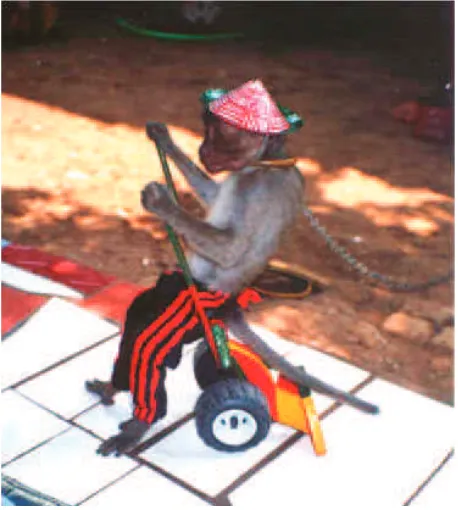
Performances can be quite elaborate and usually include music and commentary from the owners. The performing monkeys are often dressed in costume and perform acrobatics (Figure 1). Performances are typically held in
areas such as popular tourist destinations where they are likely to draw large audiences. Owners of performance monkeys may encourage monkeys to climb onto specta-tors’ shoulders and head for photos and monkeys may also come into physical contact with spectators as they collect money for their performance.
The urban environment, which performance monkeys typically inhabit, is in itself a significant aspect of the performance monkey ‘niche’. The mobility of urban populations as well as large urban centres as hubs for international travel may bring together NHPs with people who otherwise might never have contact with NHPs.
Our purpose was to investigate the prevalence of select simian pathogens in a sample of urban performance monkeys from Jakarta, Indonesia, in order to determine whether these NHPs harbor enzootic infectious agents. Specifically, we investigated the prevalence of four simian-born retroviruses known to infect humans: simian immu-nodeficiency virus, simian retrovirus, simian foamy virus, simian T-cell lymphotrophic virus, and an alphaherpes virus,Cercopithecine herpesvirus 1. Identifying the pre-valence of these simian infectious agents in the urban performance monkey population is the first step in estimating the risk of NHP-to-human transmission to a variety of people who may come into contact with these monkeys. These include the monkeys’ owners/trainers and their families, people who live in the community where the monkeys are kept, and tourists who observe performances.
Materials and methods Sampling
In July 2002, 20 performance monkeys, all long-tailed macaques (Macaca fascicularis), were identified in Kam-pung Dukuh, East Jakarta, a village known to be a community where owners of performance monkeys are concentrated. After explaining our research protocol to each monkey owner, we sedated the macaques with 3 mg/ kg of Telazol
(tiletamine HCI/zolazepam HCI). Using universal precautions, approximately 10 ml of blood were drawn from the femoral vein of each monkey. Macaques were monitored closely during anaesthesia and recovery. Approximately 6 ml of blood were placed in a separator tube and centrifuged in the field to extract the serum. The remaining whole blood was placed in a tube containing EDTA. Both the blood and serum samples were frozen and stored at)80C. Each macaque’s weight and dental
formula were collected and recorded for age assessment. Age was estimated based on the observed pattern of dental
eruption using a published dental aging protocol forM. fascicularis(Richtsmeieret al.1993). Study and data protocols were reviewed and approved by the University of Washington’s Institutional Animal Care and Use Com-mittee (3143-03).
Laboratory analyses for SRV, STLV, SIV, SFV and CHV-1
Enzyme-linked immunoabsorbent assays (ELISAs) were used to detect antibodies to SRV, STLV, SIV and CHV-1. Immunoblot assays were performed to confirm all positive SIV, STLV and SRV ELISA results.
SRV antigens for EIA
To produce large volumes of SRV-2 preparations for EIA, one vial of infected A549 cells (ATCC CCL-185) was thawed and added to a T-25 culture flask containing Dulbecco’s Modified Eagle Medium (Gibco) supplemen-ted with 10% FBS (Hyclone) and penicillin/streptomycin solution. After 2 days at 37C, the cells were trysinized and transferred to a T-175 culture flask containing 100 ml of medium. The A549 cell culture supernatant was collected and cells were passed every 3–4 days at 1·105cells/ml until a total of approximately 3–6 l of culture supernatant was harvested. The supernatant was clarified first by low speed centrifugation at 1000 rpm (Beckman GH-38 rotor) for 15 min at 4C then by filtration through 0.45lm PES filters. The clarified and
pooled supernatant was concentrated approx. tenfold by tangential flow filtration using a Pellicon R-2, PLCHK cassette filter (Millipore) with a 100 000 NMWL cutoff. The concentrate was partially purified by ultracentrifu-gation on a 20% glycerol cushion at 19 000 rpm (Beckman Type 19 rotor) for 3 h at 4C. The pellets were dispersed in Dulbecco’s Phosphate Buffered Saline (Gibco), layered on a 30% and 45% (wt/wt) sucrose step gradient, and centrifuged at 45 000 rpm (Beckman SW55Ti rotor) for 1.5 h at 4C. The SRV band at the interface of the two sucrose concentrations was aspirated and mixed with PBS, then applied to a 20% glycerol cushion. The virus was pelleted by ultracentrifugation at 45 000 rpm (Beckman SW55Ti rotor) for 1 h at 4C. Viral antigen was quantified using BCA colorimetric detection (Pierce) then frozen at )80C in aliquots of
STLV, SIV and CHV-1, purified virus for EIA
For STLV assays we relied on the considerable cross-reactivity of HTLV-1 (Zeptometrix, Buffalo, NY) (Franchini & Reitz 1994) For SIV assays we purchased SIVmac251 (Zeptometrix). For CHV-1 assays we used the considerable cross reactivity of herpes simplex-1 (HSV-1) purified viral lysate (Advanced Biotechnologies, Columbia, MD) (Hilliardet al.1989).
ELISA for SRV, STLV, SIV and CHV-1
ELISA plates (Polysorp Nunc) were coated with 100ll of
SDS-treated (0.1% SDS at 56C for 30 min) viral protein at 70 ng/well in carbonate–bicarbonate buffer pH 9.6 (Sigma) and incubated overnight at 4C in a moist chamber. The viral lysates were removed and plates were incubated with blocking buffer, 5% non-fat dry milk (BioRad) in PBS-0.1% tween, for 1 h at 37C. The plates were washed in PBS-tween four times at this step and at all subsequent wash steps. Macaque plasma samples were heat inactivated at 56C for 30 min prior to application to ELISA plate. Plasma diluted 1:100 in blocking buffer was applied in duplicate to each well in 100ll volumes and
incubated at 37C for 1 h. Positive control plasma from macaques was a pool of historical samples confirmed to be positive by producing bands to all viral proteins on Immunoblot at three or more sequential time points. Negative controls were pooled negative plasma from the Washington National Primate Research Center (WaNPRC) specific pathogen-free colonies that have tested negative on four or more consecutive time points over two or more years confirmed to be negative on Immunoblots. After the plate was washed, 100ll of secondary antibody (goat
anti-monkey IgG-peroxidase, Sigma, 1:15 000) was added and plates were incubated at 37C for 1 h. The colour reaction was run using OPD (20 mg tablets, Sigma) in phosphate-citrate buffer containing urea hydrogen peroxide (Sigma) for 20 min. The colour reaction was stopped with 1N sulphuric acid and plates were read at 492 nm. Plate cut-off values were determined for each plate on each run using the equation: Cut-off value¼ average of eight or more neg. controls + (SD·2.010) with a 95% confidence level (Freyet al.1998).
SRV STLV and SIV Immunoblots
Sucrose gradient-purified SRV was loaded onto preparative SDS-PAGE 4–15% minigels (BioRad) at 60–75 ng/mm and blots were prepared on nitrocellulose as previously des-cribed (Benvenisteet al.1993) HTLV, cross-reactive with STLV, and SIV Immunoblot strips were purchased at
Zeptometrix, Buffalo, NY. SRV strips were incubated; rocking overnight at room temperature, with heat-inacti-vated plasma diluted 1:100 in blocking buffer. STLV and SIV strips (Zeptometrix) were incubated and tested fol-lowing the manufacturer’s instructions. Positive controls (1:250) consisted of pooled historical positive samples that produce bands to all viral structural proteins [SRV- (p10/ p12, p14, p27, pp18, gp20 and gp70), (STLV – p19, p24/ 26, GD22, gp44, rgp44-HTLV-1), (SIV – p27, gp120, gp160)]. Negative controls were the same as those used for ELISA. Strips were washed in PBS-tween three times and incubated for 1 h at room temperature with goat anti-human IgG alkaline phosphatase (1:12 000, Sigma) diluted in blocking buffer. Strips were washed three times and reacted with BCIP/NBT solution (Sigma) in a dark location for 10–15 min. Reaction was stopped by washing in water, changed several times over 10 min. Reactions were deemed positive if core and envelop bands were present, indeter-minate if only core or only envelop were present and negative if bands did not appear or were not darker than negative control plasma.
Purification of DNA for PCR
DNA was purified from whole blood using the QIAamp DNA Blood Mini Kit following the manufacturer’s proto-cols (Qiagen) with modifications. Briefly 20ll protease (2·
concentrate) and 200ll buffer AL were combined with
200ll whole blood and incubated at 56C for 30 min.
After incubation 200ll ethanol was added and the entire
mixture applied to a QIAamp spin column. The purified DNA was eluted from the column with 70ll of 10 mM
Tris–HCL pH 8.0 and the concentration and purity was determined spectrophotometrically at OD260 and 280. If the 260/280 ration did not fall between 1.70 and 2.05 DNA was rejected for PCR analysis. Rejected DNA was reapplied to a new column and purified a second time.
Real-time PCR for SRV
PCR was performed using prepared master mix from the manufacturer (BioRad) containing sybrgreen or no dye. PCR reactions (50ll volume containing 0.5lg DNA +
20 pmol primer + water in BioRad master mix) and gp70 and gp20 primers were set up for each DNA sample. Each sample was run with both gp70 and gp20 primers in duplicate so all samples were tested in quadruplicate. The primers used for detection were derived from consensus sequences for SRV serotypes 1–5. The primers located in the gp70 region are conserved in serotypes 1–3
CCAGCACAGTCACAAGGCTTA) and primers located in the gp20 region are conserved in all known serotypes 1–5 (7585, CTGGWCAGCCAATGACGGG and 7695, CGCCTGTCTTAGGTTGGAGTG with probe (7621 Fam-TCACTAACCTAAGACAGGAGGGYCGTCA-BHQ1). These primers are used routinely at both WaN-PRC (R. Grant unpublished data) and California NWaN-PRC (N. Lerche personal communication) for SRV diagnostic testing. Standard controls consist of known copy number plasmids, containing SRV-2 envelop, serially diluted in negative macaque genomic DNA. In addition, positive controls were used in each PCR that consisted of 0.5lg
DNA extracted from Raji cells infected with SRV serotypes 1–5. Negative control DNA consisted of genomic DNA pooled from negative macaques in the WaNPRC specific pathogen-free colonies that have tested negative on four or more consecutive time points over two or more years. Thermal cycles were 94C – 2 min, followed by 41 cycles of 94C – 20 s, 65C – 30 s with data collection after the 65C step of each cycle. A final melt curve was run on the samples by heating from 60 to 95C in increments of 0.5C. Samples were judged positive or negative based on the threshold cycle (Tc) of the PCR product and the melting temperature of the product. To be considered positive, a sample was required to be reactive to both gp70 and gp20 primers or to be proven positive by DNA sequencing of gp70 or gp20 region. Samples with aTcless than the negative control were deemed presumptive positive. Pre-sumptive positive samples were further analysed by melting temperature profile of the PCR product. Samples were confirmed positive if the product had a melting tempera-ture peak of 84–86C, within the range of all known serotypes of SRV-1 through SRV-5. A presumptive positive with an incorrect melting temperature peak was deemed negative after DNA sequencing of PCR product and confirmation of non-viral sequences. Based on routine use of this assay for screening more than 2800 samples per year from the macaque colony at the WaNPRC since 2002 and cell culture confirmation of more than 200 samples per year, the PCR assay has a specificity of >99% and sensitivity of >98% (R. Grant unpublished data). The limit of sensitivity for the assay is 50 copies/reaction based on the optimized plasmid standard controls.
Nested PCR for detection of STLV
A protocol to amplify a wide range of primate T-lympo-tropic viruses (PTLV) using outer generic primers followed by more specific nested primers in the tax gene region of STLV was performed on DNA from peripheral blood cells as previously described (Vandammeet al.1997). Results were analyzed on 2% Nusieve (Cambrex, Inc.) agarose gels
with ethidium bromide staining. The sensitivity and spe-cificity of this assay has not been independently determined in our laboratory but plasmid copy number controls indicate a limit of sensitivity of 10–50 copies/reaction.
PCR detection of SFV
The presence of SFV DNA was determined using a nested PCR. Five hundred nanograms of purified DNA were combined with a PCR reaction mixture with a final concentration of 10 mM Tris (pH9.0), 50 mM KCI, 0.1% Triton X-100, 2 mM MgCl2 200lM each dNTP, 0.15 mg/
m BSA, 1u Taq polymerase, and 400 nM of each primer in a total volume of 50ll. The primer pairs used were: first
round, forward, 5¢CAG T GA ATT CCAGAATCTCTTC 3¢, reverse, 5¢CACTTATCCCACTAGATGGTTC 3¢and second round, forward, 5¢ CCAGAATCTCTTCATCCTA-ACTA 3¢, reverse, 5¢GATGGTTCCCTAAGCAAGGC 3¢ (Khanet al.1999). Touchdown PCR was used for both rounds with reaction conditions of: initial denaturation at 94C for 2 min, followed by seven cycles of 94C for 30 s, 60C for 30 s, and 72C for 45 s with a 2C decrease in annealing temperature per cycle to 48C, followed by 33 cycles of 94C for 30 s, 48C for 30 s, and 72C for 45 s with a final extension at 72C for 2 min. Second round conditions were the same except 19 cycles were used instead of 33.
Results
Biological samples were collected from 20 macaques. Twelve were female. Dental eruption sequences classified one macaque as an infant, 10 as juveniles, 6 as subadults and three as adults at the time of sampling. Unfortunately, serum from one of the macaques, 02TK39, was unavailable for testing and whole blood samples from three macaques, 02TK16, 02TK20 and 02TK36, yielded insufficient DNA for SFV PCR.
Serological tests
PCR tests
The majority of the animals, 9 out of 17, (52.9%) tested for SFV by PCR were positive. Repeated attempts to amplify SRV from all samples, including those positive by immunoblot, using PCR primers in two different regions of the genome were not successful. STLV was amplified from the single seropositive macaque (5.3%) but the seroinde-terminate animal was PCR negative. STLV PCR specific for common isolates was negative, however, PCR to amplify generic PTLV DNA was strongly positive, indicating a divergent STLV present in the positive macaque. DNA was of sufficient quality and control betaglobin primers amplified the correct size product (data not shown).
The results of the serological and PCR assays were analyzed quantitatively to test associations by sex and age category. The results from these tests indicate there is no statistically significant association between seropositivity (for antibodies to SRV, STLV or CHV-1) and sex (OR¼3.073, 95%CI¼19.513–0.484,P¼0.352), or age (v2¼0.04,P¼0.843). Similarly, there is no statisti-cally significant association between a positive SFV PCR
result and sex (OR¼0.818, 95%CI¼4.976–0.135, P¼1.00), or age (v2¼0.08,P¼0.778).
Discussion
This report documents the presence of antibodies to SRV, STLV and CHV-1, as well as the presence of simian foamy virus DNA in a sample of urban performance monkeys from Jakarta, Indonesia. Two of 20 urban performance monkeys had evidence of past or present infection with more than one enzootic simian virus.
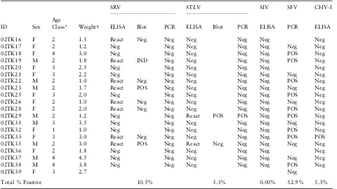
Although two of the performance monkeys were found to be seropositive for SRV, viral DNA could not be amplified from any of the 20 animals surveyed through repeated attempts using primers in conserved and non-conserved locations of the genome, gp20 and gp70, respectively. The finding of SRV seropositive macaques with no detectable virus in circulating cells has been reported in SRV infections (Lercheet al.1994). Routine surveys of macaques typically find those animals with antibody responses to the neutral-izing epitopes of gp70 are negative or express very low copy numbers of SRV in blood cells (R. Grant unpublished data). Table 1 Sample information and results for serological and molecular analyses
ID
SRV STLV SIV SFV CHV-1
Sex Age
Class* Weight ELISA Blot PCR ELISA Blot PCR ELISA PCR ELISA
02TK16 F 2 1.5 React Neg Neg Neg Neg Neg Neg
02TK17 F 2 1.2 Neg Neg Neg Neg Neg Neg Neg
02TK18 F 4 3.0 Neg Neg Neg Neg Neg POS Neg
02TK19 M 2 1.8 React IND Neg Neg Neg Neg POS Neg
02TK20 F 3 2.5 Neg Neg Neg Neg Neg Neg
02TK21 F 3 2.2 Neg Neg Neg Neg Neg Neg Neg
02TK22 M 2 1.0 React Neg Neg Neg Neg Neg POS Neg
02TK23 M 2 1.7 React POS Neg Neg Neg Neg Neg Neg
02TK25 F 3 2.0 Neg Neg Neg Neg Neg POS Neg
02TK26 F 2 1.0 React Neg Neg Neg Neg Neg Neg Neg
02TK28 F 2 2.0 React Neg Neg Neg Neg Neg POS Neg
02TK29 M 2 1.2 Neg Neg React POS POS Neg POS Neg
02TK31 M 3 3.5 Neg Neg Neg Neg Neg Neg Neg
02TK32 F 1 1.0 Neg Neg Neg Neg Neg POS Neg
02TK33 F 3 3.0 React Neg Neg Neg Neg Neg POS POS
02TK35 M 2 3.0 React POS Neg React Neg Neg Neg Neg Neg
02TK36 F 2 1.8 Neg Neg Neg Neg Neg Neg
02TK37 M 4 4.5 Neg Neg Neg Neg Neg Neg Neg
02TK38 M 4 3.8 Neg Neg Neg Neg Neg POS Neg
02TK39 F 3 2.7 Neg
Total % Positive 10.5% 5.3% 0.00% 52.9% 5.3%
POS, positive; Neg, negative; IND, indeterminate.
Although it is not unusual for animals to have long-term antibodies to SRV, it is not always possible to detect virus in these animals months or years after infection (Nick Lerche, personal communication, R. Grant unpublished data). As this study of performance monkeys used whole blood frozen at the field collection site, it is possible that some of the viral DNA was destroyed in the process of freezing and thawing many months later in the laboratory. Therefore, animals expressing low copy numbers of SRV may be undetectable in our assays and may explain the lack of any PCR positives.
The single STLV seropositive animal was positive by nested PCR. Only when using primers that amplify generic PTLV (Vandamme 1997), were we able to show a positive PCR, indicating infection with a more divergent isolate. Primers that amplify the most common HTLV/ STLV genotypes did not work for this animal. Previous reports of STLV from Indonesian macaques have des-cribed divergent isolates with variable genotypes (Rich-ardset al.1998) and animals from other islands of Indonesia have reacted to the generic primers but not specific primers (L. Jones-Engel, unpublished data). Our current results are consistent with previous findings that STLV is present in Indonesian macaques but genetic diversity is unknown.
Seroprevalence patterns of the enzootic viruses studied here generally paralleled those seen in previously studied pet macaque (Macaca nigra,Macaca nigrescens,Macaca maura,Macaca tonkeana,M. fascicularis,Macaca nemestrina) populations on Sulawesi, Indonesia, but were lower than seroprevalence rates observed among a popu-lation of free-ranging macaques (M. fascicularis) at mon-key temples in Bali, Indonesia. The percentage of performance monkeys seropositive for CHV-1 was con-sistent with the seropositivity observed among pet maca-ques in Sulawesi (15.4%;n¼104) but lower than that observed among temple macaques in Bali (82.0%;n¼38) (Engelet al.2002; Jones-Engel 2002). SFV seroprevalence among the performance monkeys was also similar to the Sulawesi pets (53.7%;n¼95) and lower than that observed among Bali temple monkeys (89.5%;n¼38) (Jones-Engelet al.2005a). Although the relatively small numbers included in the present study warrant caution in our interpretations, the seroprevalence patterns observed here may be explained by the similarity of this population to pet macaques in general, e.g. pet and performance monkeys are typically acquired when they are very young, limiting their duration of exposure to conspecifics especi-ally during sexual maturation (Jones-Engelet al.2005b). The higher seroprevalence of SFV and CHV-1 observed among free-ranging temple monkeys in Bali likely reflects intra-group transmission in more ‘natural’ contexts.
Risk of transmission
Urban performance monkeys have ongoing contact with their owners over a period of time, putting owners at risk for exposure to infectious agents that their animals may harbor. In many respects these performance monkey owners are probably like NHP pet owners (Jones-Engelet al.2005b). Contact between NHP pets and their owners is often intimate, as pets climb about and cling to their owners, especially around head and shoulders. Food sharing between pets and owners is common. Pets often groom their owners, focusing on any wounds or irregularities in the skin. These activities all have the potential to bring macaque body fluids, especially oral secretions, into contact with their owner’s mucus membranes, a potential portal of entry for infectious agents. Of the viruses examined in this research, owners may be at greatest risk for infection with SFV, which is prevalent among these macaques and which has been shown to be transmitted from macaques to humans in other contexts (Sandstromet al.2000; Brookset al.2002; Switzer et al.2004; Wolfeet al.2004; Jones-Engelet al.2005a). High levels of SFV RNA are often found in normal (non-immunocompromised) macaque buccal swabs (M. Linial and S. Murray unpublished data) as such SFV can be thought of as having opportunity of transmission given everyday activities – which have the potential to bring saliva into contact with owners’ mucous membranes, thought to be a likely route of transmission.
Transmission of the other viruses examined in this study may be relatively infrequent given their low seroprevalence among the performance monkeys. Certainly, the risk of exposure to these viruses among audience members is far less than that of owners, given their relatively fleeting contact with performance monkeys. However, monkeys that climb on audience members’ head and shoulders could potentially transmit viruses to audience members. The likely small risk to audience members must be considered multiplied by the potentially very large number of indi-viduals a monkey will come into contact with during his/ her performance ‘career’.
Conclusion
Given the emphasis placed on HIV/SIV, which has its origins in Africa, the issue of NHP to human transmission of infectious agents in Asia and South America has been largely ignored. The above research provides preliminary insight into an underappreciated context of human-NHP interaction and points the way to further study. More work in this field is needed to characterize the number and species of performing monkeys and their owners in countries throughout Asia. It is also important to know about the epidemiology of audience members as well as characterizing the types and extent of contact between performance monkeys and audience members. Finally, serostudies of performance monkey owners and their families will elucidate the rate at which viral transmission occurs in this context.
Acknowledgements
We are grateful to Drs. D. Cohn, J. Heidrich and S. Kelley and J. Froehlich, as well as L. Jorelle, L. Kuller, B. Poland, Y. Wang, C. Vinh and U. Saepuloh for facilitating this research. We also thank The Indonesian Directorate of Nature Conservation and Wildlife Management. Although this research has benefited from the contributions of the above-mentioned individuals, the authors take responsi-bility for all conclusions, interpretations and omissions. This research was supported in part by Grants from SPAWAR Grant N66001-02-C-8072 and NIH/NCRR Grant P51 RR00166 (LJ-E) and the University of Toronto, Connaught Fund (MAS) and NIH grant P51 RR013986-06 (J.A.).
References
Bell D, Roberton S & Hunter PR (2004) Animal origins of SARS coronavirus: possible links with the international trade in small carnivores.Philosophical Transactions of the Royal Society of London.Series B: Biological Sciences359, 110–1114. Benveniste RE, Kuller L, Roodman ST, Hu SL & Morton WR
(1993) Long-term protection of macaques against high-dose type D retrovirus challenge after immunization with recombinant vaccinia virus expressing envelope glycoproteins. Journal of Medical Primatology22, 74–79.
Brooks JI, Erling R, Pilon RGet al.(2002) Cross-species retroviral transmission from macaques to human beings.Lancet360, 387– 388.
Centers for Disease Control and Prevention (1989) B-virus infec-tion in humans-Michigan.Morbidity and Mortal Weekly Report
38, 453–454.
Centers for Disease Control and Prevention (1998) Fatal Cercopithecine herpesvirus1 (B Virus) infection following
a mucocutaneous exposure and interim recommendations for worker protection.Morbidity and Mortal Weekly Report47, 1073–1076.
Centers for Disease Control and Prevention (2003a) Multistate outbreak of monkeypox – Illinois, Indiana, and Wisconsin. MMWR Morbidity Mortality Weekly Report52, 537– 540.
Centers for Disease Control and Prevention (2003b) Outbreak of severe acute respiratory syndrome – Worldwide.MMWR Morbidity Mortality Weekly Report5, 226–228. Centers for Disease Control and Prevention (2003c) Update:
outbreak of severe acute respiratory syndrome – Worldwide. MMWR Morbidity Mortality Weekly Report52, 241–248. Daniel MD, Letvin NL, Sehgal PKet al.(1987) Long-term
per-sistent infection of macaque monkeys with the simian immu-nodeficiency virus.Journal of General Virology68, 3183–3189. de Arruda M, Nardin EH, Nussenzweig RSet al.(1989)
Sero-epidemiological studies of malaria in Indian tribes and monkeys of the Amazon Basin of Brazil.American Journal of Tropical Medicine and Hygiene41, 379–85.
Engel GA, Jones-Engel L, Schillaci MAet al.(2002) Human exposure to herpesvirus B-seropositive macaques, Bali, Indone-sia.Emerging Infectious Diseases8, 789–795.
Engelthaler DM, Mosley DG, Cheek JEet al.(1999) Climatic and environmental patterns associated with hantavirus pulmonary syndrome, Four Corners region, United States.Emerging Infectious Disease5, 87–94.
Franchini G, Reitz MS (1994) Phylogenesis and genetic complexity of the nonhuman primate retroviridae.AIDS Res Hum Retro-viruses10, 1046–1060.
Frey A, DiCanzio J & Zurakowski D (1998) A statistically defined endpoint titer determination method for immunoassays.Journal of Immunological Methods221, 35–41.
Gessain A, Barin F, Vernant JCet al.(1985) Antibodies to human T-lymphotropic virus type-1 in patients with tropical spastic paraparesis.Lancet2, 407–410.
Gessain A & Mahieux R (2000) Epidemiology, origin and genetic diversity of HTLV-1 retrovirus and STLV-1 simian affiliated retrovirus.Bulletin de la Societe de Pathologie Exotique93, 163–171.
Hirsch VM, Olmsted RA, Murphey-Corb Met al.(1989) An African primate lentivirus (SIVsm) closely related to HIV-2. Nature339, 389–392.
Hilliard JK, Black D & Eberle R. (1989) Simian alpha-herpesviruses and their relation to the human herpes simplex viruses.Architecture of Virology109, 83–102.
Hahn BH, Shaw GM, De Cock KMet al.(2000) AIDS as a zoo-nosis: scientific and public health implications.Science287, 607–614.
Holmes GP, Chapman LE, Stewart JAet al.(1995) Guidelines for the prevention and treatment of B-virus infections in exposed persons.Clinical Infectious Diseases20, 421–439.
Huemer HP, Larcher C, Czedik-Eysenberg Tet al.(2002) Fatal infection of a pet monkey with Human herpesvirus.Emerging Infectious Diseases8, 639–642.
Huff JL & Barry PA (2003) B-virus (Cercopithecine herpesvirus 1) infection in humans and macaques: potential for zoonotic dis-ease.Emerging Infectious Diseases9, 246–250.
Hummeler K, Davidson WL, Henle Wet al.(1959) Encephalo-myelitis due to infection withHerpesvirus simiae(Herpesvirus B): report of two fatal laboratory cases.New England Journal of Medicine261, 64–68.
Jones-Engel L (2002)Bi-directional Pathogen Transmission among Humans and Nonhuman Primates on the Indonesian Island of Sulawesi. [dissertation]. Albuquerque, University of New Mexico, NM, 118 p. Available from University Micro-films, Ann Arbor, MI.
Jones-Engel L, Engel GA, Schillaci MAet al.(2005a) Primate to Human Retroviral Transmission in Asia.Emerging Infectious Diseases11, 1028–1035.
Jones-Engel L, Engel GA, Schillaci MAet al.(2001) Detection of antibodies to select human pathogens among wild and pet macaques (Macaca tonkeana) in Sulawesi, Indonesia.American Journal of Primatology54, 171–178.
Jones-Engel L, Engel GA, Schillaci MAet al.(2004) Prevalence of enteric parasites in pet macaques in Sulawesi, Indonesia. American Journal of Primatology62, 71–82.
Jones-Engel L, Engel GA & Schillaci MA (2005b) An ethnopri-matological assessment of disease transmission among humans and wild and pet macaques on the Indonesian island of Sul-awesi. In:Commensalism and Conflict: The Primate-Human Interface. American Society of Primatology Publications, Okla-homa.
Karesh WB, Cook RA, Bennett ELet al.(2005) Wildlife trade and global disease emergence.Emerging Infectious Diseases11, 1000–1002.
Khabbaz RF, Heneine W, George JRet al.(1994) Brief report: infection of a laboratory worker with simian immunodefi-ciency virus.New England Journal of Medicine330, 172– 177.
Khan AS, Sears JF, Muller Jet al.(1999) Sensitive assays for isolation and detection of simian foamy retroviruses.Journal of Clinical Microbiology37, 2678–2686.
Lerche NW, Switzer WM, Yee JLet al.(2001) Evidence of infection with simian type-D retrovirus in persons occupation-ally exposed to nonhuman primates.Journal of Virology75, 1783–1789.
Lerche NW, Yee JL & Jennings MB (1994) Establishing specific retrovirus-free breeding colonies of macaques: an approach to primary screening and surveillance.Laboratory Animal Science
44, 217–221.
Mack TM & Noble J (1970) Natural transmission of smallpox from man to performing monkeys. An ecological curiosity. Lancet11, 752–4.
Ostrowski SR, Leslie MJ, Parrott Tet al.(1998) B-virus from pet macaque monkeys: an emerging threat in the United States? Emerging Infectious Diseases4, 117–21.
Malone N, Purnama AR, Wedana Met al.(2002) Assessment of the sale of primates at Indonesian bird markets.Asian Primates
8, 7–11.
Ohnuki-Tierney E (1987)The Monkey as Mirror. Princeton University Press, New Jersey. pp. 269.
Peeters M, Courgnaud V, Abela Bet al.(2002) Risk to human health from a plethora of simian immunodeficiency viruses in primate bushmeat.Emerging Infectious Diseases8, 451–457. Richards AL, Giri A, Iskandriati Det al.(1998) Simian
T-lymphotropic virus type I infection among wild-caught Indonesian pig-tailed macaques (Macaca nemestrina).Journal of Acquired Immune Deficiency Syndrome Human Retrovirology
19, 542–545.
Richtsmeier JT, Cheverud JM, Danahey SEet al.(1993) Sexual dimorphism of ontogeny in the crab-eating macaque (Macaca fascicularis).Journal of Human Evolution25, 1–30.
Reed KD, Melski JW, Graham MBet al.(2004) The detection of monkeypox in the Western hemisphere.New England Journal of Medicine350, 342–350.
Sandstrom PA, Phan KO, Switzer WMet al.(2000) Simian foamy virus infection among zoo keepers.Lancet355, 551–552. Shortridge KF, Gao P, Guan Yet al.(2000) Interspecies
trans-mission of influenza viruses: H5N1 virus and a Hong Kong SAR perspective.Veterinary Microbiology74, 141–147.
Switzer WM, Bhullar V, Shanmugam Vet al.(2004) Frequent simian foamy virus infection in persons occupationally exposed to nonhuman primates.Journal of Virology78, 2780–2789. Vandamme AM, Van Laethem K, Liu HFet al.(1997) Use of a
generic polymerase chain reaction assay detecting human T-lymphotropic virus (HTLV) types I, II and divergent simian strains in the evaluation of individuals with indeterminate HTLV serology.Journal of Medical Virology52, 1–7. Vandamme A, Salemi M & Desmyter J (1998) The simian origins
of the pathogenic human T-cell lymphotropic virus type I. Trends in Microbiology6, 477–483.
Weigler BJ (1992) Biology of B virus in macaques and human hosts: a review.Clinical Infectious Diseases14, 555–567. Wolfe ND, Escalante AA, Karesh WBet al.(1998) Wild primate
populations in emerging infectious disease research: The missing link?Emerging Infectious Diseases4, 149–158.
Authors
Lisa Jones-Engel(corresponding author), National Primate Research Center, University of Washington Seattle, WA, USA. Tel +1 206 221 6843; Fax: +1 206 543 7959; E-mail: [email protected]
Michael A. Schillaci, Department of Social Sciences, University of Toronto at Scarborough, 1265 Military Trail, Scarborough, Ontario, Canada M1C 1A4. Tel.: +1 416 287 7328; Fax: +1 416 287 7283; E-mail: [email protected]
Gregory A. Engel, Swedish/Providence Family Practice Residency, Seattle, WA, USA. Tel.: +1 206 320 2235; Fax: +1 206 543 7959; E-mail: [email protected]
Yasmina ParamastriandEntang Iskandar, Primate Research Center, Bogor Agricultural University, Bogor, Indonesia. Tel.: +62 251 313637; Fax: +62 251 360712; E-mail: [email protected], [email protected]
Brenda WilsonandJonathan S. Allan, Southwest Foundation for Biomedical Research, San Antonio, TX, USA. Tel.: +1 210 258 9475; Fax: +1 210 610 3332; E-mail: [email protected], [email protected]
Randall C. Kyes,Robin WatanabeandRichard Grant, National Primate Research Center, University of Washington Seattle, WA, USA. Tel.: +1 206 543 3025; Fax: +1 206 543 7959; E-mail: [email protected]; [email protected];