..
MICR
N
D
0
N
Cloning and Expression of AnsZ Gene Encoding L-Asparaginase from Local Bacillus subtilis
Diversity of Lactic Acid Bacteria Isolated from Indonesian Traditional Fermented Foods
Exploration, Isolation, and Quantification of P-carotene from Bacterial Symbion of Acropora sp.
Population and Dive-rsity ofEndophytic Bacteria Associated with Medicinal Plant Curcuma zedoaria
Effect of Micro-encapsulated Synbiotic at Different Frequencies for Luminous Vibriosis Control in White Shrimp (Litopenaeus vannamei)
SHORT COMMUNICATION
Role of Chloramphenicol Acetyltransferase (CAT) Enzyme for Early Detection of Chloramphenicol Resistant Salmonella typhi
ISSN 1978-3477, elSSN
RPXWMXセ@
Volume 8, Number 2, June 2014
Rima Azara, Is Helianti, Joni Kusnadi, and Yunianta
Apon Zaenal Mustopa and Fatimah
Naely Kurnia. Wusqy, Leenawaty Limantara, and Ferry Fredy Karwur
Tri Ratna Sulistiyani, Puspita Lisdiyanti, and Yulin Lestari
Waode Munaeni, Munti Yuhana, and Widanarni
Supiana Dian Nurtjahyani
41
48
58
65
73
81
MICR
I
N
D
0
N
Accredited at level "A" until Febroari 2019 No.040/P/ 2014
Patron
Koesnandar, 2016
Chief Editor
Debbie S Retnooiogrum, 201 6
Editorial Board Members Antonius Suwaoto, 2016 Brett Neilan, 2016
Kartini Kramadibrata, 2016 Koesoaodar, 2016 Dessy Natalia, 2016
Emawati Arifin Giri-Racbman, 2016 Friedhelm Meinhardt, 2016
Maggy Tbeoawijaya Suhartono, 2016 Maria Inge Lusida, 2016
John Acton, 2016
Managing Editor Is Helianti, 2016
Electronic Editor
Iman Rusmana, 2016
Neung Tiaamroeog, 2016 Norio Kurosawa, 2016
Astutiau Nurbasanah, 201 6
Is Helianti, 2016 Business Manager
DianaNurani, 2016 Netty W1dyastuti Sigit, 2016
Editorial Office
Indonesian Society for Microbiology (Selcretariat PERMJ)
Room 124/fMC 2 DRN, Puspiptek-Serpong, Tangerang Selatao 15314, Indonesia Phone: +62-21-7560536 ext 7119
Fax: +62-21 -7560694
E-mail: [email protected]
URL: http://jurnal.permi.or.id/iodex.php/mionlioe
Publisher
Indonesian Society for Microbiology. Published in March, June, September, and December
Subscription Prices for One Year, not including shipping and handling Individual rate
Institutional rate (institution or library) Bank
Indonesian (IDR) 150 (IDR) 240
000,-Overseas
200400
000,-Bank Mandiri Cabang Menara Thamrin. Jakarta, Ace PERMI; Ace No I 03-0002080774 Printed by: CV. lstiqom Print
ISSN 1978-3477, eISSN 2087-8575
Volume 8, Number 2, June 2014
Raija Laiho, 2016 R.atih Dewanti, 2016 Wellyzar Sjamsuridzal, 2016
Yuan Kuo Lee, 2016
GENERAL EXECUTIVE BOARD OF INDONESIAN SOCIETY FOR MJCROBIOWGY 2013-201 7
Advisory Board: Prof. Dr. Pratiwi Sudarmono, Ph.D, Sp.MK; Dr. Ir. Listyani Wijayanti; Dr. Roy A. Sparringa, M.App.Sc.; Prof. dr. Amin Soebandrio, Pb.D, Sp.MK; Prof. Dr. Ir. Betty Sri Lalcsmi Jenie, MS; Prof. Dr. Ir. Antonius Suwanto, M.Sc.; President: Dr. Ir. Koesoandar, M.Eng.; Vice Preside.DI I : Prof. Fedik A. Rantam, Pb.D; Vke President Il: dr. Mardiastuti HW, M.Sc.; General SecreWies: Diana Nurani, M.Si.; Vice General Secretaries: Ors. Nuki B. Nugroho, M.Si.; 'Ireasurer : Dr. Ni.knik Nurbayati; Scientific and PubUcation Committee: Dr. Debbie S. Retooningrum; Dr. Is Helianti; Prof. Dr. Witono
Basuki, M.Sc.; Prof. Dra. Netty Widyastuti, M.Si.; Dr. Ir. Nur Hidayat, MP; Prof. Dr. Ir. Eni Hannayani, M.Sc.; Dr. Astutiati Nwhasanah; Siti Zulaeha, S.Si.; Certification Committee: Dr. Siswa Setyahadi; Or. Ir. Agustiao; Dra. Dini Riyandini, M.Si.; Dr. k Maman Turjaman, DE; Dr. Andriansjah; Dr. Ir. Trismilah, M.Si.; Organization Advancement and Networking: dr. Purwati, SpPD, Ph.D. Prot: Dr. Ir. Endang S. R.ahayu, MS; Sri Harjanti Suhardai, Ph.D; Dr. Puspita Lisdiyanti; Dr. Retno Indrawati, drg., M.Si.; Alit Pangestu, STP; Promotion and Advocation Committee: Ora. Harmastuti Sukiman, M.Agr.; Dra. MG. lsworoRukmi, M.Kes.;Jimmy Hariantono,Pb.D; Ir.Dwi Kusuma lodriani, MP; SugeogPSugiharto, M.Sc.; Rosdyana Salim, A.Md.
• I
MICR@bialagy
l l O O N £ S I A ISSN 1978-3477, elSSN 2087-8587 Vol.8, No.2, June 2014, p 65-72Available onhne at
http://Jumal.penn1.or.id/index.phplmionline
DOI: 10.5454/IIU.8.2.4
Population and Diversity of Endophytic Bacteria Associated
with Medicinal Plant
Curcuma zedoaria
TRI RAINA SULISTIYANI
1.i,
PUSPITA LISDIYANTl
3, ANDYULIN
LESTARJ
1• 4
•
1
Department of Biology, Faculty of Mathematic and Natural Sciences, Institut Pertanian Bogor, Dramaga Campus, Bogor 16680, Indonesia;
'Research Center for Biology, Indonesian Institute of Sciences, Cibinong Science Center,Cibinong 169JJ, Indonesia;
J Research Center for Biotechnology, Indonesian Institute of Sciences, Cibinong Science Center, Cibinong 169 II, Indonesia;
'Biopharmaca Research Center. lnstitut Pertanian Bogar, Bogor l 6151, Indonesia
Traditionally Curcuma zedoaria (white turmeric) known as herbal medicine which possessing many biological activities. Many endophytic bacteria live in association with their host and may play an important biological roles. The main interest of this study was to investigate the endophytic bacterial diversity associated with white turmeric. White turmerics were collected from three locations in Bogor, West Java, Indonesia. The isolation of endophytic bacteria セ。ウ@ carried out using 4 kinds media (Nutrient Agar (NA), NA contained white turmeric extract (NAT), Water Yeast Extract Agar (WYEA), WYEA contained white turmeric extract (WYEAT)), and 2 methods of spread plate and plant piece methods. The identification of selected isolates was
conducted by molecular analysis based on l 6S rDNA. The suitable media and method of isolation endopbytic bacteria were NA and spread plate method. A total of 207 bacterial colonies were isolated from rhizomes, stems, and leaves and 73 endopbytic bacteria were selected based on morphological characteristics. From them, 32% isolates from Bojong Gede, 22% isolates from Cibioong and 46% isolates from Dramaga were obtained. Endophytic bacteria were predominated 38% in the rhizomes, 32% of stems, and 30% of leaves. Based on 16S rDNA sequence analysis, the isolates were belonging to the cluster Alphaproteobacteria, Betaproteobacteria, Gammaproteobacteria, Firmicutes, and Actinobacteria, with twenty three different genera includes Stenothropomonas, Pseudomonas, Enterobacter, Providencia, Klebsie//a, Dickeya, Pantoea, Bacillus, Acinetobacter, Citrobacter, Mycobacterium, Cellulomonas, Microbacterium, Methylobacterium, Penylobacterium, Roseomonas, Agrobacterium, Bosea, Xanthobacter, Rhizobium, Burlcholderia, Ralstonia, and Alcaligenes. The plant location, age, part of plant, media and method of isolation seem to influence the endophytic bacterial communities.
Key words: l 6S rDNA, Curcuma zedoaria, diversity, eodophytic bacteria, population
Secara tradisional kunyit putih merupakan tanaman herbal yang banyak digunakan untuk pengobatan penyakit terutama yang berhubungan dengan kanker. Bakteri endofit banyak ditemukan hidup dalamjaringan tanaman inang dan memainkan peran biologi yang penting. Penelitian ini bertujuan untuk mengkaji keragaman bakteri endofit yang berasosiasi dengan tanaman kunyit putih (Curcuma zedoaria). Tanaman kunyit putih
diambil dari tiga lokasi yang berbeda di Bogor, Jawa Barat, Indonesia. lsolasi bakteri endofit dilakukan menggunakan 4 macam media (Nutrient Agar (NA), NA dengan ekstrak kunyit putib (NAT), Water Yeast Extract Agar (WYEA), WYEA dengan ekstrak kunyit putih (WYEAT)) dan 2 metode yaitu metode sebar dan potongan tanaman. Identifikasi isolat terseleksi menggunakan analisis molekuler berdasarkan I 6S rDNA. Media dan metode isolasi bakteri endofit yang cocok adalab media NA dan metode sebar. Sebanyak 207 isolat telab berhasil diisolasi dari bagian akar, batang dan daun. Sebanyak 73 isolat yang berbeda dari 207 isolat dipilih berdasarkan perbedaan karakteristik morfologi. Tiga puluh dua persen isolat diperoleh dari Bojong Gede, 22% isolat dari Cibinong dan 46% isolat berasal dari Dramaga. Bakteri endofit didominasi oleb bakteri dari rimpang yaitu sebanyak 38%, 32% dari batang, dan 30% dari daun. Berdasarkan basil analisis sekuen 168 rDNA, bakteri yang diperoleh termasuk dalam kluster Alphaproteobacteria, Betaproteobacteria, Gammaproteobacteria, Firmicutes, dan Actinobacteria, dengan 23 genus yang berbeda yaitu Stenothropomonas, Pseudomonas, Enterobacter, Providencia, Klebsie/Ja, Dickeya, Pantoea, Bacillus, Acinetobacter, Citrobacter, mケ」ッ「。セエ・イゥオュL@
Cel/u/omonas, Microbacterium. Methy/obacterium, Penylobacterium, Roseomonas, Agrobacterium, Bosea, Xanthobacter. Rhizobium, Burkholderia, Ralstonia, and Alcaligenes. Lokasi, usia, bagian tanaman, media dan metode isolasi berpengaruh terbadap komunitas bakteri endofit yang diketahui dari suatu tanaman.
Katakunci : l 6S rDNA, bakteri endofit, Curcuma zedoaria, keragaman, populasi
The endophytic microbes have been known as secondary metabolites producers which have several
•Corresponding author; Phone: +62-251-8622833, E-mail:
66 SULISTIYANI £T AL.
compounds, such as anticancer, antibiotic, antimycotic · and antiviral (Christina et al. 2013). Population and profile of endophytic microbes are influenced by location of the host plants (Procopio et al. 2009), environmental conditions, plants species, and plants age (Dalal and Kulkarni 2013). Plants that used by human as a traditional medicine with high etnobotanical history are possessing great biodiversity of endophytic microbes.
Zingiberaceae is a family of the important medicinal plants. Many compounds have been detected in Zingiberaceae species, such as, turmerin, sesquiterpenes, steroid and essential oils (Joy et al.
1998). Several important genera belong to the ginger family are Curcuma, Kaempferia, Hedychium, Amomum, Zingiber, Alpinia, Elettaria, Costus, and each having different compounds which can be used in
pharmaceutical industry. One of the ginger family that interesting to be investigated for their endophytic bacteria is Curcuma spp. Curcuma zedoaria known as white turmeric is traditionally used as herbal medicine to treat diseases related to cancer. In addition, several studies showed that white turmeric has pharmacological effects of antibacterial (Banisalam et al. 2011 ), anticancer and antioxidants (Muthu-kumar
et al. 2012). According to Lakshmi et al. (2011), essential oil from the rhizome of white turmeric has activity to inhibit the proliferation of cancer cells.
Several endophytic microbes have been isolated which are able to produce various bioactive compounds. Pseudomonas, Bacillus, and
Burkholderia were the most commonly isolated bacterial genera and potential as a bioactive
compounds producer. Taxol as the world's first billion
dollar anticancer drug was produced by fungus
Taxomyces andreanae from the yew Taxus brevifolia
(Ryan et al. 2007). Bacillus amyloliquefaciens from
Microbiol Indones
Ophiopogon japonicus showed antitumor activity against gastric carcinoma cell lines (Chen et al. 2013), endopbytic microbes from the rhizome of C. zeodaria
produced antirnicrobes compound (Srikandace et al.
2007). Actinomycetes which was isolated from rhizome of Curcuma aeruginosa had alpha glucosidase inhibition activity (Pujiyanto et al. 2012).
Natural products produced by endophytic bacteria can be applied as a foundation for the development of therapeutic agents. Many researches have explored the potential of endophytic bacteria as source of bioactive compounds producer, however, information on the diversity of endophytic bacteria of a particular plant has not been studied in depth. In Indonesia, the study on the diversity and activity of endophytic bacteria to
produce several bioactive compounds in white turmeric bas not been done. The main objective of the study was to explore the diversity of endophytic bacteria in white turmeric from three locations based on molecular identification ofl 6S rDNAsequences.
MATERIALS AND METHODS
Plant Materials. White turmerics (C. zedoaria)
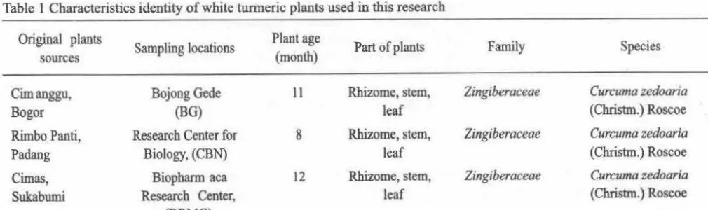
were collected from three locations in Bogor, West Java, Indonesia. The plants materials collected from private garden in Bojong Gede (BG), experiment garden of Research Center for Biology, Indonesian Institute of Sciences, Cibinong (CBN) and garden of medicinal plants collection of Biopharmaca Research Center, Bogor Agricultural University, Dramaga (DRMG) (Table 1 ). White turmeric plants were identified based on the morphological characteristics at
the Herbarium Bogoricnse, Indonesian Institute of
Sciences, Cibinong, Indonesia. From each plant materials, rhizome, stem, and leaf were collected for endophytic bacteria isolation.
Table l Characteristics identity of white tunneric plants used in this research
Original plants
Sampling locations Plant age Part of plants Family Species
sources (month)
Cilnanggu, Bojong Gede II Rhizome, stem, Zingiberaceae Curcuma zedoaria
Bogor (BG) leaf (Christm.) Roscoe
Rimbo Panti, Research Center for 8 Rhizome, stem, Zingiberaceae Curcuma zedoaria
Padang Biology, (CBN) leaf (Christm.) Roscoe
Cilnas, Biopbarm aca 12 Rhizome, stem, Zingiberaceae Curcuma zedoaria
Sukabumi Research Center, leaf (Christm.) Roscoe
[image:4.621.57.562.634.784.2]Volume 8, 2014
Surface Sterilization of Rhizomes, Stems, and Leaves. Rhizomes, stems, and leaves were thoroughly washed to remove external soil and microbes using running tap water for 5-10 min. Surface sterilization was done by stepwise soaking using 70% etanol solution for 3 min, 3% (v/v) sodium hypochloride for 5 min, 70% etanol solution for 30 s, and followed by three times rinsing with sterile distilled water. The samples were dried using sterilized towel tissue.
Isolation of Endophytic Bacteria. Endophytic bacteria isolation was done by plant piece and spread plate methods. Rhizomes, stems and leaves were cut using sterile knife approximately into 4-6 mm pieces.
In the plant piece method, pieces of samples were placed on four different media, Nutrient Agar (NA), NA contained 2% white turmeric plant extract (NAT), Water Yeast Extract Agar (WYEA), and WYEA contained 2% white turmeric plant extract (WYEAT). The media were supplemented with cycloheximide 50 µg mL"1 to avoid the growth of fungi. Plates were incubated at 28°C for 2-15 days. ln spread plate method, isolation of endophytic bacteria was done by grinding 1 g of samples in 9 mL of sterilized distilled water and 100 µL of 10-1 and 10-2 serial dilutions were spreaded on four different media same as above. The plates were incubated at 28°C for 2-15 days. Bacterial colonies which appeared on the media in a spread plate method were counted and expressed in colony forming units (CFU) per gram, and population data were transfonned to log (CFU per gram sample (CFU g·1
).
Some endophytic bacterial isolates which grew both on the media using spread plate and plant piece methods were picked up based on several phenotypic characteristics and then purified to obtain a single colony. Based on their different morphological characteristics, the endophytic bacteria were selected for further studies. Phenotypic characteristics which were used to observe the colony were: color, surface, the margin of the colony and gram reaction using KOH test.
DNA Extraction and Amplification of 168
rDNA. DNA extraction was conducted by colony PCR method (Packeiser et al. 2013) using Gradient PCR machines (Eppendorf Mastercycler Gradient PCR System 5331). Amplification of l 6S rDNA was performed by PCR using primer pair of 27F AGAGTTTGATCCTGGCTCAG-3') and 1492R (5'-GGTTACCTTGTIACGACTT-3') (Palaniappan et al.
2010). The l 6S rDNA amplification was carried out in a total volume of 25 µL containing U1trapure water, GoTaq Green Master Mix, 10 µM of each primer,
Microbiol Indones 67
dimethyl sulfoxide (DMSO), and DNA template. The PCR conditions was set as follows: initial denaturation at 95 °C for 90 s, followed by 30 cycles of denatura-tion at 95°C, for 30 s; annealing at 50°C, for 30 s; elongation at 72 °C, for 90 s and final extension at 72 °C for 5 min, finally at 4 °C for 20 min. PCR products were analyzed using l % agarose gel. Gel was soaked in ethidium bromide solution (5 µgmL1) for 30 min, rinsed with lX
TAE buffer, and the results were detected using a UV transilluminator.
DNA Sequencing and Phylogenetic Analysis. The amplified DNA were partially sequenced using forward primer 27F by automated DNA sequencer (ABI PRISM 3130 Genetic Analyzer) (Applied Biosystems). The sequenced data were processed using Bioedit programme. The homology of l 6S rDNA sequence were searched using BLAS1N at the NCBI website and the references sequence was obtained from the GenBank (www.ncbi.nlm.nih.gov). Constructions of phylogenetic tree was done using neighbor-joining tree method (NJT) implemented in MEGA 5.05 software (Tamura et al. 2011 ). Model of K2+G+I (Kimura2-parameter and Gamma distributed) was selected as the best-fit substitution model for the current analysis. Strength of internal branches of the phylogenetic tree was tested with boostrap analysis using l 000 replications.
RESULTS
Plant Materials Identity . Observation of morphological characters of white turmeric plant was done referred to the plant identification book of Flora of Java and compared to herbarium specimen in the Herbarium Bogoriense. Based on the morphological characteristics, all samples were identified as C.
zedoaria (Christm.) Roscoe, the member of the genus
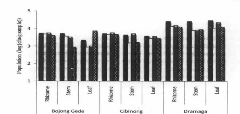
Curcuma in the family of Zingiberaceae (Table 1 ). Population of Endophytic Bacteria Associated with C. zedoaria. The population of endophytic bacteria contained in white tunneric plants differed between location, age, rhizome, stem and leaf, and also influenced by media and method of isolation. Based on the data presented in Fig 1, three plant materials showed to have different endophytic bacterial population ranging from 2 to 4 log (CFU g·1
). The
68 SuusTIY ANl ET AL.
endophytic bacteria was found in samples that collected from DRMG and among the part of plant, rhizome showed the highest population of endophytic bacteria. Two hundred and seven endophytic bacteria were isolated from different parts of white turmeric plants.
Based on the morphological characteristics of207 isolates, 73 were selected for further studies. Among the 73 selected isolates, 23 isolates from BG (32%), 16
5
cu E
-
Cl.IE Cl.I
..,
cu Ejセ@ v; _. 2
...::
セ@
a:
Bojong Gede
Microbiol lndones
from CBN (22%) and 34 from DRMG (46%). Furthermore, 28 isolates were obtained from rhizomes (38%), 23 isolates from stems (32%) and 22 isolates from leaves (30%). The Gram reaction results showed that 49 isolates were Gram-negative and 24 isolates were positive bacteria (Table 3). Both of Gram-positive and negative bacteria were found in all samples and bacteria from the rhizomes, stems and leaves were dominated by Gram negative bacteria.
セ@
-
Cl.Iセ@
-..,
E
..,
cu cu
...
_, 0 _,.,,
セ@
.,,
Clblnor.g Dramaga
[image:6.626.101.562.213.433.2]Locations
Fig 1 The population of white turmeric endophytic bacteria from three sampling locations based on spread plate method, • NA, 0 NAT, • WYEA, m WYEAT.
Table 2 The number of selected endophytic bacterial isolates obtained from white turmeric using spread plate and plant piece methods
NA NAT WYEA WYE AT
s
ps
ps
ps
pRhizome
6 1 3 2 1 1
Bojong Stem
7 2
Gede 2 3 2 2 3
Leaf
4 4 7 3 4 2 8 2
Rhizome 2 3 3 1
Cibinong Stem 3 5 3 4 2 2
Leaf
5 6 6 4 3
Rhizome 4 4 4 5 5 2 4 2 Dramaga Stem 5 4 3 4 3 2 4 3
Leaf 3 2 2 4 3 2 5
39 27 24 34 28 12 31 12
Total 207
[image:6.626.69.564.531.786.2]Volume 8, 2014
Molecular Identity of Endophytic Bacteria
Based on Partial Sequencing of 16S rDNA.
Basedon
the results of partial sequencing of about 700 - 1200 bp
and analysis of 16S rDNA, the 73 isolates showed high
similarities between 97% to 100% with the data bases
in GenBank. The molecular identification of all
isolates into species level were presented in Table 3.
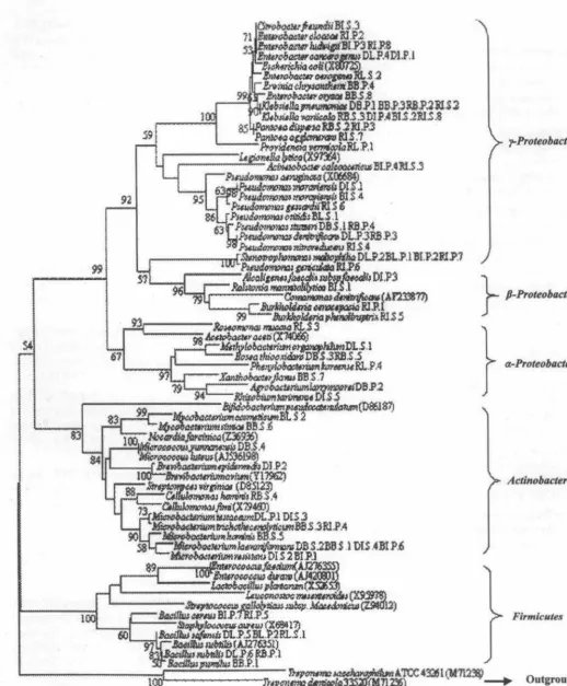
According to phylogenetic tree analysis, the isolates
widely distributed to the cluster of Alphaproteobacteria,
Microbiol Indones 69
Betaproteobacteria, Gammaproteobacteria, Firmicutes, andActinobacteria,
with twenty three different genera,
including Stenothropomonas, Pseudomonas,
[image:7.618.38.537.220.804.2]Enterobacter, Providencia, K/ebsiella, Dickeya, Pantoea, Bacillus, Acinetobacter, Citrobacter, Mycobacterium, Cellulomonas, Microbacterium, Methylobacterium. Penylobacleriwn, Roseomonas, Agrobacterium, Bosea, Xanthobacter, Rhizobium, Burkholderia. Ra/stonia. andA/caligenes (Fig 2).
Table 3 Diversity of endophytic bacteria from white turmeric plants based on 16S rDNA analysis KO
Research Center for Biology, Biophannaca Research Center,
H Bojong Gede Total
test Cibinong Dramaga
1. Bacillus subtilis I. Bacillus safensis I. Microbacterium
+ 2. Cellulomonas triclwthecenolyticum 5
lwminis 2. Bacillus cereus
I. Klehsiella I. Providencia vermicola I. Burkholderia cenocepacia pneumoniae 2. Phenylobacterium Jwreense 2. Burlcholderia phenoliruptrix
2. Pseudomonas 3. Enterobacter aerogenes 3. Enterobacter cloacae denilrificans 4. Roseomonas mucosa 4. Enterobacter ludwigii
3. Pseudomonas 5. Pantoea dispersa
Rhizome stutzeri 6. Pantoea agg/omerans
4. Pantoea dispersa 7. Pseudomonas geniculata 5. Klebsiella variicola 8. Pseudomonas gessardii 23 6. Bosea thiooxidans 9. Pseudomonas nitroreducens
10.Stenotrophomonas maltophilia
1 1. Klebsiella pneumoniae
12. Klehsiella variicola
13. Acinetobacter calcoaceticus
I. Microbacterium 1. Bacillus safensis 1. Microbacterium resistens laevaniformans 2. Mycobacterium cosmeticum 2. Bacillus cereus
2. Microbacterium 3. Microbacterium trichothecenolyticum laevaniformans
+ 3. Microbacterium 10
hominis
4. Mycobacterium simiae
Stem 5. Bacillus pumilus
1. Klebsiella l . Stenotrophomonas I . Stenotrophomonas pneumoniae maltophilia maltophilia
2. Erwin la chrysanthemi 2. Pseudomonas otilidis 2. Enterobacter /udwigii
3. Xanthobacter jlavus 3. Acinelobactcr calcoaceticus
13 4. Enterobacter oryzae 4. Ralstonia mannitoli/ytico
5. Klebsiella variicola
6. Citrobacter freundii
7. Pseudom()nas moraviensis
l. Microbacterium l. Microbacterium testaceum I. Mu:robacterium resistens laevaniformans 2. Bacillus safensis 2. Microbacterium testaceum
+ 2. Micrococcus 3. Bacillus subtillis 3. Microbacterium 9
yunnanensis laevaniformans
I. Pseudomonas stutzeri 1. Stenotrophomo111JS
4. Brevibacterium epidermidis
1. Enterobacter cancerogenus
2. Klebsiella maltophilia 2. Alcaligenes faeca/is subsp.
2. Pseudomonas denitri.ficans faecal is
13 3. Enterobacter cancerogenus 3. Klebsiella variicola
4. Methylobacterium 4. Pseudomonas moraviensis organophilum 5. Rhizobium tarimense
16 34 73
70 SUL!STIY AN1 ET AL.
DISCUSSION
The population of endophytic bacteria differed between location, age, rhizome, stem and leaf, and their diversity was also influenced by growth media. Io
this study, isolation of endophytic bacteria of white turmeric plant from three locations in West Java, Indonesia was done using two methods and four different media. Endophytic bacteria were succesfully isolated using four kinds of media, but addition of extract of white turmeric plants seemed to decrease population and diversity of endophytic bacteria. Among the four kinds of media used, NA was the suitable media for the endophytic bacteria isolation compared to WYEA. Based on the isolation method, the number of endophytic bacteria obtained using spread plate method was higher compared to plant piece method. This may caused by the differences in size and preparation of samples between the two methods. In the spread plate method, sample ( 4-6 mm2
)
were firstly ground to pieces and spread over the plate for bacterial growth. When the sample extracted using water, more microbes inside a plant moved to the water. This method seems to give more chance for endophytic bacteria to grow. While for the plant piece method the chance for endophytic bacteria to grow is limited because the sample ( 4-6
mm)
were directly put on media.Several genera of bacteria can be found in three different locations, i.e. Microbacterium, Pseudomonas, Enterobacter, Bacillus, Stenothropomonas, Klebsie/la, Mycobacterium, and Pantoea. Pseudomonas sp. and
Bacillus sp. are the most abundant endophytic bacteria found in the plants. They are considered easy to be cultured (Seghers et al. 2004). The presence of microbes in a host plant can be affected by the compounds contained in the host plants (Strobel and Daisy 2003). The plants of the same species may produce relatively similar bioactive compounds (Bernhoft 2010).
Rhizome, stem and leaf from CBN had the lowest abundance of endophytic bacteria. The plant materials originally come from tissue culture which was subsequently domesticated in that place. It could be possible reason for limited numbers of endophytes. Endophytic bacteria of white turmeric plant from DRMG was more diverse compared to other samples. The differences may also be influenced by the different
in
ecological niche condition of the plant. The fact that more than one hundred of medicinal plants can beMicrobiol lndones
found inDRMG, may also influence the soil microbial diversity. Another reason which can influence the endophytes diversity is age of host plant. Sample taken from DRMG was the oldest (12 months), followed by sample from BG (11 months), and sample from CBN (8 months). As
a
mature plant developed, all the nutrients for the endophytic bacteria may be more available and abundance thus stable endophytic population can be obtained. Age of plants has been reported to influence the variation of endophytic community in the gingseng plants (Vendan et al. 2010).Three plant materials of white turmeric plants showed different population of endophytic bacteria. The population on the rhizome of the plant was higher than those of the stems and leaves. The greater population found in the rhizome may be caused by the content of rhizome compounds. The plant uses the rhizome to store starch, protein, fat and other nutrients which are useful for the plant and its endosymbionts. Dalal and Kulkarni (2013) reported that population of endophytic microbes in roots or rhizome were the highest compared other part of plants, due to root is the earliest place for microbes entering the plant.
Among the 73 selected isolates, isolates belongs to the cluster of Gammaproteobacteria was the most dominant, followed by Actinobacteria, Alphaproteo-bacteria, Firmicutes, and Betaproteobacteria, they were 37, 16, 8, 8, 4 respectively. The genera of
Microbacterium was dominant, followed by
Pseudomonas, Bacillus, Klebsiella and Enterobacter.
In the present study, 73 endophytic bacteria which represented 46 species were belonging to 23 different bacterial genera have been identified from variously locations, aged, and part of plant of white turmerics. Cho et al. (2007) isolated 13 different bacterial genera of 63 endophytic bacteria from gingseng roots cultivated in three different areas. Vendan et al. (2010) isolated four clusters, 9 genera in 51 isolates from variously aged gingseng plants. Germida et al. (1998) reported that isolated 18 endophytic bacterial genera in
-
.
•Volume 8, 2014 Microbiol Indones 71
.,..Proteobacteria
92
} P-Proteobacteria
61
} a-Proteobacteria
Actinobacteria
l Firmicutes
Outgroup
[image:9.622.32.550.54.680.2]O.OS
72 SuuSTTY AN!
er
AL.REFERENCES
Banisalam B, Sani W, Philip K, lmdadul H, Khorasani A. 2011. Comparison between in vitro and in vivo
antibacterial activity of Curcuma zedoaria from Malaysia. Afr J Biotech. 10(55):11676-11681. doi:l0.5897/AJBl0.962.
BernhoftA. 2010. A briefreview on bioactive compounds in plants. In: Bioactive compounds in plants -benefits and risks for man and animals. Symposium held at The Norwegian Academy of Science and Letters, 2008 Nov
13 - 14.0slo. pll-17.
Chen YT, Yuan Q, Shan LT, Lin MA, Cheng DQ, Lil CY. 2013. Antitumor activity of bacterial exopolysaccharides from the endophyte Bacillus amyloliquefaciens sp. isolated from Ophiopogon japonicus. Oncol Lett. 5:1787-1792. doi :
10.3892/ol.2013.1284
Cho KM, Hong SY, Lee SM, Kim YH, K.abng GG, Lim YP, Kim H, Yun HD. 2007. Endopbytic bacterial communities in ginseng and their antifungal activity against pathogen s. Microb Ecol. 54:341 -351. doi: 10.1 007/s00248-007-9208-3.
Christina A, Christapber V, Bbore SJ. 2013. Endophytic bacteria as a source of novel antibiotics: An overview. Pbarmacogn Rev. 7( 13):11 -16. doi: 10.4103/0973-7847.112833.
Dalal J and Kulkarni N. 2013. Population dynamics and diversity of endophytic bacteria associated with soybean (Glycine max (L) Merril). Brit Microbiol Res J. 3(1):96-105.
Germida JJ, Siciliano SD, de Freitas JR, Seib AM. 1998. _ Diversity of root-associated bacteria associated with field-grown canola (Brassica napus L.) and wheat
(Triticum aestivum L.). FEMS Microbiol Ecol. 26:43-50.
Joy PP, Thomas J , Mathew S, Skaria BP. QYセX N@ _ Zingiberaceous Medicinal and Aromatic Plants. India:
Aromatic and Medicinal Plants Research Station, Odakkali,Asamannoor P.O.
Lakshmi S, Padmaja G, Remani P. 2011. Anti tumour effects of isocurcumenol isolated from Curcuma zedoaria
rhizomes on human and murine cancer cells. Int J Med Chem. 2011: l-13. doi: 10.1155/2011/253962.
Muthu-kumar T, Christy AMV, Mangadu A, Malaisamy M, Siviraj C, Arjun P, Raaman N, Balasubramanian K. 2012. Anticancer and antioxidant activity of Curcuma zedoaria and Curcuma amada rhizome extracts. J Acad IndusRes.1(2):91-96.
Microbiol lndones
Packeiser H, Lim C, Balagurunathan B, Wu J, Zhao H. 2013. An extremely simple and effective colony PCR procedure for bacteria, yeasts, and microalgae. Appl Biochem Biotech. 169:695-700. doi:l0.1007/s12010-012-0043-8.
Palaniappan P, Chauhan PS, Saravanan VS, Anandbam R, Sa T. 2010. Isolation and characterization of plant growth promoting endopbytic bacterial isolates from root nodule of Lespedeza sp. Biol Fert Soils. 46:807-816. doi: I 0.1007/s00374-010-0485-5.
Procopio REL, Araujo WL, Maccberoni JW, Azevedo JL. 2009. Characterization of an endophytic bacterial community associated with Eucalyptus spp. Gen Mot Res. 8(4): 1408-1422.
Pujiyanto S, Lestari Y, Suwanto A, Budiarti S, Darusman LK. 2012. Alpba-glucosidase inbt"bitor activity and characterization of endophytic Actinomycetes isolated from some indonesian diabetic medicinal plants. Int J Phann Phann Sci. 4( 1):327-333.
Ryan RP, Germaine K, Franks A, Ryan DJ, Dowling DN. 2007. Minireview: Bacterial endopbytes: recent developments and applications. FEMS Microbiol Lett. 278:1-9. doi:lO. l l l l /j. 1574-6968.2007.00918.x.
Seghers D, Wittebolle L, Top EM, Verstraete W, Siciliano SD. 2004. Impact of agricultural practices on the Zea
mays L. endophytic community. Appl Environ Microbiol. 70(3):1475-1482. doi :
10. l 128/AEM.70.3.1475-1482.2004.
Srikandace Y, Hapsari Y, Simanjuntak P. 2007. Seleksi mikroba endofit Curcuma zedoa ria dalam memproduksi senyawa kirilia antimikroba. JIFI. 5(2):77-84.
Strobel G, Daisy B. 2003. Bioprospecting for microbial endophytes and their natural products. Microbiol Mol Biol Rev . 67(4) : 491-502 . doi : 10. l 128/MMBR.67.4.491502.2003.
Tamura K, Peterson D, Peterson N, Stecher G, Nei M, Kumar S. 20 11. MEGA5 : Molecular evolutionary genetics analysis using maximum likehood, evolutionary distance, and maximum parsimony methods. Mot Biol Evol. 28(10):2731-2739 . doi: 10.1093/molbev/msrl 21.
Vendan RT, Yu YJ, Lee SH, Rhee YH. 2010. Diversity of endopbytic bacteria in ginseng and their potential for plant growth promotion. J Microbiol. 48(5):559-565.doi: I 0 .1 007/s l 2275-010-0082-1.