Med J Indones
246
LesmanaEndoscopic management of
difficult
common
bile duct stones
L.A. Lesmana
Abstrak
Tetah diselidiki 45 pasien dengan batu saluran empedu (BSE) sulit yang tidak dapat dikeluarknn dengan kombinasi sfingterotomi s e dur endo s kopik tambahan ; Iitotip si koledokoskopi peroral pada 7 ( l5%)' ri dari pasien lanjut usia dengan umur n empedu didapatkan pada seluruh 22 pasien yang menjalnni LM. Pemecahan dan pengeluaran batu
dip
LEH atau LL' Pada satu pasien' batu timbul kembali d.ua tahun sesudah LEH dan memerlukantindalan
berhasilan drainase empedu didapatkan pada 16 pasien dengan endoprostesis. Lima pasien mengalami penyumbatanendop
lama pemantauan 24 bulan; tiga berhasil menjalani LM dan dua lainnya mendapatkan endoprostesis baru. Tilak ditemakan komplikasi dalam studi ini' Prosedur endoskopik tambahan bermanfaat dalam tatalnksana BSE sulit.Abstract
We have studied 45 patients with dfficult common bile d sphincterotomy combined with stone removal using a Dormia ba àechanical lithotrtpsy (ML) in 22 (49Vo| electrohydraulic (EHL) and biliary stenting in the rema
72-85) who had cholangitis and
extraction were successful in
al
later a'fterofch
16 stentedste.n
successfult.
No
beneficialin treating dfficult CBD stones.
Keywords : biliary stenting, Iithotripsy, endoprosthesis, choledochoscopy
Endoscopic management of common bile duct (CBD) stones hàs become the treatment of choice, especially
successfully removed by endoscopic sphincterotomy follofued with stone removal using a Dormia basket or a balloon catheter.3-S However, in patients with
dif-ficult
CBD stones which include large stones, im-pacted stones,or
stones located above the ductal narrowing, several additional techniques have been developedfor
endoscopic removalof
the
stones through the incised PaPilla.be ac-
lectro-'P"'1:îl sy.
If
all these attempts fail to extract or crush large CBD stones and the patient has a high surgical risk, then i nsertion of an endoscopi c bili ary-prosthesi sl alloTn g theimpacting concrements has been suggested.'
We report herein our experience with additional endo-scopi- procedures
in
patientswith difficult
CBD stones.METHODS
patients doscopic
Vol 8, No 3, July - September 1999
biliary unit, Husada hospital, Jakarta. All patients un-derwent endoscopic sphincterotomy (ES) and
at-tempted removal of the stones using a Dormia basket with a diameter of 22 mm and length of 40 mm (FG 22
Q, Olympus optical Co., Tokyo, Japan).
Successful ES and stone extraction were obtained in 262 (98Eo) and 197 (7 4Vo) patients respectively. Of the
65 patients with Dormia extraction failure, 20
under-went surgery prior to the availability of lithotryptor and biliary stent at our unit. The remaining 45 patients were
defined to have difficult CBD stones (large, impacted,
or stones located above the distal narrowing) and
in-cluded in this study.
There were 26 females and 19 males with a mean age of 62 years (range 34-85 years). Patients with difficult CBD stones underwent mechanical, electrohydraulic
or pulse dye laser lithotrypsy and biliary stenting ac-cording
to
the following algorithm: a) mechanicallithotripsy for stones which could not be removed after grasping in the Dormia basket, b) electrohydraulic or
pulse dye lithotripsy
for
stones which could not be engaged in the Dormia basket (impacted stones), orc) biliary stenting for patients who had cholangitis and
surgical risk.
Additional endoscopic procedures
Mechanical lithotrtpsy
Mechanical lithotripsy
(ML) was performed
eitherwith
Soehendra lithotryptor (Wilson-Cook Medical Inc., Winston Salem, North Carolina, USA) or BMLlithotryptor (Olympus Optical Co., Tokyo, Japan).
The Soehendra lithotryptor consists
of
a cranking device and a 6 mm metal coil sheath. First, the handleof the Dormia basket containing the entrapped stone was clipped off and the endoscope was removed from the patient. A metal coil sheath was then advanced over
the plastic catheter up to the basket under fluoroscopic
guidanie. The basket wire and the sheath were then
connected to the cranking device and the stone was crushed by drawing the basket forcefully into the metal sheath.
The Olympus BML lithotryptor consists of coil sheath, tube sheath, basket wire, and BML handle. First, the instrument was introduced into the bile duct through
the incised papilla. The basket was opened by pushing the holder of
BML
and the stone was then captured. After grasping the stone, the knob of the BML handleDfficult conlmon bile duct
stones
247was rotated to crush the stone by squeezing with the basket.
Electrohydraulic and pulse dye laser lithotripsy
Electrohydraulic or pulse dye laser lithotripsy were carried out via peroral choledochoscopy using the
"mother-baby" endoscope system (Olympus TJF M20
mother scope and Olympus CHF 820 babyscope).
Electrohydraulic lithotripsy. Prior to performing EH,L, a 7FR nasobiliary catheter was inserted above the stone
for
bile duct irrigation with physiological saline to provide a fluid medium for effective generation and conduction of EHL shock wave and to allow controlcholangiography. After positioning the motherscope in front of the incised papilla, the babyscope was passed through the operating channel of the motherscope and
introduced under direct vision into the common bile
duct. The bile duct was examined endoscopically and
impacted stone was localized.
A
4.5 FR EHL probe, connected to the shock wave generator unit (Lithotron F,L23, Walz Electronic Inc., Germany), was insertedthrough the operating channel of the babyscope and placed directy to the stone surface. The shock wave generator was set to discharge twelve impulses per
second in volleys of 3 impulses; the energy was set at 0.46 Joule per impulse. Using a foot pedal to activate
the generator, spark discharges were applied over one
to two seconds. This was repeated until all stone
frag-ments were passable through the incised papilla. Duct clearance was confirmed by cholangiography.
Pulse dye laser lithotripsy. The flashlamp pulsed dye laser (Pulsolith, Technomed International, Lyon, France) was used in this study. The dye was Coumarin
green, producing a laser pulse wave length at 504 nm
with a pulse duration of 2.4 microseconds.
The laser beam was transmitted to the impacted stone
by direct contact via a 3-m long, 200 pm diameter
flexible quarts fiber inserted into the common bile duct through the operating channel of the babyscope. The procedure was repeated until fragments of stones could
be removed through the incised papilla.
Biliary stenting
A
straight 7or
l0 FR
endoprosthesis with side flaps(Wilson Cook Medical Inc., Winston-Salem, North Carolina, USA) were inserted into the common bile
248 Lesmana
stone(s) and, when possible, wedged
in
the intra hepatic ducts to prevent dislodgement. The distal end of the endoprosthesis was placed in the duodenum.stone-free with ML
22 patients
Med J Indones
RESULTS
The outcome of endoscopic management of 45 patients with
difficult
CBD stones is summarized in Figurel.
stone-fiee with EHL
or LL
7 patients
I
I
recurrent stone
1 patient
ML
was performedin
22
(49Vo) patients,9
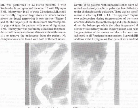
with Soehendra lithotryptor and the other 13 with Olympus BML lithotryptor. In all of these 22patients, ML could successfully fragment large stones or stones located above the ductal narrowing in one session (Figure 2 and 3). The majority of the stones weremacroscopical-ly
pigment type. In patients with several big stones,BML lithotryptor was preferably used since the proce-dure could be repeated several times without the
neces-sity to remove the endoscope from the patient. No complications were found with both of the techniques.
stent clogging Iost of follow-up
I I patients
Seven (157o) patients with impacted stones were
sub-mitted to electrohydraulic or pulse dye laser lithotripsy under cholangioscopic guidance. There was no spesific
reason in selecting EHL or LL. This approach required two endoscopists during fragmentation of the stones; one would handle the motherscope and simultaneously direct the babyscope while the other fragmented the
stones with electrohydraulic shock wave or laser beam.
Fragmentation of the stones and duct clearance were achieved in allT patients in one session: five with EHL
and two with LL (Figure 4). One patient with markedly stone-free new stent
with
ML
2 patients [image:3.595.85.582.137.352.2]3 patients
Figure.l. Outcome of difficult CBD stones using mechanical lithotripsy (ML), electohydraulic lithotrophy (EHL), pulse dye Laser lithotripsy (LL) and biliary stenting (BS).
45 patients
[image:3.595.85.572.347.726.2]Vol 8, No 3, JuLy - September 1999 Dfficult common bile duct
stones
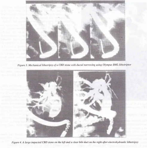
249Figure 3. Mechanical lithotipsy of a CBD stone with ductal narrowing using Olympus BML lithotriptor
Figure 4. A large impacted CBD stone on the lelt and a clear bile duct on the right after electrohydraulic lithotripsy
dilated CBD developed recurrent stone two years after
EHL. This patient underwent successful surgical
ex-ploration of the CBD, and the stone was brown and soft being characteristically of calcium bilirubinate type.
Minor bleeding occurred
in
two patients when theelectrohydraulic shock waves contacted the bile duct
wall. The bleeding stopped spontaneously in both of
the patients. No other complications were found.
The remaining 16 elderly patients with an average age
of 79 years (range 72-85) presented with cholangitis, defined as the combination of fever, leucocytosis and
stones in the biliary tract. Successful biliary drainage,
defined as resolution
of
jaundice and fever, was achieved in alll6
patients (l)OVa) after placement ofan endoprosthesis in the CBD (Figure 5).
During 24 month follow-up, five patients developed
clogging of the stent; three of these subsequently
un-derwent successful
ML
and the othertwo
patientsreceived new stents. Longterm complications
of
theremaining 11 patients could not be monitored due to
lost of contact. There were no complications related to
250 lzsmana
Figure 5. Biliary stenting in an elderly patient with a large CBD stone (arrow) associatedwith cholangitis
DISCUSSION
The present study supports the benefit and safety of mechanical lithotripsy in-treating difficult CBD stones as previously reported./'ô
In
contrastto
the
pre-dominantly cholesterol type of gall-stones found inWestern countries, most of CBD stones in our country are belonged to calcium bilirubinate type which are often soft-and fragile.ls Therefore, ML is highly effec-tive to crush and remove large CBD stones. This might
also
explain
that
stone fragmentation and duct clearance could be successfully obtained in one session in these series.It should be noted that ML with Soehendra lithotryptor was performed only to stones which could be captured in the Olympus Dormia basket. The disadvantage of
using Soehendra lithotryptor is the need to reinsert the endoscope in patients with several large stones. The patients with impacted stones in the CBD which could
not
be capturedin
the Dormia basket underwent electrohydraulicor
pulse dye laser lithotrypsy. Our results have shown that EHL is very effective to frag-ment impacted stone(s), which are in agreement withMed J Indones
earlier studies.8-10 One patient in this study, however, developed recurrent stone two years after successful EHL which required surgery. Two recent studies have analysed the risk factors for the formation of recurrent
stones after endoscopic sphincterotomy for choledo-cholithiasis.le'20
In itre
iirst
study,
multivariateanalysis
of 223 patients
after sphincterotomy has shown that a bile duct of 15 mm or greater, and the presence of papillary diverticulum are associated with recurrent CBD stones.lgTh"
second study revealed that recurrent stones occurred in 12.37o of 410 patients after endoscopic sphincterotomy during a meanfol-low-up of 122 months.
All
of these recurrent stones were calcium-bilirubinate type. Since the activity ofp-glucuronidase released from bacteria have been shown
to contribute to
the formationof
calcium-bilirubinate stones, this finding suggestedthe
role ofascendins biliarv infection in the recurrent stone for-mation.26ln fact, ou, patient with recurrent stone had
markedly dilated CBD and the stone was
macroscopi-cally calcium-bilirubinate type.
As suggested by others, EHL was carried out under
peroral choledochoscopic guidance to avoid complica-tions, since EH shock wave may injure the wall of the bile duct.2l'22 Our limited experience in two patients
with pulse dye laser lithotripsy revealed the effective-ness of this technique in fragmenting impacted stones. The efficacy and safety of laser lithotrypsy appeared
to
be comparableto
EHL
when performed under cholangiosôopic guidanc".la' ls Lur"i lithotripsy might also be useful as an alternative treatment for irretriev-able CBD stones in our country, since pigment stones are more susceotibleto ablation
than cholesterol stones.23Longterm follow-up after ES has shown that about l07o
of patients may develop symptoms,including
cholangitis, stenosis and recurrent stones.'*'"There-fore, careful follow-up
of
patients is mandatory to investigate late biliary tract complications of ES.Our results also support previous studies that biliary
stenting is an alternative endoscopic method for the treatment of irretrievable CBD stones.16-18 The
place-ment of a biliary stent results
in immediate biliary
drainage, which is essential for treatment andpreven-tion of cholangitis. This may enable elective surgical
and endoscopic treatment later, when the condition of the patient is better.
Three of our 5 patients with stent-clogging underwent
Vol 8, No j, July - September 1999
the other two patients which required exchange of
stent, the endoprosthesis could serve as a definitive treatment. Long term complications
in
the other ll
patients with permanent stent were difficult to monitor, since most of them were sent from other centres. In one recent study of 114 patients with irretrievable CBD stones using standard techniqqg, 55 received per-manent and 59 temporary stents.'" Thirty one of these59 patients underwent subsequent successful surgery and25 became stone free after repeating ERC, whereas the remaining three patients received permanent stent. Long term complications occurred
in
40Voof
those with permanent stent, with cholangitis being the most prevalent. Therefore, it has been suggested that per-manent biliary stenting should be restricted to patients who have high risk for elective treatment at a latterstage.
CONCLUSION
Additional endoscopic procedures including mechani-cal, electrohydraulic, pulse dye laser lithotripsy and
biliary stenting are beneficial in treating difficult CBD stones:
Acknowledgement
The author wishes to acknowledge the collaboration of Drs. Nanik Tjokrosetio and Aries Bunyamin.
REFERBNCES
1. Classen M, Demling L. Endoscopische sphinckterotomine
der papilla Vateri. Deutsch Med Wochenschr 1974; 99:
496-',7.
2. Kawai K, Akasaka Y, Murakami M, Tada M, Koli Y. Endo-scopic sphincterotomy of the ampulla of Vater. Gastrointest Endosc 197 4;20:148-51.
3. Cotton PB. Non-operative removal of bile duct stones by duodenoscopic sphincterotomy. Br J Surg 1980;67:1-5. 4. Viceconte G, Viceconte GW, Pietropaolo V, Montori A.
Endoscopic sphingterotomy: indications and results. Br J
Surg. 1981;68:376-80.
5. Mee AS, Vallon AG, Croker JR, Cotton PB. Non-operative removal of bile duct stones by duodenoscopic sphinctero-tomy in the elderly. Br Med J l98l; 283:.521-3.
6. SchneiderMU, MatekW, BauerR, DomschkeW. Mechani-cal lithotripsy of bile duct stones in 209 patients- effect of technical advances. Endoscopy 1988; 20: 248-53.
7. Siegel J, ben-Zvi JS, Pullano W. Mechanical lithotripsy of
common duct stones. Gastrointest Endosc 1990; 36: 351-6.
8. Binmoeller KF, Bruckner M, Thonke F, Soehendra N.
Treat-ment of difficult bile duct stones using mechanical, electrohydraulic and extracorporeal shock wave lithotripsy. Endoscopy 1993 ; 25 : 201 -6.
9. Liquory CL, Bonnel D, Canard JM, Cormud F, Dumont JL. Intracorporeal electrohydraulic shock wave lithotripsy of
Dfficult common bile duct
stones
251common bile duct stones: preliminary results in 7 cases.
Endoscopy 198'l : 237 -40.
10. Leung JWC, Chung SCC. Electrohydraulic lithotripsy with peroral choledochoscopy. Br Med J 1989; 299:595-8.
I l. Sauerbruch T, Stern M. Fragmentation of bile duct stones by extra-corporeal shock waves. Gastrointerology 1989; 96:
146-52.
12. Staritz M, Rainbow A, Grosse A, Hurst A, Floth A, Milden-berger P, et al. Electromagnetically generated extra
cor-poreal shockwaves for fragmentation of extra and intra hepatic bile duct stones: indications, success and problems during a l5 month clinical experience. Gut 1990; 3l:222-5. 13. Ponchon T, Gagnon P, Valette PJ, Henry L, Chavaillon A,
Thieulin F. Pulsed dye laser lithotripsy of bile duct stones'
Gastroenterology l99l ; 100: 1730-6.
14. Dawson SL, Mueller PR, Lee MJ, Saine S, Kelsey P,
Nishioka NS. Treatment of bile duct stones by laser lithotrip-sy: results in l2 patients. AJR 1992; 158: 1007-9.
15. Cotton PB, Forbes A, Leung JWC, Dineen L. Endoscopic
stenting for long-term treat'ment of long bile duct stones: 2-to 5-year follow-up. Gastrointest Endosc 1987; 33: 411-2. 16. Siegel JH, Yatto RP. Biliary endoprosthesis for the
manage-ment of retained common bile duct stones. Am J
Gastroenterol 1984;'l 9 : 5O-4.
17. Soomers AJ, Nagengast FM, Yap SH. Endoscopic
place-ment ofbiliary endoprosthesis in patients with endoscopical-ly unextractable common bile duct stones.
A
long-term follow-up study of 26 patients. Endoscopy 1990;22: 24-6.18. Lesmana LA. Clinical and biochemical aspects of
choledocholithiasis [Dissertation]. Amsterdam: University of Amsterdam; 1989.
I 9. Pereira-Lima JC, Jakobs R, Winter UH, Benz C, Martin WR, Adamek HE, et al. Long-term results (7 to
l0
years) ofendoscopic papillotomy for cholidocholithiasis.
Multi-variate analysis of prognostic factors for the recurrence of
biliary symptoms. Gastrointest Endosc 1998; 48: 457-64. 20. Tanaka M, Tahakata S, Konomi H, Matsunaga H, Yokohata
K, Takeda T, et al. Long-term consequence of endoscopic sphincterotomy for bile duct stones. Gastrointest Endosc 1998; 48: 465-9.
21. Horrison J, Morris DL, Haynes J, Hitchlock A, Womack C,
Wherry DC. Electrohydraulic lithotripsy of gallstones in
invitro and animal studies. Gut 1987; 28:26'1-71.
22. Sievert CE, Silvis SE. Evaluation of electrohydraulic lithotripsy on human gallstones. Am J Gastroenterol 1985;
80:854.
23. Nishioka NS, Levins PC, Munay SC, Parrish JA, Anderson
RR. Fragmentation of biliary calculi with tunable dye lasers' Gastroenterolo gy 1987 ; 93:. 250-5.
24. Leese T, Neoptolemos JP, Can-Locke DL. Success, failures, rarely complications and their management following endo-scopic spheinecterotomy: results in 394 consecutive patients
from a single centre. Br J Surg 1985; 72; 215-9'
25. Escourrou J, Cordova JA, Lazorthes F, Frexinos J. Early and late complications after endoscopic sphincterotomy for biliary lithiasis with and without the gallbladder "in situ". Gut 1984; 25:598-602.
26. Sergman JJGHM, Rauws EAJ, Tijssen JGP, Tytgat GNJ' Huibregtse K. Biliary endoprosthesis in elderly patients with
endoscopically irretrievable common bile duct stones.
Vol 8, No 4, October - December 1999 Widal test for typhoid
fever?
237Is Widal test
still
a
usefull method
as
a
routine early
diagnostic
for
typhoid
fever in hospitals
?Sylvia Y. Muliawan*, Lucky Hartati Moeharioi
Abstrak
Telah dilakukan penelitian uji Widal untuk mengetahui apakah uji Widal sebagai alat diagnostik dini demam tifoid yang rutin dikerjakan di Rumah Sakit masih bermanfaat pada saat
ini.
Pada penelitian ini digunakan tiga grup spesimen darah tunggaL, yaitu 2 grup spesimen berasal dari pasien rawat inap di Rumah Sakit dengan demam yang telah dikonfirmasikan dengan kultur sebagai positif (grup I) dan negatif (grup II), dan satu grup spesimen orang sehat yaitu donor darah. Hasil uji Widal dinyatakan positif bila titer aglutinin O Salmonella typhi lebih dari 80. Pada uji Widal sensitivitas yang diperoleh adalah 37Vo, spesifisitas 97Vo, nilai prediksi positif 90Vo, dan nilai prediksi negatif 73Vo. Akhir-akhir ini berkembang berbagai tes serologi, antara lain metode ELISA dengan menggunakan protein membran luar Salmonella typhi sebagai antigen, yang menunjukkan nilai sensitivitas dan spesifisitas lebih tinggi dibanding dengan uji Widal. Dengan demikian, penggunaan uji Widal secara rutin sebagai alat diagnostik dini demam tifuid perlu dipertimbangkan kembali.Abstract
In this study, we carried out Widal test to establish whether it is still signifcant as a routine early diagnostic tool of typhoidfever in the present time. We used three groups of patients: the first two Broups were hospitalized patients with fever, of which singLe blood specimen were taken and conjirmed by culture as positive (group I) and negative (group II), and the third group was healthy blood donors. AntiOagglutinintitersregardedasposilivewerethoseexceedingl:S0.ThesensitivityofsingleWidaltestwas3TVo,speciflcity gTVo,positivepredictivevaLueglVo,andnegativepredictivevalueT3Vo. Otherserologytestsdevelopedcurrently,amongotherstheuse of outer membrane protein preparation of Salmonella typhi as an antigen in the ELISA test, have shown higher sensitivity and specifcity thantheWidaltest. Therefore,theuseoftheWidaltestasarouîineearlydiagnostictoolfortyphoidfevermightneedtobereconsidered.
Keywords: Salmonella typhi, ELISA test, outer membrane protein, anti O agglutinin
Typhoid fever
is
an endemic acute systemic illness caused by infection of Salmonella typhi ( S. typ hi).
The disease remains a public health problem in developingcountries, including Indonesia.
In
several countriesannual epidemiological data for typhoid fever are ob-tained from clinical or laboratory results, and as the disease mimics several infectious diseases, it is
there-fore difficult to obtain a true picture of it.
The best method to detect an infection caused by S.
typhi
is
roorganism fromclini-cal
spec
is, how_ever, timecon-suming,
days,2-1 and it depends on laboratory facilities available in the areas. Among*
Department of Microbiology, Faculty of Medicine,
IJniver-,
sity ofTrisakti, Jakarta, Indonesia'
Department of Microbiology, Faculty of Medicine, Univer-sity of Indonesia, Jakarta, Indonesiathe many methods
of obtaining
clinical specimens,blood culture is the one routinely used for the isolation
of
the microorganism. Although culture may show specificity,it lacks sensitivity mainly
if
the patientshave already taken antibiotic treatments. On the other hand, routine serology test, i.e. Widal test,8 although
widely used, lacks sensitivity and specificity. For a
meaningful interpretation of the test, demonstration of a 4-fold rise in antibody titers between acute and
con-valescent sera
in
a7-14 days interval is essential.3'6Due to those reasons, during the last decade efforts have been carried out in developing newer methods for
early diagnosis of typhoid fever. One of the diagnostic
methods to
be
developedis ELISA
using outermem,brane protein (OMP) preparation as an anti-gen.e-l I Previous studies have indicated the suitability
of S.
typhi
OMPs as immunogensin
stimulatinganti-S. typhi antibodies formatio"n. I 2- I 4.
Earlier study
240 Istiantoro et al. Med J Indones
Six-month follow-up
of laser
in-situ
keratomileusis
for
myopia
Istiantoro, Tjahjono D. Gondhowihardjo, Johan Hutauruk
Abstrak
Penelitian retrospektif untuk menilai hasil refraksi dari laser in-situ keratomileusis (lnsik) menggunakan excimer Laser untuk
menentukan ketepatan, prediktabilitas, keamanan dan stabilitas koreksi miopia. lnsik dilakukan pada 475 mata pada 302 penderita
miopia dengan ekwivalen sferis (ES) dari 450 sampai -27 diopti (D). Diba7i dalam grup A (ES kurang dari 4.00D), B (SE 4.00
sampai
-l
1.99) dan C (ES -12.00 D atau lebih). Pengamatan dilakuknn sampai 6 bulan. Hasil: pada semua grup hanya 122 matamendapat pengamatan 6 bulan. Rerata pra-bedah ES -8.45D t4.66 dan rerata pra-bedah silinder -1.10D + 1.07 (dari plano sampai
4.00 D). Pada 6 bulnn, hasil koreksi dalam kisaran 2.00 D dari koreksi seharusnya tercapai pada 95.6 Vo grup A, 97.7 Vo grup B dan
78. I7a grup C. Rerata regesi kurang dari 1.00 D terdapat pada semua grup pada pengamatan 6 buLan. Komplikasi terdapat pada 29
mata (6 Vo) untuk seluruh grup. Kesimpulan: Lasik sangat efektif dan tepat untuk koreksi miopia rendah dan sedang. Untuk miopia tinggi
(>-12,00 D) efektivitas danprediktabilitas cukup baik. Stabilitas refraksisampai6 bulantampaknyabaik, namundiperlukanpengamatan
Lebih Lama lagi.
Abstract
Retrospective study to evaluate the refractive results of laser in-situ keratomileusis (LASIK) using excimer laser performed on
myopic
eyes;
correct myopia. I'ASIK wasperformed
on
ranging from 4.50 to -27.00diopters
(D).
to -11.99 D), or C (SE -12.00D or
higher).
2 eyes at 6 months. The meanpreoperative spherical equivalent was -8.45D 4.66 [SD], and the mean preoperative cylinder was -l.I1D
!
1.07 [SD] (range: planoto -6.00D). At 6 months, 95.6Vo of the eyes in Group A, 97.7Vo in Group B, and 78.lVo in Group C were within 2.00 D of intended
correction. The mean regression at 6 months was less than 1.00 D in all Broups. Complications were observed in 29 eyes (6Vo) of all
groups. Conclusion: LASIK was found to be very effective and predictable in the coruection of low and moderate myopia. For high
myopia (> - l 2.00 D| the effectiveness and predictability of LASIK were fairly good. Results after 6 months tend to suggest the stability
of postoperative refraction, but longlerm follow-up will be required to make further conclusions.
Keywords: Insik, excimer laser, comealflap technique
The use of ultraviolet 193-nm excimer laser for corneal
surgery has been suggested in several studies since the
first study by Trokel et all in 1983. Radiation emitted
by the argon fluoride excimer laser has demonstrated
a
unique abilityto
ablate corneal tissuewith
sub-accuracy, le
Its appticat
1989 * with
ogy known as photorefractive keratectomy (PRK) has
evolved, there are 2 major problems encountered. The
first problem is subepithelial haze in the visual axis,
Department of Ophthalmology, Faculty of Medicine,
Universitt of Indonesia, Jakarta, Indonesia
and the second is the predictability and stability of the
refractive results in high myopia.
Since PRK did not prove its safety, effectivene_ss, and
predictabilig/
in
myopia higher ihan 6.00 D.5'6Pal-likaris et
al'
suggested the corneal flap technique for"laser in situ keratomileusis" (LASIK) for high levels
of myopia. LASIK is a refractive surgery technique
that uses microkeratome to raise a corneal flap and
followed by an excimer laser to ablate the stromal bed.
8,9
This study analyzes thè refractive results of 475
con-secutive
LASIK
procedures performed on normal,sighted myopic eyes;
to
determineits
efficacy and