\b
ISSN 2087-3840
WOOD RESEARCH Journal
Journal of Indonesian Wood Research Society
Volume 1, Number 2, October 2010
•
•
•
•
•
•
•
•
Control of Ory-wood Termite Infestation by Bait System
Development of Bio-control Technology for Subterranean Termites
Coptotermes curvignathus Holmgren Using Electromagnetic Waves
Molecular Identification of Decay Fungi in Xylem of Yellow Meranti
(Shoreo gibboso) Canker
Relationship between Wood Properties and Developed Qrving Schedule of Inferior Teak (Tectono grondis L.F) and Mahogany
(Swietenio mocrophylla King)
VuJiati lndrayani
Farah Oiba, Ferry Hadary, Seno Darmawan Panjaitan, and Tsuyoshi Yoshimura
Erwin, Shuhei Takemoto, and Yuji Imamura
Tomy Ustyanto, Ganis Lukmandaru, Chandra Pramadya, Dwi Siswanto, and Nobuaki Hattori
Hypoglycemic Effect of Mahogany (Swietenia macrophyl/a King) Bark Syamsul Falah, Mega Safithri, Takeshi Katayama, Extracts in Alloxan-induced Diabetic Rats and Toshisada Suzuki
Precise Structure of Acidic Polysaccharide Present in Salvia Hygrogels Rike Vudianti, Myrtha Karina, Masahiro Sakamoto, and Jun-ichi Azuma
Wood Characteristic of Superior Sengon (Paroserianthes falcotoria) Sri Hartati, Enny Sudarmonowati, Widya Collection and Prospect of Wood Properties Improvement through
Genetic Engineering .
Micropropagation and Protoplast Culture in Paraserianthes fo/catoria
Fatriasari, Euis Hermiati, Wahyu Owianto, Rumi Kaida, Kei'ichi Baba, and Takahisa Hayashi
Miyuki Chujo, Shinso Yokota, Futoshi lshiguri, Kazuya lizuka, Dody Priadi, Nurul Sumiasri, and Nobuo Yoshizawa
65
71
78
83
89
95
103
108
Volume 1, Number 2, October 2010
ISSN
RPXWセSXTP@WOOD RESEARCH Journal
Journal of Indonesian Wood Research Society
Bogar Agricultural University
Prof. Wasrin Syafii Prof. Fauzi Febrianto Dr. Naresworo Nugroho
Indonesian Institute of Sciences
Dr. Myrtha Karina Dr. Sulaeman Yusuf Dr. Puspita Lisdiyanti
Mufawarman University
Prof. Sipon Muladi Dr. Rudianto Amirta Dr. lrawan W. Kusuma
Gajah Mada University
Dr. Sri Nugroho Marsoem
Chief Editor
Dr. Wahyu Dwianto, M.Agr.
Editorial Board Members
Prof. Muh. Yusram Massijaya Prof. Imam Wahyudi
Dr. Subyakto Dr. I Nyoman J. Wistara Dr. I Wayan Darmawan
Advisory Board Members
Hasanuddin University
Prof. Musrizal Muin
Research Institute for Human Settlement
Dr. Anita Firmanti
Kyoto University
Prof. Toshiaki Umezawa Prof. Kohei Komatsu
Kangwon National University
Prof. Nam-Hun Kim
Universiti Putra Malaysia
Prof. M. Hamami Syahri Dr. Edi Suhaimi Bakar
University of Melbourne
Dr. Barbara Ozarska
Supporting Staffs
Oklahoma State University
Prof. Salim Hiziroglu
The University of Tokyo
Prof. Naoto Ando
FPRDI Philippine
Dr. Dwight Eusebio
Paris Tech Cluny
Prof. Remy Marchal
Dresden University
Dr. Christian Gottleber
Tokyo University of Agriculture
Prof. Takahisha Hayashi
Dr. Sasa Sofyan Munawar, M.Si Karnita Yuniarti, M.Sc.
Hypoglycemic Effect of Mahogany
(Swietenia macrophylla
King) Bark Extracts
in Alloxan-induced Diabetic Rats
Syamsul Falah, Mega Safithri, Takeshi Katayama, and Toshisada Suzuki
Abstract
In this study, in vivo hypoglycemic activity of mahogany ( Swietenia macrophylla) bark extracts was evaluated against alloxan-induced diabetic rats. The hypoglycemic effect was compared to that of standard glibenclamide. Ornl administration of hot water and methanol extracts at a dose of 250 mg/kg body weight for thirteen days of daily treatment to diabetic rats was found to possess significant dose dependant hypoglycemic effect in diabetic rats. It less active than that of glibenclamide at dose of 3.22 mg/kg. However, the hot water extract showed significant hypoglycemic activity compared to that standard drug. Phytochemical analysis of hot water and methanol extracts has shown posistive test for the presence of alkaloids, fiavonoids, tannins, saponins, dan terpenoids. Histopathological studies of pancreas revealed its significant effect of セM」・ャャ@
count. Therefore, the hot water extract could serve as good adjuvant to other oral hypoglycemic agents and seems to be promising for the development of phytomedicines for diabetes mellitus.
Key words: Swietenia macrophyl/a, bark extract, hypoglycemic activity, alloxan-induced diabetic rats. Introduction
Diabetes mellitus is a group of metabolic diseases characterized by hyperglycemia resulting from defects in insulin secretion, insulin action, or both. The chronic hyperglycemia of diabetes is associated with long-term damage, dysfunction, and failure of various organs, especially the eyes, kidneys, nerves, heart, and blood vessels (Bowman and Russel 2001 ). Statistical projections mentioned that number of diabetics in the world wili increase from 151 million in the year 2000 up to 221 million in the year 2010, and hence it become Indonesia is the fourth in the highest number of diabetics in the world after India, China, and USA (King et al. 1998; Boyle et al. 2001; Zimmetetal. 2001).
Traditional medicinal plants have been employed successfully by the local communities since long time to treat diabetes without adverse effects. Researches in traditional medicine ·for appropriate hypoglycemic agents have been focused on plants due to traditional medicine gives better treatments than drugs (Rates 2001). Seeds of mahogany ( Swietenia macrophy!la ) have been used for treatment of diabetes as a folk medicine in Indonesia (Kadota et al. 1990). The seed also have been used for leishmaniasis and abortion medicine by an Amazonian Bolivian ethnic group (Bourdy et al. 2000) and for treatment of hypertension and malaria (Kadota et al. 1990). However, bioactivities from the bark have not been investigated extensively. The bark of mahogany, collected from Indonesia, contain ftavonoids with high antioxidant activity, namely swietemacrophyllanin, catechin, and epichatechin (Falah et al. 2008). In this study, the hypoglycemic effect of mahogany bark extracts were evaluated, and phytochemicals compounds of the extracts were examined. The effect of hot water and methanol extracts were evaluated on diabetic rats and its effects were compared
with glibenclamide, a standard hypoglycemic agent. Materials and Methods Plant Material
Mahogany bark was collected from Sumedang, Indonesia since March 2009. A dried bark poWcler of mahogany (500 g) were boiled in 1 liter of water for 4 h to give a hot water extract (29 g). The extract was filtered with filter paper (Whatman, no. 1) and evaporated with rotary evaporator at 60°C, and the crude extract was used in biological assay. Another 3000 g of the dried bark powder was extracted with acetone for 48 h at room temperature to give acetone extract (237 g), and then the residue was extracted again by methanol to yield methanol extract (184 g). The acetone was used for extraction of non polar substances, i.e. fatty acid, wax. The methanol extract was evaporated, and the extract was used in biological assay. Chemicals and Drugs
The solvents were of analytical grade and purchased from Merck, Germany. Alloxan and glibenclamide were obtained from Sigma Chemical, USA and Daonil Aventis Pharmacy, USA, respectively. All other chemicals were of analytical grade.
Qualitative Phytochemical Analyses (Harborne 1987) Alkaloid Test. The hot water and methanol extracts of 0.1 g each were added with 3 ml of chloroform and 3 drops of ammonia. The chloroform fraction was separated and acidified with 10 drops of H2S04 2M. The H2S04 fractions were taken and added separately with Dragendof, Meyer, and Wagner reagents. The alkaloids content was indicated by white precipitant upon addition of Meyer reagent, orange precipitant upon Dragendorf reagent, and brown precipitant upon addition of Wagner reagent.
Hypoglycemic Effect of Mahogany (Swietenia macrophyl/a King) Bark Extracts in Alloxan-induced Diabetic Rats
Saponin Test. The extracts of 0.1 g were added with 2 ml of H20 and heated for 5 min. The mixtures were cooled down, stirred up until foamy appearance can be observed to indicate the presence of saponin.
Flavonoid Test. The extracts of 0.1 g were soaked with 2 ml of 30% methanol and heated. The filtrates were added with 1 drop of concentrated H2S04. The presence of ftavonoid was indicated by the formation of red pigment.
Phenolic Hydroquinone Test. The extracts of 0.1 g were soaked with 2 ml of 30% methanol, heated and filtered. The filtrates were added with 1 drop of NaOH 10%(b/v). The presence of phenolic hydroquinone was indicated by the formation of red color.
Triterpenoid Test. The extracts of 0.1 g were added with 2 ml of 30% ethanol, heated and filtered . The filtrates were evaporated and then diethyl ether was added. The Lieberman Burchard reagent (3 drops of acetic acid anhydride and 1 drop of concentrated H2SQ4) was added to the ether layer. The presence of triterpenoid was indicated by the formation of reddish-violet pigment.
Tannin Test. The extracts of 0.1 g were added with 2 ml of H20 and heated for several minutes. The mixtures were filtered and the filtrates were added with FeCb 1 % (b/v). The presence of tannin was indicated by the formation of dark-blue or greenish-black color.
Animals
Male Sprague-Dawley rats of 3 weeks old were obtained from The National Agency of Drug and Food Control of Indonesia. They were fed with a standard laboratory diet and allowed food and water ad libitum for an acclimatization periods of 2 weeks prior to experiments. The animals were divided into five groups of seven each and housed individually during the experimental period.
Experimental Design
All the rats (Sprague dawley albino male rats) were randomly divided into the five groups.
Group A: Normal rats administered NaCl 0.9% by intraperitoneal and orally aquades 1 ml daily for 13 days.
Group B: Diabetic control rats administered alloxan 150 mg/kg by intraperitoneal and orally aquades 1 ml daily for 13 days.
Group C: Diabetic rats administered standard drug glibenclamide (3.22 mg/kg, orally) daily for 13 days.
Group D: Diabetic rats administered hot water extract (250 mg/kg, orally) daily for 13 days.
Group E: Diabetic rats administered methanol extract (250 mg/kg, orally) daily for 13 days.
90
Alloxan was injected to all rat groups on 1st day. Treatment with the extracts and glibenclamide was started · 48 h after alloxan injection. Blood samples were obtained from the tail vein in fasting rats for 18 h and blood glucose levels were measured using an electronic glucometer (Miles Inc, USA). Fasting blood glucose and body weight were measured on 1st, 3rd, and 15th days.
Statistical Analysis
All the values of body weight and fasting blood sugar were expressed as mean
±
standard error of mean (S.E.M) and analyzed for ANOVA and Duncan's t-test. Differences between groups were considered significant at P < 0.05.Histopathological Studies
All the animals were sacrificed on 15th day by cervical dislocation. Pancreases were excised, isolated, and were subjected to histopathological studies and microscopical finding were noted. The pancreas tissues were removed immediately and washed with ice-cooled saline, and then fixed in 10% of neutral formalin. The sections stained in haemetoxylin and aeosin and mounted were observed
under microscope.
Results and Discussion
Phytochemicals assay of hot water and methanol extracts of mahogany bark revealed the presence of the flavonoids, tannins, triterpenoids, saponins and alkaloids (Table 1). Phytochemical compounds such as, ftavonoids , triterpenoids, alkaloids, and phenolics are known to be bioactive antidiabetic principles (Nagappa et al. 2003; Battu
et al. 2007; Safithri and Fahma 2008).
The effect of treatment on rat body weight on 1st day
showed that rats body weight in all groups did not differ significantly (P<0.05) (Table 2). Rats body weight decreased on 3rd day after alloxan (B, D, and E group) induction; the highest degradation occurred at group D (4.4% from body weight of 1st day). However, body weight degradation on 3rd day in B, D, and E group did not different significantly with A and C group (P<0.05). On 15th day, the rats body weight was measured to evaluate the effect of hot water and methanol extracts of mahogany bark which orally administered at the dose of 250 mg/kg body weight (D and E group). During 13 days treatment (from 3rd day till 15th day), hot water and methanol extracts reduced the body weight by 8.54% and 7.36%, respectively. D and E groups did not differ with B and C group (P<0.05). It means that mahogany bark extracts reduced rat body weight the same as B and C group. Body weight reduction was also indicated by aqueous extract of Terminalia catappa at the dose of 42 mg/kg for 12 days treatment in alloxan-induced diabetes up to 63.09% (Nagappa et al. 2003) and decoction of Piper
crocatum of 322 mg/kg for 10 days treatment in
alloxan-induced diabetes up to 17.28% (Safithri and Fahma 2008). The decrease of body weight in diabetes is due to ·
continuous excretion of glucose and glycogen synthesis (Defronzo et al. 1992).
Measurement of blood glucose level was carried out on the 1st, 3rd, and 15th day to observe the effect of aquades, glibenclamide, and mahogany bark extracts orally administration and induction of NaCl or alloxan. The induction influences blood glucose rats during experiment. On 1st day (before treatment), rats blood glucose in all groups resulted not significant different (P<0.05) (Table 3) and performed at normal range (60-110 mg/di). However, after NaCl and alloxan induction (on 3rd day), rats blood glucose increased. Induction of alloxan (150 mg/kg) (B, C, D, and E group) increased blood glucose up to 0.5-1.0 folds. Blood glucose levels data showed B group has the highest increasing by 150.9%. Increasing of blood glucose rats on 3rd day after induction of alloxan, showed significantly different (P<0.05) with A group (Table 3). Rats blood sugar was reduced up to 45.73% and 25.80% in thirteen days after treated with hot water and methanol
extracts at a dose of 250 mg/kg. It is less active than that of glibenclamide which reduced blood sugar level by 48.42%. · It was indicated that the hot water extract showed significant antihyperglycemic activity as compared to that of standard drug.
Antihyperglycemic activity from decoction of
P
crocatum at 322 mg/kg body weight reduced blood glucose
level up to 10.46% after ten days given to diabetic rat. The extract contained flavonoids, alkaloids, and tannin (Safithri and Fahma 2008). Blood glucose reduction up to 3.68% occured from alcoholic extract of Chinese squash
(Benincasa hispida) at 200 mg/kg after 24 h given to
diabetic mice. The extract contained alkaloids, flavonoids, saponins, and steroids (Battu et al. 2007) . The alcohol extract of gopher plant (Euphorbia leucophy/lum) at 500 mg/kg in diabetic mice showed that it possessed an antihyperglycemic activity to reduce blood glucose up to 21.54% after 24 h the extract was given (Satyanarayana et al. 2006).
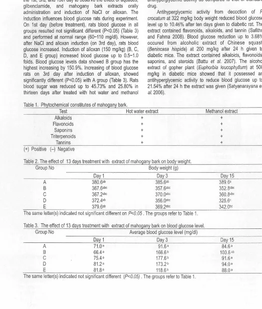
Table 1. Phytochemical constitutes of mahogany bark.
Test Hot water extract Methanol extract Alkaloids +
Flavonoids +
Saponins +
T riterpenoids +
Tannins + (+) Positive (-) Negative
Table 2. The effect of 13 days treatment with extract of mahogany bark on body weight. Group No Body weight (g)
Day 1 Day 3 A 38Q.6ab 385.6ab B 367.6abc 357.6abc C 367 .2abc 370.0abc D 372.4ab 356.0abc E 379.6ab 369.2abc
The same letter(s) indicated not significant different on P<0.05 . The groups refer to Table 1. Table 3. The effect of 13 days treatment with extract of mahogany bark on blood glucose level.
Group No Average blood glucose level (mg/di) Day 1 Day 3
A 71.0 a 91.6 a B 66.4a 166.6b C 75.4a 177.6b
D 81.2 a 173.2 b E 81.8a 118.6 b
The same letter(s) indicated not significant different (P<0.05) . The groups refer to Table 1.
Hypoglycemic Effect of Mahogany (Swietenia macrophylla King) Bark Extracts in Alloxan-induced Diabetic Rats Syamsul Falah, Mega Safithri, Takeshi Katayama, and Toshisada Suzuki
+ + +
+
+
Day 15 389.6a 352.8abc 360.8abc 325.6C 342.0bc
Day 15 84.6a 103.6 ab 91 .6 a
94.Qa 88.Q a
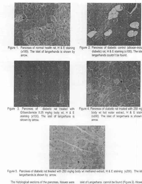
[image:5.595.11.534.75.692.2]Figure 1. Pancreas of normal health rat, H & E staining (x100). The islet of langerhands is shown by arrow.
Figure 3. Pancreas of diabetic rat treated with Glibenclamide 0.25 mg/kg body wt, H & E staining (x100). The islet of langerhans is shown by arrow.
Figure 2. Pancreas of diabetic control (alloxan-induced diabetic) rat, H & E staining (x100). The islet of langerhands couldn't be found.
Figure 4. Pancreas of diabetic rat treated with 250 mg/kg body wt hot water extract, H & E staining (x200). The islet of langerhans is shown by arrow.
Figure 5. Pancreas of diabetic rat treated with 250 mg/kg body wt methanol extract, H & E staining (x200) . The islet of langerhands is shown by arrow.
The histological sections of the pancreas, tissues were observed to know the effect of extract of mahogany bark in alloxan diabetic rats . The cellular integrity and architecture were intact in the A group. Besides that, in group A there was no specific abnormalities, and easy to find the islets of Langerhans (Figure 1 ). Pancreatic sections stained with hematoxylin and eosin (H & E) showed that alloxan caused fat necrosis; acinar cell necrosis, and hemorrhage and the
92
islet of Langerhans cannot be found (Figure 2). However, administration of glibenclamide at the dose of 3.22 mg/kg/day orally in alloxan diabetic rats showed no necrosis. The size and the number of islets of Langerhans is smaller and fewer than normal group, respectively (Figure 3). Meanwhile, administration of hot water extract of mahogany bark (250 mg/kg/day, orally) in alloxan diabetic rats showed fat necrosis, acinar cell necrosis, the number.
[image:6.602.43.513.20.628.2]and size of islets of Langerhans smaller than normal group (Figure 4). Furthermore, administration of methanol extract of mahogany bark (250 mg/kg/day, orally) in alloxan diabetic rats showed necrosis and easy to be found the islets -0f Langerhans (Figure 5).
In this study, the pancreatic
f1
cells were destroyed with the help of alloxan. Alloxan is one of the usual substances used for the induction of diabetes mellitus apart from streptozotocin. Alloxan has a destructive effect on thef1
cells of the islets of Langerhans (Szkdelski 2001 ). The alloxan produce permanent hyperglycemia by selective destruction of the
f1
cells of the islets of Langerhans are in agreement with those of Singh and Gupta (2007a). The histopathological study of diabetic treated with the extracts indicated the increasing of volume density of islets and percentage off1
cells, in the diabetic rats that received the extracts, which may be a sign of regeneration. Signs of regeneration off1
cells, potentiation of insulin secretion from survivingf1
cells of the islets of Langerhans and decrease of blood glucose have been reported following consumption of some plant extracts (Yadav et al. 2008; Singh and Gupta 2007b). Hot water and methanol extracts of mahogany bark may have some chemical components that exert regenerative effects onf1
cells, stimulate these cells to produce more insulin (pancreatotrophic action) or may have some insulinlike substances. Induction of regenerative stimulus in diabetic state triggers pancreatic regenerative processes, thereby restoring functional activities of the pancreas (Adewole and Ojewole 2007). A higher dose of the extract has a greater restorative effect on the islet cells of diabetic rats than a lower dose of extract.Conclusions
The hot water and methanol extracts of mahogany bark contained fiavonoids, tannins, triterpenoids, sapcnins and alkaloids. Oral administration of hot water and methanol extract at a dose of 250mg/kg for thirteen days of daily treatment led to reduce blood sugar level by 45.73% and 25.80%, respectively. It is less active than that of glibenclamide which reduce blood sugar level by 48.42%. However, the hot water extract showed significant antihyperglycemic activity as compared to that of standard drug . Histophatological study indicated the extracts exert
regenerative effect on [l, cells , stimulate to produce more
insulin. Further research is needed to explore different mechanisms to reduce blood glucose levels.
References
Adewole, S.O.; J.A.O. Ojewole. 2007. Insulin-induced lmmunohistochemical and Morphological Changes in Pancreatic セM」・ャャウ@ of Streptozotocin-treated Diabetic Rats. Methods and Findings in Experimental and Clinical Pharmacology 29: 447-455.
Battu, G.R.; S.N. Mamidipalli; R. Parimi; R.K. Viriyala; R.P.
p。エセィオャ。[@ L.R. Mood. 2007. Hypoglycemic and Anti-hyperglycemic Effect of Alcoholic Extract of Benincasa
hispida in Normal and in Alloxan Induced Diabetic
Rats. Pharmacognosy Magazine 3: 101-105.
Bourdy, G.; S.J. De Walt; L.R. Chavez de Michel; A. Roca; E. Deharo; V. Munoz; L. Balderrama; C. Quenevo; A. Gimenez. 2000. Medicinal Plants Uses of the Tacana, An Amazonian Bolivian Ethnic Group. J. Ethnopharmacol 70: 87-109.
Bowman, B.A.; R.M. Russel. 2001. Present Knowledge in Nutrition. and ed. Washington. DC: International Life Sciences Institute.
Boyle, J.P.; A.A. Honeycutt; K.M. Narayan; T.J. Hoergoer; L.S. Geiss; H. Chen; T.J. Thompson. 2001. Projection of Diabetes Burden through 2050: Impact of Changing Demography and Disease Prevalence in the U.S. Diabetes Care 24: 1936-1940.
Defronzo, R.A.; R.C. Bonadonna; I. Ferrannini. 1992. Pathogenesis of the Type 2 (Non-insulin Dependent) Diabetes Mellitus: A Balanced Overview. Diabetologia 35: 389-397.
Falah, S.; T. Suzuki; T. Katayama. 2008. Chemical Constituents from Swietenia macrophylla Bark and their Antioxidant Activity. Pakistan Journal of Biological Science 16: 2007-2012.
Harborne, J.B. 1987. Phytochemical Methods. London: Chapman and Hall.
Kadota, S.; L. Marpaung; T. Kikuchi; H. Ekimoto. 1990. Constituents of the Seed of Swietenia mahagoni
JACQ. I. Isolations, Structures and 1H and 13C Nuclear
Magnetic Resonance Signal Assignment of New Tetranortriterpenoids Related to Swietenine and Swietenolide. Chem Pharm Bull 38: 639-651 .
King, H. ; R.E. Aubert; W.H. Herman. 1998. Global Burden of Diabetes 1995-2025: Prevalence, Numerical Estimates and Projections. Diabetes Care 21 : 1414-1431 . Nagappa, A.N .; P.A. Thakurdesai; N.V. Rao; J. Singh. 2003.
Antidiabetic Activity of Terminalia catappa Linn Fruits. J. Ethnopharmacol 88: 45-50.
Rates, S.M. 2001 . Plants as a Source of Drugs. Toxicon 39: 603-613.
Safithri, M.; F. Fahma. 2008. Potency of Piper crocatum
Decoction as an .Antihiperglycemia in Rat Strain
Sprague Dawley. Hayati Journal of Bioscience 15:
44-48.
Satyanarayana, T.; B.M. Katyayani; E. Hema Latha; A.M. Anjana ; E.M. Chinna . 2006 . Hypoglycemic and Antihyperglycemic Effect of Alcoholic Extract of
Euphorbia leucophylla in Normal and in Alloxan
Induced Diabetic Rats. Pharmacognosy Magazine 2: 244-255.
Singh , N.; Gupta M. 2007a. Effect of Ethanolic Extract of
Syzygium cumini (Linn.) Seed Powder on Pancreatic
Islets of Alloxan Diabetic Rats. Indian Journal of Experimental Biology 45: 861-867.
Singh, N.; M. Gupta. 2007b. Regeneration of セM」・ャャウ@ in Islets ·
Hypoglycemic Effect of Mahogany ( Swietenia macrophylla King) Bark Extracts in Alloxan-induced Diabetic Rats
of Langerhans of Pancreas of Alloxan Diabetic Rats by Acetone Extract of Momordica charantia (Linn.) (bitter gourd) Fruits. Indian J. Experimental Biol 45: 1055-1062.
Szkdelski,
T.
2001. The Mechanism of Alloxan and Streptozotocin Action in セ@ Cells of the Rat Pancreas. J. Physiol. Res. 50: 536-546.Yadav, J.P.; S. Saini; A.N. Kalia; A.S. Dangi. 2008. Hypoglycemic Activity of Ethanolic Extract of Salvadora
oleoides in Normal and Alloxan-induced Diabetes Rats.
Indian J. Pharmacol 40: 23-27.
Zimmel,
P.;
K.G. Alberti; J. Shaw,. 2001 . Global and Societal Implications of the Diabetes Epidemic. Nature 414: 782-787.94
Syamsul Falah, Mega Safithri Department of Biochemistry,
Faculty of Mathematics and Natural Sciences, Bogor Agricultural University
Kampus Dramaga, Bogor 16680, Indonesia Tel./Fax. : +62-251-8423267
E-mail : [email protected]
Takeshi Katayama, Toshisada Suzuki Department of Applied Biological Science, Faculty of Agriculture, Kagawa University, Japan Tel. : +81-87-891-3083
Fax. : +81-87-891-3021
E-mail : [email protected]
Precise Structure of Acidic Polysaccharide Present in
Salvia
Hydrogels
Rike Yudianti, Myrtha Karina, Masahiro Sakamoto, and Jun-ichi Azuma
Abstract
Precise structures of acidic B-(1,4)-xylan in the hydrogels from three species of Salvia (S. miltiorrhiza (SM}, S. sclarea
(SS) and
S.
viridis (SV)) were characterized. SS and SV contained two different acidic residues (4-0-methylglucuronic acid (MeGlcA) and glucuronic acid (GlcA)) substituted at 02 of B-(1,4)-linked xylopyranose residues, whereas MeGlcA is absent in SM. Molar ratios of xylose to uronic acid are 2.0 : 1.0 (SM), 1.7 : 1.0 (SS), 1.4 : 1.0 (SV). Distribution of acidic residues in the B-(1,4)-xylan chains was analyzed by Matrix-Assisted Laser Desorption/Ionization (MALDl)/Time of Flight (TOF) mass spectroscopy after reduction and partial hydrolysis. The results showed that many series of ions appeared as sodium adducts [M+Na]+, indicating that uronic acid residues are randomly and mixed distributed in xylo-oligosaccharide chains in the SS and SV xylans. All species showed presence of oligosaccharides in ranges of m/z 833.3-2561.2 (SM), 657.2-1655.5 (SS) and 731 .2-1421 .5 (SV). Acidic residues in SS and SV are distributed in shorter xylo-oligosaccharides than those in SM, although complicated substituted profiles with MeGlcA and GlcA were similarly detected in SS and SV. Presence of long free xylan chains in the SM oligosaccharides supported lower number of substituent in its xylan backbone.Key words: Salvia, hydrogel, MALDl/MS, 4-0-methylglucuronic acid, glucuronic acid, xylan.
Introduction
Development of new utilization method of bio-based materials for replacement of materials based on fossil resources is one of the tasks of "Green Chemistry". Utilization of cellulosic materials is a key step to achieve the final goal. We have been interested in cellulosic hydrogels produced from nutlets of three different Salvias (S.
miltiorriza (SM),
S.
sclarea (SS),S.
viridis (SV)) (Yudianti etal. 2009a; 2009b; 2009c). As the importance of natural resource increased, characterization of the hydrogels is becoming a challenging work to access new field of technology. These hydrogels contain acidic polysaccharides which contain about 25-30% of uronic acid (Yudianti et al.
2005; 2007). High xylose content (above 88% of neutral sugars) in all acidic polysaccharides indicated that framework of acidic fraction was composed of xylan. Carboxyl groups (COOH) of acidic residues substituted in xylan backbone promote electrostatic repulsion and calcium bridge formation which contribute hydrogel formation (Yudianti et al. 2009a; 2009b; 2009c), suggesting importance of acidic polysaccharides in these Salvia
hydrogels.
On account of chemical properties of the Salvia
hydrogels, only a few preliminary research works were published (Weber et al. 1991; Lin et al. 1994). Weber et al.
(1991) reported carbohydrate contents in the nutlets of three
Salvia species (S. columbariae, S. carduacea and S.
hispanica) as 35.4-43.8 % (Weber et al. 1991 ). Lin et al.
(1994) isolated an acidic xylan consisting of xylose: glucose : 4-0-methylglucuronic acid in a molar ratio of 2 : 1 : 1 from the hydrogels produced from three Salvia species (S.
hispanica, S. columbariae and S. polystachya) (Lin and
Daniel 1994). They also succeeded to isolate an aldobiouronic acid,
2-0(4-0methyl-a-o-Precise Structure of Acidic Polysaccharide Present in Salvia Hydrogels Rike Yudianti, Myrtha Karina, Masahiro Sakamoto, and Jun-ichi Azuma
glucopyranosiduronic acid)-0-xylose, from this xylan. So far no chemical properties have been characterized on account of the present Salvia hydrogels. Because chemical properties are basically important for further characterization of their physicochemical properties, in this paper we intended to focus on the precise chemical properties of their acidic polysaccharides. These Salvia species were selected as the starting materials because of differences in sugar composition and commercial availability of their nutlets.
Recently, MALDl-TOF/MS spectroscopy has been widely used for structural analysis of high molecular weight compounds such as synthetic polymer (Choi et al. 2007; Janiak and Blank 2006), protein (Taranenko et al. 2003), several oligosaccharides containing acidic residues (Hsu et al. 2007) in fruit xylan (Reis et al. 2002; 2003), hardwood and softwood xylans (Jacob et al. 2001). This approach has been desirably applied to structural analysis of other type of polysaccharides. Deep analysis of acidic xylan as oligosaccharides conducted by MALDl-TOF/MS will make a great contribution to the characterization of the type of substituent and degree of substituent pattern distributed in B-( 1,4 )-xylopyranose backbone.
In this paper, precise structural characterization of the acidic xylans present in the hydrogels from three species of
Salvias was carried out by thorough carbohydrate analysis
including reduction of uronic acid carboxyl groups followed by methylation analysis and MALDl-TOF/MAS analysis of oligosaccharides obtained by partial acid hydrolysis.
Materials and Methods
Isolation of Hydrogel from Salvia Nutlets
Nutlets of three species of Salvias (S. miltiorrhiza, S.
sclarea and
S.
viridis) in Lamiaceae family used as origin ofhydrogels were purchased on March, 2009, from Richters
Co., Ontario, Canada. After soaking in water, the hydrogels expanded out from exocarp layer of seeds were isolated by treatment of electric mixer for 7 sec and subsequently filtrated through 180 µm screen.
Chemical Analysis
Alkaline-soluble portion of each hydrogel was recovered by extraction with 17.5% sodium hydroxyde solution containing 3% of boric acid and separated into neutral and acidic fraction by Anion Exchange Chromatography on Toyopearl DEAE-650M. Adsorbed fractions were recovered by a linear gradient elution of sodium chloride to 1.2 M in 5.0 mM sodium phosphate buffer, pH 6.8.
Reduction of uronic acid in the acidic fraction was performed following the procedure described by Taylor and Conrad (1972). The acidic fraction (50 mg) was dissolved in 10 ml of distilled water. N-cyclohexyl-N-(2-morpholinoethyl) carbodiimide-methyl-p-toluenesulfonate (CMC) (250 mg) was added under maintaining pH at 4. 75 with 0.1 N hydrochloric-acid for 1 h by automatic titrator, Hiranuma COM-1600. After stirring for 1 h, 1 g of sodium borohydride (NaBH4) was added portion wisely under maintaining pH at 7.0 with 1.0 N hydrochloric acid for 1 h. The reduced material was recovered by dialysis against water, concentrated by vacuum evaporator and finally freeze-dried . The process was repeated once again.
For partial acid hydrolysis of the reduced acidic xylans, each polysaccharide (100 mg) was hydrolyzed with 0.1 N sulfuric acid (50 ml) for 2 h at 100°C. After cooling down, the hydrolyzed solution was neutralized with barium carbonate, filtrated to remove barium sulfate and subsequently treated with joint columns of Dowex 50x8 (H+
form) and Dowex 1x8 (acetate form). Eluted solution contain ing neutral partially degraded materials was concentrated to a small volume by evaporator. Finally, all degraded materials were recovered by freeze drier.
MALDl-TOF/MS spectroscopy and MAlDl-TOF/TOF MS including collision induced dissociation (CID) was done by Ultraftex Ill (Bruker Daltonics Co.) equipped with Smart Beam (YAG laser, 355 nm) and 2,5-dihydroxybenzoic acid (DHB) was used as the matrix. For CID argon was used as the collision gas at 8 kV.
Molecular Weight Analysis
Analysis of Molecular Weight of a!kali soluble portion was estimated by Size Exclusion Chromatography on a column of YMC-Pack Diol-300 S-5 (8.0 mm x 50.0 cm) using 5.0 mM sodium phosphate buffer, pH 6.8, containing 0.1 M sodium chloride as an elution solvent at 0.6 mUmin . Elution was monitored by refractive index detector (TOSO Rl-8) and recorded by Waters 7 41 Data Module.
Methylation Analysis
Permethylation of polysaccharides was carried out according to the Hakomori method (Hakamori 1964). The
96
permethylated polysaccharides were subjected to two step hydrolysis by treatment with 90% formic acid for 2 h at 100°C and 0.5 N sulfuric acid for 12 h at 100°C. After neutralization with barium carbonate, the hydrolyzate was reduced with sodium borohydride and acetylated with mixture of acetic anhydride and pyridine ( 1: 1, v/v). The resulting mixture of partially methylated alditol acetates was analyzed by GC/MS with a Shimadzu Parvum 2 (70 eV) using a column of CBP-1 (0.25 µm, 0.25 mm x 25 m) and a.
linear temperature gradient from 140°C to 220°C at 2°C/min.
Results and Discussion
Carbohydrate Compositional Analysis of Salvia
Hydrogels
The results of carbohydrate compositional analyses summarized in Table 1 showed that the SV hydrogel contained the lowest hemicellulose (63.8%) and the highest cellulose (36.2%). Conversely, the SM hydrogel contained the highest hemicellulose (81.2%) and the lowest cellulose (18.75%). Intermediate cellulose (25.6%) and hemicellulose (74.3%) contents were observed in the SS hydrogel.
Hemicellulosic polysaccharides were further fractionated into neutral and acidic fractions by Anion Exchange Chromatography with a linear gradient elution of sodium chloride up to 1.2 M. Acidic fraction adsorbed on a column was recovered at 0.6 M sodium chloride in all hydrogels. The proportion of the acidic fraction decreased from 43.5 (SM) to 17.6% (SV). The molecular weights of the isolated fractions were estimated to be in the order of 1 os-1 os as shown in Table 1. Carbohydrate compositions of all isolated polysaccharides were listed in Table 2. Generally, galactose and xylose are major sugars except glucose in the native hydrogels and hemicellulosic polysaccharides. Neutral sugar analysis of the separated fractions , however, revealed that xylose was the major neutral sugar in the acidic fraction (89.2-93.0%), while localized distribution of galactose (29.2-53.5%) together with arabinose and glucose was observed in the neutral fraction . Uronic acids present in the hydrogels were further analyzed by comparison of the neutral sugar compositional data given before and after reduction. The results showed that both SS and SV hydrogels contained mixtures of glucuronic acid (GlcA) and 4-0methylglucuronic acid (MeGlcA), recovered as glucose (Glc) and 4-0 methylglucose (MeGlc), respectively. However, only GlcA was detected in the SM hydrogel. Molar ratios of xylose to MeGlcA to GlcA were estimated as in 2.1 : 0.0 : 1.0 (SM), 5.2 : 1.0 : 2.2 (SS) and 4.3 : 1.0 : 1.9 (SV), respectively, corresponding to molar ratios of xylose to uronic acid could be 2.0 : 1.0 (SM), 1.7 : 1.0 (SS) and 1.4 : 1.0 (SV), respectively. Presence of MeGlcA as 4-0methylglucitol pentaacetate was confirmed by GC/MS analysis.
,,
Table 1. Carbohydrate composition of hydrogels and fractionated fractions of Salvia hydrogels.
Salvia spp.
Contents of cellulose and hemicellulose in hydrogels (%)
Composition of hemicellulose
(%)
Molecular weight
( x
1Q-5)Cellulose Hemicellulose Neutral fraction Acidic fraction Neutral fraction Acidic fraction
S.
mi/tiorrhizaS.
sclareaS.
viridis18.7 81.2 25.6 74.3 36.2 63.8
56.6 43.5 10.7 7.9 77.9 22.1 13.6 12.7 82.4 17.6 13.6 16.7 Table 2. Carbohydrate composition of polysaccharides isolated from Salvia hydrogels (SM : S. miltiorrhiza, SS : S. sclarea,
SV :
S.
viridis .Polysaccharides Sa/viaspp. Relative neutral sugar composition (%) Increment after reduction
Native Hemicellulose Neutral fraction Acidic fraction Reduced acidic fraction SM SS
sv
SM SSsv
SM SSsv
SM SSsv
SM SSsv
Ara 7.0 1.6 0.6 7.3 1.0 0.5 19.3 1.5 0.5 3.6 1.2 0.8 1.4 0.5 0.2 Rha 2.7 0.8 0.4 2.4 1.3 0.3 0.0 0.0 0.0 3.8 2.2 1.0 2.4 3.1 0.9 Gal 8.5 23.1 27.2 8.5 30.6 41.4 29.6 59.1 53.5 2.1 4.5 13.1 2.8 4.2 4.1 Glc 27.4 40.4 53.8 9.7 18.2 31 .3 37.7 37.5 44.5 1.3 0.9 8.4 0.5 0.4 2.0 Xyl 51.9 34.1 18.0 72.0 48.8 23.2 8.8 1.3 0.7 89.2 91.2 93.0 62.6 57.2 55.2 Man 2.4 0.0 0.0 0.0 0.0 3.4 5.4 0.6 0.9 0.0 0.0 0.0 0.0 0.0 0.0MeGlc Glc
30.5 10.9 23.6 12.9 24.5 (Ara : arabinose ; Rha : rhamnose; Gal : galactose; Glc : glucose; Xyl : xylose ; Man : mannose ; MeGlc : 4-0-methylglucopyranose)
Methylation Analysis of Reduced Acidic Polysaccharides
The results of methylation analysis of the reduced acidic polysaccharides were listed in Table 3. The overall profiles agreed with presence of a common backbone structure of (1,4)-linked xylans. Similarity of the amount of 2,3,4,6-tetra-O-methylated glucopyranose residues to that of 3-0-methylated xylopyranose residues indicates attachment of all glucose derivatives at 0-2 of xylopyranose residues , confirming the presence of (1,4)-linked xylan with highly substitution at 0-2 positions. Ratios of substituted (3-0-methylated xylopyranose) to unsubtituted xylopyranose (2,3-di-O-methylated xylopyranose) residues in xylan backbone are 1.0:1.75 (SM), 1.0:1.0 (SS) and 1.2:1.0 (SV) respectively. The results presented above indicate that the xylan backbones in the acidic polysaccharides of all hydrogels, SM, SS and SV, had abnormally high substitution with uronic acids, confirming the previous results of Lin dan Daniel (1994) . The present results indicate for the first time that the xylans in the SS and SV hydrogels have mixed substitutions with MeGlcA and GlcA, while GlcA was exclusively substituted in the SM hydrogel.
Precise Structure of Acidic Polysaccharide Present in Salvia Hydrogels Rike Yudianti, Myrtha Karina, Masahiro Sakamoto, and Jun-ichi Azuma
Table 3. Mode of linkages present in the acidic fractions of hemicellulosic polysaccharides of Salvia
hydrogels after reduction . Origin of the Methylated
acidic fractions sugar Mode of linkage % 2,3 - Xyl 4)-Xylp-(1 40.2 2,3,4,6 - Glc Glcp-(1 25.2
S. miltiorrhiza 3 - Xyl 2,4)-Xylp-(1 22.8
2,3,6 - Glc 4)-Glcp-(1 5.3 2,3,4 - Xyl Xylp-(1 6.6 2,3 - Xyl 4)-Xylp-(1 30.7 2,3,4,6 - G!c Glcp-(1 33.6
S. sclarea 3 - Xyl 2,4)-Xylp-(1 31.2
2,3,6- Glc 4)-Glcp-(1 1.2 2,3,4 - Xyl Xylp-(1 3.3
2,3 - Xyl 4)-Xylp-(1 26.5
2,3,4,6 - Glc Glcp-(1 36.9
S. viridis 3 - Xyl 2,4)-Xylp-(1 34.0
2,3,6 - Glc 4)-Glcp-(1 0.5 2,3,4 - Xyl Xylp-(1 0.8 (Xyl : xylose ; Glc : glucose ; Glcp : glucopyranose ; Xylp : xylopyranose)
MALDl-TOF/TOF MS Analysis of Oligosaccharides Prepared from Reduced Acidic Polysaccharides
Distribution of uronic acid residues along the xylan backbone was examined by partial acid hydrolysis. Because the linkages between xylopyranose and MeGlcA or GlcA substituents are more stable toward acid hydrolysis than
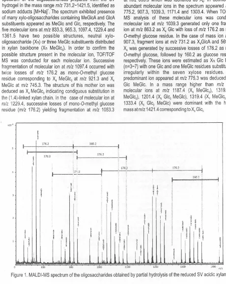
11-(1,4) linkages between xylose residues in xylan backbone, analysis of oligosaccharides prepared from reduced acidic fractions was carried out. Figure 1 showed MALDI mass spectrum of the oligosaccharides prepared from the SV hydrogel in the mass range
m/z
731 .2-1421 .5, identified assodium adducts [M+Na]'. The spectrum exhibited presence of many xylo-oligosaccharides containing MeGlcA and GlcA substituents appeared as MeGlc and Glc, respectively. The five molecular ions at
mlz
833.3, 965.3, 1097.4, 1229.4 and1361.5 have two possible structures, neutral xylo-oligosaccharide
(Xn}
or three MeGlc substituents distributed in xylan backbone(Xn
MeGlcJ In order to confirm the possible structure present in the molecular ion, TOF/TOF MS was conducted for each molecular ion. Successive fragmentation of molecular ion atm/z
1097.4 occurred withtwice losses of
m/z
176.2 as mono-0-methyl glucoseresidue corresponding to
X
4 MeGlc2 atm/z
921.3 andX
4 MeGlc atmlz
745.3. The structure of this mother ion wasdeduced as X4 MeGlc3 indicating condiguous substitution in
the (1,4)-linked xylan chain. In the case of molecular ion at
m!z
1229.4, successive losses of mono-0-methyl glucoseresidue (m/z 176.2) yielding fragmentation at
m!z
1053.3セ@
3
I 176.2 I 160.2
I I
I
-i
•
176.2セ@ 176.2
.
.
セ@
I
I
II
17". 2'
i
II
セ@I
;
a
l
l
I
9セ@
I
l
I
I
I
I
l
セ@
I
セ@ セ@ セ@ I l
I I
identified as X5 MeGlc2 and
m/z
877.2 as X5 MeGlc,respectively. From the molecular ion at
mlz
1361.5, threefragment ions were also generated with
a
loss ofmlz
176.2from the reducing end to form fragment セ@ MeGlcA2
appeared at
mlz
1185.4, followed by loss ofmlz
176.2 asfragment X6 MeGlc at
mlz
1009.2 and finally loss ofmlz
176.2 to form セ@ at
m/z
833.3. These analytical resultsindicate that the five mother ions have structures strongly corresponded to
Xn
MeGlc3 (n=3-6), except a molecular ionat
m!z
833.3 which was identified asX
6. In addition, fiveabundant molecular ions in the spectrum appeared at
m/z
775.2, 907.3, 1039.3, 1171.4 and 1303.4. When TOF/TOF MS analysis of these molecular ions was conducted, molecular ion at m/z 1039.3 generated only one fragment ion at·
mlz
863.2 asX
4 Glc with loss of
mlz
176.2 asmono-0-methyl glucose residue. In the case of mass ion at
m/z
907.3, fragment ions at
mlz
731 .2 asX
4GlcA and 569.2 asX
4 was generated by successive losses of 176.2 asmono-0-methyl glucose, followed by 160.2 as glucose residues, respectively. These ions were estimated as
Xn
Glc MeGlc (n=3-7) with one Glc and one MeGlc residues substituted in irregularly within the seven xylose residues. Most predominant ion appeared atmlz
775.3 was deduced as X3Glc MeGlc. In a mass range higher than
m/z
1100,molecular ions at
m!z
1187.4 Hセ@ MeGlc2), 1319.4 (X7
MeGlc2) , 1201.4 (X5 Glc2 MeGlc), 1319.4 (X7 MeGlc2} , and
1333.4 Hセ@ Glc1 MeGlc) were dominant with the highest
mass at m/z 1421.4 corresponding to X8 Glc2
- I
176.2 176.2 I
.
I
160.2I
I
!
l'
I
I
f
¥I
I
I
I
I
セ@!
I
iI
II
' 1
I
l
·i
1.
セ@
' セ@!
i
i
gi
セ@
§i
I
l
i
セ@ セ@セ@
i
セ@
d
i
セ@
I
§
I
セ@•
1
l
セ@
'. l
l
.il
i
u.l
J
. l I,,
L
セ@ l .I800 900 1000 1100 1200 1300 1400 m/z
Figure 1. MALO I-MS spectrum of the oligosaccharides obtained by partial hydrolysis of the reduced SV acidic xylan.
[image:12.595.49.509.156.732.2]1.0
,_!
176.2
I 176.2
I
I
176. 2
':
160.2I •
i
I •
I
I
1I
I
176.2l
176.2 ,I
176.2I
I I
I
t
I
I
176.2 176.2 II
di
I
iI
II 60.2 I
I
I
I
セ@
I
i
2 I II
I
I
i
セ@
1
l
t
§3
!
セ@i.
セ@!
I
'J
i
I
1
I
I
!I.
I ii
t
I
i
'
I
i \
I
li
II
セ@
i
セ@
I
セ@
セ@I
セ@セ@
t
. I ..
lu
... II I t ll .L1.I
lU
1.1,
.111d.l
セi@
10.8
0.6
0.4
0.2
700 800 900 1000 1100 1200 1300 !400 1500 1600 m/z
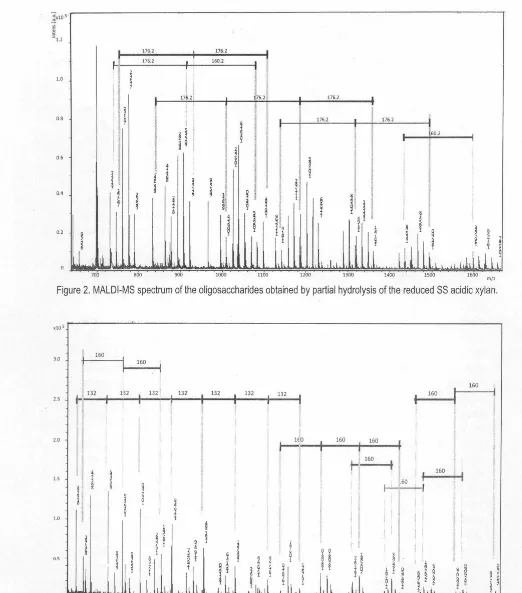
Figure 2. MALDl-MS spectrum of the oligosaccharides obtained by partial hydrolysis of the reduced SS acidic xylan.
160
3.0
160
. I
160
132 u2
I
1321 132
I
132 132 1322.5 t---+---+·--l·---l·i---+---t-j---1
I
160P XPP セセセセセセセセNAZANAAZAyAZセ、AAN、AANセセセセセセセセセN]TセセLGNOLjjLAセセセセセ@
Figure 3. MALDl-MS spectrum of the oligosaccharides obtained by partial hydrolysis of the reduced SM acidic xylan.
Precise Structure of Acidic Polysaccharide Present in Salvia Hydrogels Rike Yudianti, Myrtha Karina, Masahiro Sakamoto, and Jun-ichi Azuma
ml•
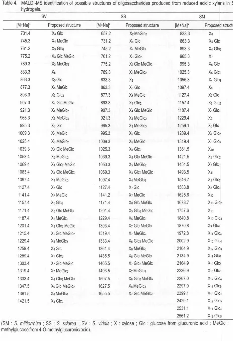
[image:13.602.11.533.27.620.2]Table 4. MALDl-MS identification of possible structures of oligosaccharides produced from reduced acidic xylans in Salvia h dro els.
sv
SS SM[M+Na]• Proposed structure [M+Na]• Proposed structure [M+Na]• Proposed structure
731.4 X4 Glc 557.2 X2MeGlci 833.3 Xs
745.3 X4 MeGlc 731 .2 X4 Glc 863.3 Xs Glc
761.2 X3 Gle2 745.2 X4 MeGlc 893.3 X4 Glc2
775.2 X3Glc MeGlc 761 .2 X3 Gle2 965.3 X1
789.3 X3 MeGlc2 775.2 XJGle MeGle 995.3 Xs Gle
833.3 Xs 789.3 XJMeGlci 1025.3 Xs Glc2
863.3 XsGlc 833.3 Xs 1055.3 X4 Gle3 877.3 Xs MeGlc 863.3 Xs Gle 1097.4 Xa
893.3 X3 Gle2 877.3 Xs MeGle 1127.4 X1Gle
907.3 X4 Gle MeGlc 893.3 X4 Gle2 1157.4 Xs Glc2
921 .3 X4 MeGlci 907.3 X4 Gle MeGlc 1187.4 Xs Gle3
965.3 X3 MeGlc3 921.3 X4 MeGlc2 1229.4 Xg
995.3 Xs Gle 965.3 X3 MeGle3 1259.1 XaGle
1009.3 Xs MeGlc 995.3 Xs Glc 1289.4 X1 Glc2
1025.4 Xs MeG!ci 1009.3 Xs MeGlc 1319.4 Xs Gle3
1039.3 Xs Gle MeGlc 1025.3 Xs Glci 1361.5 X10
1053.4 Xs MeGlc2 1039.3 Xs Gle MeGlc 1421 .5 Xa Glc2
1069.4 X4 Glci MeGlc 1053.3 Xs MeGle2 1451.5 X1Gle3
1083.4 X4 Glc MeGle2 1069.3 X4 Glc2 MeGlc 1493.5 X11
1097.4 X4 MeGlc3 1097.4 X4 MeGle3 1546.7 Xg Glc2
1127.4 X1 Glc 1127.4 X1Glc 1583.8 Xa Glc3
1141.4 X1 MeGlc 1141.2 X1 MeGle 1625.6 X12 1157.4 XsGlc2 1171.4 Xs Gle MeGlc 1678.7 X10 Gle2
1171 .4 Xs Gle MeGle 1201.4 Xs Glc2 MeGlc 1757.6 X13
1187.4 XsMeGlc2 1229.4 Xs MeGle3 1840.8 X10 Gle3
1201.4 Xs Glci MeGlc 1303.4 X1 Glc MeGle 1870.8 Xg Gle4
1215.4 Xs Glc MeGlci 1319.4 X1 MeGic2 1972.8 X11 GicJ
1229.4 XsMeGlc3 1333.4 Xs Glc2 MeGlc 2002.9 X10 GIC4
1259.4 XaGlc 1361.4 Xs MeGlc3 2104.9 X12 Glc3 1289.4 X1 Glc2 1435.5 XaGlc MeGlc 2134.9 X11 GIC4
1303.4 X1Glc MeGlc 1465.5 X1 Gle2 MeGlc 2164.9 X10 Gies 1319.4 X1 MeGlc2 1493.5 X1MeGlc3 2236.9 X13 Gle3
1333.4 Xs Glc2 MeGlc 1597.5 Xa Glci MeGlc 2267.0 X12 GIC4 1347.5 Xs Gle MeGlci 1627.5 XaMeGle3 2297.0 X11 Gies 1361.5 XsMeGlc3 1655.5 X1 Glc MeGicJ 2399.1 X13 GIC4
1421.5 Xa Gle2 2429.1 X12 Gies
2531.1 X14 GIC4 2561.2 X13 Gies
(SM : S. miltiorrhiza ; SS : S. sc/area ; SV : S. viridis ; X : xylose ; Glc : glucose from glucuronic acid ; MeGlc : 4-0-methylglucose from 4-0-methylglucuronic acid).
[image:14.600.55.518.37.714.2]\
'
Basically, the structures proposed for the SS oligosaccharide are similar to the oligosaccharides given from SV. As shown in the MALDI mass spectrum of the oligosaccharides derived from SS hydrogel (Figure 2), molecular ions appeared in a range from m/z 657.2 to 1655.5. The highest mass at mlz 1655.5 generated successive ions at rrlz 1493.5, 1319.4 and 1141.2 corresponding to
X1
MeGlcJ,X1
MeGb,X1
MeGlc with losses of glucose residue and mono-0-methyl glucose from the reducing end.MALDI mass spectrum of oligosaccharides given after reduction and partial hydrolysis of the acidic polysaccharide from SM (Figure 3) showed profiles different from the SS and SV hydrogels. The molecular ions were also identified as sodium adducts [M+Na]+ in a mass range of mlz
833.3-2561.2 including ions assignable as the xylo-oligosaccharides contained various degrees of Glc residues. Presence of mass ions corresponding to xylo-oligosaccharides (Xn, n=6-13) were detected and mass ion appeared at m/z 1625.6 resulted in formation of several fragment ions with successive losses of six xylose residues at m/z 1493.5, 1361.4, 1229.4, 1097.4, 965.3 and 833.3. These ions were more abundant than other mass ions in a mass range m/z 800-1800. The highest mass ion appeared at mlz 2561.2 was proposed to have a structure of X13 Gies based on successive generation of fragment ions at m/z 2399.1 as X13 Glc.i and 2236.9 as X1 3 Glc3 with losses of glucose residues. Two fragment ions occurred at mlz
2267.4 and 2104.9 were similarly deduced to have structures corresponding to X12 Glet and X12 Glc3, respectively, with the losses of successive glucose residues . The ion mass at mlz 2429.1 was suggested to have a structure of X12 Gies. Similar fragmentation also occurred for ions which have mass numbers at mlz 2297.4 and 2164.9 with successive losses of glucose residues corresponding to have a possible structure of Xn Gies (n=10-13). A low abundant ion at m/z 2164.9 produced fragment ions at mlz
2002.9, 1840.8 and 1678. 7 with lose of one glucose residue corresponding to oligosaccharides which have structures of X10Glc.i, X10Glc3 and X10Glcz, respectively. Other ion at m/z
1187.4 generated fragmentation with successive losses of glucose residue to form Xs GicJ (m/z 1025.3) and Xs Glc2
( m/z 863.3). The ion mass at mlz 2429.1 was suggested to have a structure of X12 Gies. Similar fragmentation also occurred for ions which have mass numbers at m/z 2297.4 and 2164.9 with successive losses of glucose residues corresponding to have a possible structure of Xn Gies (n=10-13). A low abundant ion at m/z 2164.9 produced have fragment ions at m/z 2002.9, 1840.8 and 1678.7 with loss of glucose residue corresponding to oligosaccharides which have structures of X10GIC4, X10Glc3 and X10Glc2, respectively. Other ion at m/z 1187.4 generated fragmentation with successive losses of glucose residue to form Xs GicJ (m/z 1025.3) and Xs Glc2 (m/z 863.3). All deduced structures of the oligosaccharides produced from the present Salvia hydrogels were listed in Table 4.
Precise Structure of Acidic Polysaccharide Present in Salvia Hydrogels Rike Yudianti, Myrtha Karina, Masahiro Sakamoto, and Jun-ichi Azuma
Presence of random and contiguous substitutions was a new finding for acidic xylans in the Salvia hydrogels.
Conclusions
Acidic polysaccharides present in the hydrogels produced from three species of Salvia (S. miltiorrhiza (SM),
S. sclarea (SS) and S. viridis (SV)) were commonly
composed of セMHQ L TIMクケャ。ョウ@ highly substituted at 02 positions with uronic acid in molar ratios of xylose to uronic acid of 2.1 : 1.0 (SM), 1.7 : 1.0 (SS), 1.4 : 1.0 (SV), respecitively. Mixed substitutions with MeGlcA and GlcA occurred in both of the SS and SV hydrogels, while GlcA was exclusively substituted in the SM hydrogel. The precise chemical composition analysis and MALDl-TOF/TOF MS analyses elucidated random and contiguous substitutions of GlcA and MeGlcA at 0-2 of xylopyranosyl residues. In addtion, SM oligosaccharides contained higher degree of free xylopyranose residues than those in SS and SV in agreement with the lowest content of uronic acid among three Salvias.
References
Choi,
H.;
E.K. Choe; E.K. Yang; S. Jang; C.R. Park. 2007. Characterization of Synthetic Polyamides by MALDl-TOF Mass Spectrometry, Bull. Korean Chem. Soc., 28: 2354-2358.Hakamori, S. 1964. A Rapid Permethylation of Glycolipid and Polysaccharide Catalyzed by Methylsulfinyl Carbanion in Dimethyl Sulfoxyde. J. Biochem., 55: 205-208.
Hsu, N-Y.; W.B. Yang; C.H. Wong; Y.C. Lee; R.T. Lee; Y.S. Wang; C.H. Chen. 2007. Matrix-assisted Laser Desorption/Ionization Mass Spectrometry of Polysaccharides with 20,40,60-Trihydroxyacetophenone as Matrix. Rapid Commun. Mass Spectrom., 21 : 2137-2146.
Jacob, A. ; P.T. Larsson; 0. Dahlman. 2001 . Distribution of Uronic Acid in Xylans from Various Species of Soft-and Hardwooda Determined by MALDI Mass Spectrometry, Biomacromol., 2: 979-990.
Janiak, C.; F. Blank. 2006. Metallocene Catalysts for Olefin Oligomerization. Macromol. Symp., 236: 14-22. Lin, K.Y; J.R. Daniel. 1994. Structure of Chia Nulle!
Polysaccharide Exudates. J. Carbohydr. Polym ., 23: 13-18.
Reis, A.; M.A. Coimbra; P. Domingues; J. Ferrer-Correla; M.R.M. Domingues. 2002. Structural Characterization of Underivatised Olive Pulp Xylo-Olygosaccharides by Mass Spectrometry Using Matrix-Assisted Laser Desorption/Ionisation and Electrospray Ionisation. Rapid Commun., Mass Spectrom. 16: 2124-2132 .. Reis, A.; M.R.M. Domingues; P. Domingues; A.J.
Ferrer-Correla; M.A. Coimbra. 2003. Structural Characterisation by MALDl/MS of Olive
Oligosaccharides Obtained by Partial Acid Hydrolysis, Carbohydr. Polym., 53: 101-107.
Taranenko, N.I.; A.V. Pashkova; V.M. Doroshenko. 2003. Negative and Positive AP-MALDI Analysis of Synthetic Phosphopeptides and Bovine セMc。ウ・ゥョ@ using Immobilized Metal Affinity Chromatography Ga(lll) IMAC. Proceedings of the 5151 ASMS Conference on
Mass Spectrometry and Allied Topics, Canada. Taylor, R.L.; H.E. Conrad. 1972. Stoichiometric
Depolymerization of Polyuronides and Glycosaminoglycuronans to Monosaccharides following Reduction of Their Carbodiimide-Activated Carboxyl Group. Biochemistry, 11 : 1383-1388. Weber, C. W.; H.S. Gentry; E.A. Kohlhepp; P.R. McCrohan.
1991. The Nutritional and Chemical Evaluation of Chia Seeds. Ecol. Food Nutr., 26;119-125.
Yudianti, R.; L. lndrarti; M. Sakamoto;
J.
Azuma. 2005. Cellulose-Hemicellulose Composite Present in Hydrogel from Salvia spp., Proceedings of The 6th International Wood Science Symposium, JSPS-LIPI Core University Program in the Field of Wood Science : 199-204.Yudianti, R. ; L. lndrarti; M. Karina; M. Sakamoto;
J.
Azuma. 2007. Chemical Composition of Salvia Hydrogel,J.
Trop. Wood Sci. Technol., 5: 12-16.
Yudianti, R. ; M. Karina;
J.
Azuma. 2009a. Rheological Behavior of Salvia Hydrogel at Temperature and pH Variation. IndonesiaJ.
Technol. , 30: 332-338.102
Yudianti, R. ; M. Karina; M. Sakamoto;
J.
Azuma. 2009b. Effects of salts on rheological behavior of Salviahydrogels. Macromol. Res. , 17: 332-338.
Yudianti , R., M. Karina; M. Sakamoto;
J.
Azuma. 2009c. DSC Analysis on Water State of Salvia Hydrogels.Macromol. Res., 17:1015-1020. ,
Rike Yudianti*, Myrtha Karina
Research Center for Physics, Division of New Material JI. Cisitu/Sangkuriang, Kompleks LIPI
Bandung 40135, Indonesia
*Corresponding Author Tel. : +62-22-2503052 Fax. : +62-22-2503050 E-mail : [email protected]
Jun-ichi Azuma, Masahiro Sakamoto Laboratory of Forest Biochemistry, Division of Environmental Science,
Graduate School of Agriculture, Kyoto University Kitashirakawa Oiwake-cho, Kyoto 606-8502, Japan Tel. : +81-75-753-6465
Fax. : +81-75-753-6471
E-mail : [email protected]
Wood Research Journal Vol. 1 • No. 2 • 2010