Free-Living Ice-Nucleating Active Bacteria from
High Mountain Lake Habitats
MUNTI YUHANA1*ANDKURT HANSELMANN2
1Department of Aquaculture, Faculty of Fisheries and Marine Sciences,Institut Pertanian Bogor, Darmaga Campus, Bogor 16680, Indonesia; 2Microbial Ecology Group, Institute of Plant Biology,
University of Zurich, Switzerland
We collected the culturable heterotrophic bacteria from oligotrophic high mountain lake habitats and tested their capability to induce ice formation. Direct plating was carried out using low-nutrient medium at a temperature of between 3 and 4°C. As many as 84 isolates were recovered from glacial ice and natural biofilm growing on granite rocks surface. Six out of 84 isolates were capable of expressing the ice-nucleation phenotype. After autoclaving the cell suspension at 121°C for 15 min, isolate J78 was still able to retain the ability for ice formation. Heat-stable ice nuclei produced by ice-nucleating active bacteria have potential applications in biotechnology. Characterization of INA bacteria was performed employing live-dead Gram staining and molecular methods. Universal primers for Bacteria (S-D-Bact-0008-b-S-20 and S-D-Bact-1524-a-A-18) were used for PCR to amplify almost the full length of the 16S rRNA genes of selected INA isolates. Restriction fragment length polymorphism analysis resulted in 2 unique patterns, as represented by J43 and J83, respectively. Based on DNA sequencing of 16S rRNA gene, isolate J43 (GeneBank accession no. AJ864852) was closely related to Pseudomonas mephitica (99.2% sequence similarity) and Janthinobacterium lividum (99% similarity), whereas isolate J83 (GeneBank accession no. AJ864859) showed 100% sequence identity to Pseudomonas fluorescens.
Key words: high mountain lake habitats, ice nucleation, 16S rRNA gene, free-living bacteria _____________________________________________
________________________
*Corresponding author, Phone: +62-251-8628755,
Fax: +62-251-8622941, E-mail: [email protected]
Volume 2, Number 3, December 2008
ISSN 1978-3477 p 131-134
Various Gram-negative bacteria are capable of catalyzing ice formation at temperatures of -2 to -12°C in nature (Lindow et al. 1982; Hirano et al. 1985). Most of ice-nucleating-active (INA) bacteria are associated with plants (Lindow et al. 1978; Lindermann et al. 1982; Loper and Lindow 1994; Waturangi et al. 2008) or animals (Lee et al. 1995; Worland and Block 1999). Other microorganisms, including several genera of fungi (Pouleur et al. 1992) and lichens (Kieft 1988) have been reported in their ability to induce ice formation. Ice-nucleating active (INA) bacteria have been recovered from various geographical areas, such as Antarctica, temperate, subtropical or tropical regions. INA bacteria, which induce frost damage in various plants, can contribute to a very devastating loss in agricultural crops production (Lindow et al. 1982).
INA bacteria produce outer surface membrane protein that can act as a catalyst for the unusual transition of water from liquid to its solid phase (Gurian-Sherman and Lindow 1993). Droplets of pure water (INA-free water) remain in its liquid state (supercool) up to a temperature of -40°C. The higher temperature of ice catalysis conferred by bacterial ice-nuclei makes them useful in ice-nucleation-limited processes such as artificial snow production, the freezing of some food products and possibly in future weather modification schemes (Gurian-Sherman and Lindow 1993).
Up to today, reports on free living INA bacteria recovered from high mountain lake habitats remain limited. Microorganisms that survive and actively grow in these habitats might be good sources of cold-active-proteins, such as ice nucleation active protein, which are catalytically efficient at low temperatures (Gerday et al. 1997). In this study, we report our study on free living INA bacteria recovered from these extreme habitats.
MATERIALS AND METHODS
Samples Collection and Treatments. Samples were collected from the Joeri lakes catchment, located in the south-eastern Swiss Alps at an altitude of approximately of 2 750 m a.s.l (Yuhana et al. 2006).Lake water and biofilm samples were aseptically collected during the snow-free season. Subsurface water samples were obtained in sterile glass bottles and patches of biofilm samples were collected from the surface of submerged rocks. Aliquots of 50 and 100 µl of water were spread-plated and subcultured onto minimal growth agar medium (pH 7.1), whereas biofilm samples were spread aseptically onto the medium after mechanical disruption of the biofilm with a sterile loop. Growth temperature for all cultures was 4±1°C. The minimal growth medium (designated as MM) contained the following ingredients (final pH 7.1): 6 µM MgSO4, 10 µM CaCl2, 20 µM Na2CO3, 14 µM NaNO3, 10 µM NH4Cl, 1.75 µM K2HPO4, 2.7 µM EDTA Na salt, 10 mg l-1 yeast extract, 10 mg l-1 peptone, 15 g l-1 washed-bacteriological agar and 1 x trace elements, (added to the autoclaved basal medium). The concentrated trace elements stock solution (10 000 x) contained the following: 850 µM ZnSO4·7H2O, 7 100 µM MnCl2·4H2O, 86 µM Co(NO3 )2·6H2O, 1 600 µM Na2MoO4·2H2O, 29 750 µM Citrate·H2O and 21 480 µM Ferric ammonium citrate.
When growth occurred (after 14 to 21 days of incubation in the cold), single colonies of visibly dominant and different colony morphotypes were subcultured onto new minimal medium. Isolates were maintained in 10 x diluted Luria Bertani (LB) agar medium (pH 7.2) containing the following ingredients: 0.5 g l-1 Bacto tryptone, 1.0 g l-1 yeast extract, 0.5 g l-1 NaCl and 15 g l-1 bacteriological agar.
distinguish dead cells from living ones. Staining was carried out as described by the manufacturer (Molecular Probes, Inc.) using the Viability Gram Staining Kit (V-7023 Molecular Probes Inc.). The treated samples on slides were observed with a Zeiss Axioplan microscope (Carl Zeiss, Oberkochen Germany) employing 3 different excitation filters (365-395, 450-490 and 546-580 nm) and photographed with an Optronic digital camera.
Ice Nucleation Assay. The ice nucleation capacity of all 84 isolates was tested qualitatively by the tube assay (modified from Hirano et al. 1985). Isolates were grown in 10 ml liquid MM and incubated at 4°C until the stationary phase was reached. Cells were then suspended in 10 ml autoclaved PBS solution (10 mM potassium phosphate-buffered saline, pH 7.0) and kept in a cooling-bath at -2 to -10°C for 5 to 10 min. A suspension of E. coli cells containing plasmid pJL1703 (Loper and Lindow 1994) was used as a positive control, whereas a cell-free PBS solution served as a negative control. The ice nucleation activity of each INA isolate was observed by droplet-freezing assay and the ice nucleation frequency was calculated by the following formula: N(t) = [- (ln f) ] / V, (Vali 1971) where N(t) is the frequency of ice nucleation at T temperature, f indicates the proportion of droplets unfrozen and V is the volume of individual droplets. Genomic DNA Extraction. Total genomic DNA from pure isolates was extracted with cetyltrimethyl ammonium bromide (CTAB) (modified from Murray and Thompson 1980). This provided a simple, non toxic and inexpensive method and yielded enough DNA template for PCR amplification. After pelleting the cells, extraction buffer (2% w/v CTAB, 100 mM Tris-Cl pH 8.0, 1.4 M NaCl, 20 mM EDTA) was added and mixed. The solution was incubated at 60°C for 30 min and centrifuged at 12 000 xg(4°C) for 10 min. After transferring the supernatant into a fresh tube, an equal volume of chloroform:isoamyl alcohol (24:1) was added, mixed gently and the solution was centrifugated at 12 000 xg(4°C) for 10 min, these steps were repeated one more time. The resultant supernatant was transferred into a new tube and 1/10 volume equivalent of 7.5 M ammonium acetate was added. DNA was then collected by precipitation in ethanol. DNA extracts were checked using the following electrophoresis conditions: agarose gel (1% w/v), [Tris-Acetate-EDTA] (TAE) buffer 0.5 x (20 mM Trizma base, 10 mM glacial acetic acid, 0.5 mM EDTA), running time 30 min at 5 V/cm. Successful extraction of DNA was verified by staining the gels in a 1 µg ml-1 ethidium bromide solution.
16S rRNA Gene Amplification by PCR. Nearly full-length 16S rRNA genes were amplified by PCR using the bacterial primers: S-D-Bact-0008-b-S-20 (5'-AGA GTT TGA TCC TGG CTC AG-3') and S-D-Bact-1524-a-A-18 (5'-AAG GAG GTG ATC CAR CCG-3'). PCR amplification was performed in a 25 µl reaction volume with a Techne Thermocycler (Techne LTD, Oxford, UK). Each reaction mixture contained (final concentration) Taq buffer (1x) (Sigma), 1.5-2.0 mM MgCl2, 0.1 mg ml-1 DNase-free Bovine Serum Albumin (Amersham, Pharmacia Biotech Inc.), 0.2 mM dNTPs, 200 nM of forward and reverse primer, respectively, 40 U Taq Polymerase (Sigma), ddH2O and approximately of 20-100 ng of template DNA. The following touch-down PCR program was used: initial denaturation at 94°C for 2 min; 20 cycles of 94°C for
20 sec, 63°C for 30 sec with lowering temperature by 0.5°C in every cycle, 72°C for 80 sec with increasing duration by 1 sec in every cycle; another 20 cycles of 94°C for 20 sec, 53°C for 30 sec, 72°C for 100 sec with increasing period by 1 sec every cycle; followed by a final extension step at 72°C for 10 min. PCR products were analyzed by electrophoresis in 1% w/v agarose gels in 0.5x TAE running buffer, stained with ethidium bromide (1 µg ml-1) and photographed under UV. For RFLP analysis, 8 µl PCR products were double-digested with 1 U restriction enzyme HinfI (5'-G/ANTC) and HaeIII (5'-GG/CC) in a total reaction volume of 10 µl.
Sequencing. The PCR products of the 16S rRNA genes from different RLFP patterns were purified through microcon centrifugal filter devices (Microcon YM 100, Millipore, Bedford, Mass., USA). Nearly full-length 16S rRNA genes were bidirectionally sequenced with a DNA sequencer (ABI Prism 377), using ABI Prism® Big DyeTM v2.0 (Applied Biosystems) as described by the manufacturer with the following 6 primers: S-D-Bact-0008-b-S-20 (5'-AGA GTT TGA TCC TGG CTC AG-3'), S-*-Univ-0519-a-A-18 (5'-GWA TTA CCG CGG CKG CTG-3'), S-*-Univ-0519-a-S-18 (5'-CAG CMG CCG CGG TAA TWC-3’), S-D-Bact-1099-b-S-16 (5’-GYA ACG AGC GCA ACC C-3’), S-D-Bact-1099-b-A-16 (5’-GGG TTG CGC TCG TTR C-3’) and S-D-Bact-1524-a-A-18 (5'-AAG GAG GTG ATC CAR CCG-3'). For a 10 µl-single PCR reaction, 5 to 20 ng DNA template, 3 µl Big Dye (Applied Biosystems) and 3 µl of 1.5 µM primer were used. After the sequencing PCR, the products were purified with Sephadex G-50 (Amersham, Pharmacia Biotech AB) and loaded onto a sequencing machine (ABI Prism 377 DNA Sequencer). The BLAST search tool available from NCBI (http://www.ncbi.nlm.nih.gov/blast) was used to list the closest neighbors of the sequences.
Phylogenetic Tree Construction. The phylogenetictree was analysed online using the Phylip Interface, available at the ribosomal database project (http://rdp8.cme.msu.edu/ html/analyses.html). Sequence data was imported and aligned to their closest neighbors by an automated alignment. The distance matrix was calculated by maximum likelihood method and a phylogenetic tree was constructed based on neighbor-joining analysis.
RESULTS
Eighty-four isolates were recovered from lake water, glacier and biofilm growing on granite rocks surface. Generally they are cream-pigmented morphotypes and some of them were capable of producing water-soluble yellow-greenish fluorescent pigments. Test tube assay showed that 6 out of 84 isolates were capable of catalyzing the ice nucleation in the tubes at temperatures between -2 to -5°C. Fluorescent microscopy determination after live-dead Gram staining, showed that all of the 6 INA isolates were Gram negative and rod shaped (Fig 1).
After autoclaving the cell suspension at 121°C for 15 minutes, isolate J78 still showed its ability to initiate the ice formation at lower temperatures, ranging from -8 to -10°C. Other INA isolates (J43, J71, J77, J83 and J84) and autoclaved-positive control, were no longer capable of catalyzing ice formation even at lower temperatures (Fig 2).
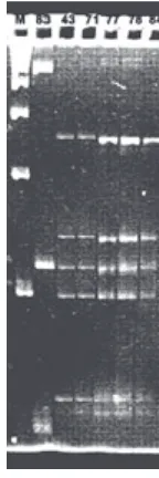
isolates J43, J71, J77, J78 and J84 (Fig 3). 16S rRNA genes of isolates J83 and J43 (as a representative of other 5 INA isolates) were selected for sequencing.
The DNA sequence of isolate J43 (1484 bp) showed 99.2% similarity to Pseudomonas mephitica or Janthinobacterium lividum. The nucleotides have been submitted to the GenBank with accession no. AJ864852. DNA sequence of isolate J83 (1491 bp) showed 100% similarity to P. fluorescens CCM 2115 and has been submitted with accession no. AJ864859.
DISCUSSION
All INA isolates from high mountain lake habitats showed ice nucleation activity at temperatures warmer than -5°C. Similar results were shown in a previous study (Kieft 1988) of INA associated with lichens isolated from high mountain habitats. His study showed that several epilithic lichen samples of the genera Rhizoplaca, Xanthoparmelia and Xanthoria expressed ice nucleation activity at temperature as warm as -2.3°C. According to the ice nucleation proteins classification described by Turner et al. (1990), all of our INA isolates are characterized as class A, which showed ice nucleation activity between temperature range of -2 to -5°C. Waturangi et al. (2008) reported that
Fig 1 Fluorescent micrographs of isolate J43 and J83. Live cells fluoresce blue, whereas dead cells fluoresce green. Both isolates were Gram negative.
J 4 3
J 8 3
19 µµµµµm
The highest ice nucleation activity among INA isolates was shown by J84 with frequency of 60 nuclei ml-1 and the lowest was indicated by isolate J71 (32 nuclei ml-1). Isolates J43, J77 and J83 had the same ice nucleation frequency of 38 nuclei ml-1 and isolate J78 was 46 nuclei ml-1 (Table 1). The temperature of the assay was warm (-2°C). Ice nucleation activity of isolate J78 after autoclaving was 46 nuclei ml-1 at -8°C.
PCR amplifications of the 16S rRNA gene of selected INA isolates yielded products of approximately 1500 bp. By using double restriction with HinfI and HaeIII, two different RFLP patterns were obtained. J83 has a unique pattern, whereas J43 shows corresponding band patterns with
Fig 2 Ice nucleating assay tubes. (a) Assay carried out before autoclaving the cell suspension at the temperature ranging from from -2 to -5°C. Assay showed ice nucleation capability of isolate J43, J71, J77, J78, J83, and J84. J57 represents a non ice-nucleating-active isolate, remaining unfrozen after 30 minutes incubation at temperatures ranging from -8 to -10°C. Cell-free PBS solution (-) serves as a negative control, while cell suspension of E. coli containing plasmid pJL1703 was used as a positive control; (b) Assay carried out after autoclaving the cell suspension. Suspension of the isolate J78 remaining capable of inducing the ice nucleation at lower temperatures ranging from -8 to -10°C.
a
b
Table 1 Ice nucleation activity of free living INA bacteria from high mountain lake habitats
No of frozen droplets/ total droplets Isolate
J43 J71 J77 J78 J83 J84
J78 (after autoclaving)
17/20 16/20 17/20 18/20 17/20 19/20 18/20
38 nuclei/ml (-2°C) 32 nuclei/ml (-2°C) 38 nuclei/ml (-2°C) 46 nuclei/ml (-2°C) 38 nuclei/ml (-2°C) 60 nuclei/ml (-2°C) 47 nuclei/ml (-8°C) N (t), ice nucleation activity; where N, frequency of ice nucleation at temperature t.
N(t) (at temperature)
Fig 3 RFLP patterns of 16S-rRNA genes from the INA isolates. Lane 1, molecular marker 100 bp ladder (Pharmacia, Biotech); Lane 2-7, J83, J43, J71, J77, J78, and J84. Running conditions on Spreadex EL-800 gel (Elchrom Sci.): 70 Vcm-2 for 90 min, running temperature
55°C in 30 mM TAE buffer.
tropical INA bacterial isolates showed ice nucleation activity at cooler temperature of -8°C and categorized as class B. At this temperature, the ice nucleation activity values of tropical INA isolates were between 38 and 60 nuclei ml-1
.
Isolate J78 showed the existance of heat-stable INA
protein, since the heat treatment of 121°C for 15 min did not
diminish its capability to initiate ice formation. Previous
studies also reported heat-stable INA protein isolated from a lichen, Rizoplaca chrysoleuca (Kieft 1988) and from Fusarium (Pouleur et al. 1992). Kieft (1988) reported that control cell suspension (no heat treatment) and 10 min treatment at temperatures of 40 to 70°C, showed the ice nucleation activity at temperatures between -2.3 to -3.3°C, whereas the ice nucleation activity at a lower temperature of -14.6°C was detected after cell suspension exposed by heat treatment at temperature range of 80 to 95°C. Our isolate J78 showed its ice nucleation ability at warmer temperatures than that of INA expressed by R. chrysoleuca, i.e. at temperatures ranging from -8 to -10°C. This might indicate a more stable INA protein structure present in this isolate, however, further studies are required to determine the uniqueness of the J78 INA protein.
Based on their DNA sequences, our INA isolates phylogenetically fall into the â- and ã-subgroups of Proteobacteria (Fig 4). Isolate J83, which is closest related to P. fluorescens strain IAM12022, belonging to the gamma-subdivison. P. fluorescens has been reported to belong to the bacteria which are able to catalyze ice formation (Lee et al. 1995) while isolate J43 falls into the â- subdivison. This isolate is closely related to P. mephitica ATCC 33665T or J. lividum DSM 1522T. To our knowledge, there are no other reports on the ability of the bacteria belonging to P. mephitica or J. lividum in catalyzing the ice formation.
Bacteria which are able to catalyze water crystallization may have an advantage over those which cannot. Those which can will be able to survive in frozen environments through slow cellular dehydration (Baertlein et al. 1992). This is a very important property for microorganisms to survive in this cold and extreme habitat. Ice formation on the outside of the cell allows water molecules to move from the cytoplasm across the cell membrane to join crystals of pure water nucleated extracellularly. This increases the osmotic potential inside the cells, thereby preventing freezing and cell damage by internal ice crystals. The ice nucleation process allows
for ordered propagation of ice throughout the cell rather than a rapid freezing, which can result in membrane rupture and cell death (Baertlein et al. 1992). Since this ability was only found in 6 out of the 84 isolates, we must assume that other bacteria use different strategies to survive periods of freezing, for instance by producing anti-freeze proteins (Feller et al. 1996) or other osmolytes.
ACKNOWLEDGEMENTS
This work was supported by an annual grant from the Microbial Ecology Group, Insitute of Plant Biology, University of Zurich. Authors would like to thank to Steven Lindow, UCLA-Berkeley, USA for positive control sample of E. coli (pJL1703).
REFERENCES
Baertlein DA, Lindow SE, Panopaulos NJ, Lee SP, Mindrinos MN, Chen THH. 1992. Expression of bacterial ice nucleation gene in plants. Plant Physiol 100:1730-1736.
Feller G, Narinx E, Arpigny JL, Aittaleb M, Baise E, Genicot S, Gerday C. 1996. Enzymes from psychrophilic organisms. FEMS Microbiol Rev 18:189-202.
Gerday C, Aittaleb M, Arpigny JL, Baise E, Chessa JP, Garsoux G, Petrescu I, Feller G. 1997. Psychrophilic enzymes: a thermodynamic challenge. Biochim Biophys Acta Prot Struct Mol Enzymol 1342:119-131.
Gurian-Sherman D, Lindow SE. 1993. Bacterial ice nucleation: significance and molecular basis. FASEB J 7:1338-1343. Hirano SS, Baker LT, Christen DU. 1985. Ice nucleation of individual
leaves in relation to population sizes of ice nucleation active bacteria and frost injury. Plant Physiol 77:259-265.
Kieft TL. 1988. Ice nucleation activity in Lichens. Appl Environ Microbiol 54:1678-1681.
Lee MR, Lee RE, Strong-Gunderson JM, Minges SR. 1995. Isolation of ice-nucleating active bacteria from the freeze-tolerant frog,
Rana sylvatica.Cryobiology 32:358-365.
Lindermann J, Constantinidou HA, Barchet WR, Upper CD. 1982. Plants as sources of airborne bacteria, including ice nucleation bacteria. Appl Environ Microbiol 44:1059-1063.
Lindow SE, Arny DC, Upper CD. 1978. Distribution of ice nucleation-active bacteria on plants in nature. Appl Environ Microbiol
36:831-838.
Lindow SE, Arny DC, Upper CD. 1982. Bacterial ice-nucleation: a factor in frost injury to plants. Plant Physiol 70:1084-1089. Loper JE, Lindow SE. 1994. A biological sensor for iron available to
bacteria in their habitats on plant surfaces. Appl Environ Microbiol
60:1934-1941.