14
Kiharaetal. Med J IndonesCurrent
Approaches in the Management of Emissional
Dysfunction
Kazunori Kihara, Kenji Sato, Masao Ando, Hiroyuki Oshima
Abstrak
Tinjauan pustaka ini nengupas nekanisme kontrol syaraf pada traktus seninal dan transpor spenna uelalui traktus tersebut,
untuk menemukan penanganan rasional dari disfungsi emisional. Makalah ini menbahas tentang identifikasi syaraf simpatis retroperitoneal dan intrapelvikyang mengatur pancaran setnen dari saluran ejakalatorius (PSSE), jaras kontpensatori untuk PSSE sesudah pentotongan syaraf hipogastrika, mekanisnre transpor sperrna nelalui vas deferens, dan cara baru untuk metnicu pancara,t sernen.
Abstract
This revieuw article explored the nechanism of neural control of the senûnal tract and sperril transport through it, to approach the rational managenent of enissional dysfunction. The identification of retroperitoneal and intrapelvic syntpathetic nerves conrrolling senûnal enûssion frotn the ejaculatory duct (SEED), the conrpensatory pathways for SEED afier rransection of hypogastric nerve, the nechanisn of sperm transport through the vas deferetrs, and a new ,nethod to generate sentinal enûssiott were discussed.
Keywords : Senûnal enission, ejaculatory duct, lunbar splanchnic nerve, co,npensatory pathways
Introduction
Ejaculation consists of seminal emission, bladder neck closure and protrusion. Seminal emission
from the
ejaculatory duct (SEED)is regarded
to
be a spinal reflex thatis
further modifiedby
the upper central nervous system. Loss of SEED is a cause of infertilityin
menwith
spinal cord
or
other nerve injuries. Stimulating a siteof
the above reflex arch has beentried
to
induceartificial
seminal emissionin
such patients,but success
has beenlimited
because of serious side effects,low
effectiveness, necessity of invasive operation and/or necessityof
generalanes-thesia. To approach the rational management of emis-sional dysfunction, we explored the mechanisms
of
neural control of the seminal tract and sperm transportDepartment of Urology and Allied Health Science, Tokyo Medi-cal attd Dental University, School of Medicine, Tolcyo, Japan
Corresponding address : H- Oshima, M"D., Ph.D.,
Departntent of Urology, Tol<yo Medical and Dental University, School of, Medicine,
I-5-45,
hitna, Bunkyo-ku, Tokyo 113, Japanthrough
it.
The findings lead us to a new method to induce artificial seminal emission.Identification of retroperitoneal and intrapelvic sympathetic nerves controlling seminal emission from the ejaculatory duct
Seminal emission from the ejaculatory duct (SEED) to the posterior urethra occurs by efferent signals via the retroperitoneal and intrapelvic sympathetic nervous system, and the main signals are regarded
to
pass through the superior lrypogastri" plexus and bilateral hypogastric nerves.r-r The proximal pathway tg the superior hypogastric plexus has remained controver-sial;it
has been assumed that SEED is controlled by the splanchnic nervesthat
branchfrom the
lower thoracic and upper lumbar ganglia of the sympathetic trunk but not those from the lower lumbar ganglia.a Our recent study, however, has indicated that not the thoracic splanchnic nerves but each lumba splanchnic nerve_(L2-5) has the ability to generate SEED as fol-lows.5Animals
compara-tive anatomy
of
retroperitoneal sympathetic nervous system between the human and dog.As
shown in Figurel,
the retroperitoneal sympathetic nervous sys-tem was similar except for the inferior mesenteric and superior hypogastric plexusesin the human, which
fuse as the caudal mesenteric plexus in the dog.Stimulution of the splanchnic nerve
Operations were carried out under general anesthesia. An exposed nerve was severed and electrical stimula-tion was given to its distal end using a wire electrode for 5 minutes with stimulus parameters of 8V, 2msec duration and l0 Hz provided by a stimulator (Model DPS-06, Daiya Medical System Co.p., Tokyo). The experiment of each dog was discontinued once SEED occurred. Seminal emission was observed by exposing verumontanum under vision. Bladder neck contraction was detected with a pressure sensitive balloon catheter inserted
into
the bladder neck and connectedto a
pressure transducer (DISA 21 G Ol type cystometer).6 Electrical stimulationof the
intermesenteric plexus which composed of nerve fibers from thoracic and Lr ganglia caused emission in none of the dogs examined.In contrast,
the
samestimulation
of eaeh lumba
splanchnic nerve branchedfrom the
sympathetic trunks below the Lz level generated SEED except fora
veryfew
cases (Figure2).r
Similar results were obtainedfor
bladder neck closure.6 Therefore,it
is concluded that efferent signals via the nerves from thoracis andLr ganglia
do not generate seminal emis-sion or bladder neck closure at ejaculation, while those via each lumbar splanchnic nerve (L2-5) have ability to generate seminal emission and bladder neck closure.Control of bilateral seminal emissions
from
ejaculatory ducts by lumba splanchnic nerve Since the presenceor
absenceof SEED
by signals
through
the
lumbar
splanchnic nerve and
the hypogastric nerve has been found as mentioned above, lateralityof efferent signals
for SEED
was inves-tigated. The routeof signals
for
theright
and left SEEDs was investigated by electrical stimulationof
each lumbar splanchnic nerve in the dog.7 The results indicated that each lumba splanchnic nerve generatesbilateral
SEEDsby
sendingsignals
to bilateral
epididymal tails and therefore we concluded that someof the signals
through each lumbar splanchnic nerve cross to the other side at the caudal mesenteric plexus and/or the prostatic plexus as shown in Figure 3. The contraction pressureof each epididymal
tail
was measured by inserting a fine elastic needle, which wasconnected
to
a transducer
(Statham plOF;Z, Nihon Kohden Cotp., Tokyo), into the vas lumen near the distal endof each epididymal
tail.
In intact
dogs, electrical stimulationof
a
lumbar splanchnic nerve caused bilateral SEEDs with a greater volume at the stimulatedside.
After'. trânsectionof
a
unilateral hypogastric nerve,bilateral
SEEDs occurred by electrical stimulationof
thecontralateral
lumbar splanchnic nerve with a greater volume at the stimu-lated side andby
the stimulationof the
ipsilateral lumbar splanchnic nerve with a greater volume at the contralateral side. Stimulation of a lumbar splanchnicnerve
causedintraluminal
pressuresof
bilateralepididymal
tails
regardlessof
the laterality of
hypogastric nerve transection (Figure 4). The pressure increase in the epididymal tail was the greateslat the stimulated side in the presencê of an intact ipsilateral hypogastric nerve, which was assumed to be generatedby noncrossing
signals. The secondary pressure in-crease in the epididymis occurred by the stimulationof
the other sidein
the presenceof an
intact ipsilateral hypogastric nerve, indicating the presenceof
signals crossing to the other side only at the caudal mesenteric plexus. The contraction pressure at the side of a tran-sected hypogastric nerve increased with the weakest level regardless of the side stimulated, indicating the presence of signals crossing to the other side at the site between the hypogastric nerve and the epididymal tail, probably at the prostatic plexuslCompensatory pathways
for
SEED after transec-tion of the hypogastric nerve : a mechanismof
neurogenic retrograde ejaculationl6 Kihara et al. Med J Indones
Figure l. Ànatonical dissectiott of thoracoluntbar splanchnic nerves in the dog (lefi) and hunan (right). Cattine caudal nesenreric
pleus coittcides v'ith both inferior ilrcsenteric and superior hl,pogastric pletu.ses in hu.nan. CMA
:
caudal ntesenreric arterl', CMP:
caudal nesenteric plerus, HN:
hlpogastric nerve, IMP:
inlernesenreric pletus, InMP:
inferior nesenleric ple.rus, ItrMA:
inferionnesenteric artery, RA:
renal artery', RV:
renal vein, SA:
sper,ilalic artery', SHP:
superior hypogastric ple.rus, ST:
sy'npathetic trunk.o/3
l/L
2/2
Figure 2. Seninal enissionfron the ejaculatory duct b1' electrical sîitttulation of the splanchnic nerves. Variatiotrs of rhe righr luttt-bar splanchnic nerves and rhe sites of electical stinularions v,ere presented. (A) cranial two o.f three lutnbar spl.anchnic nen'es unite to one, (B) cranial tv,o of three lunbar splanchnic nen'es unite, (C) three separated and (D).four separated lunbar splrurchnic ner-yes. Ccses of senrinal enùssiotr occurred/nunrber of dogs e.rantined u,ere indicated in thefig,ure. The sinilar results were obktined itr the left side. CA
:
celiac artery, CrMP:
cranial nesenreric plerus, CrMA:
cranial nrcsenteric artery. See leg,entls to FiS4ure I .for other abbreviatiotrs.Figure 3' schenatic presentatiotl of canine relroperitoneal and intrapelvic sl,nrpathctic nervous s),stettt.for setnitnl entissiotrfrottt rhe
eiacula
d that of 4I
tn each lunùar splanchnic nerve to bilarirat vasa deferetttia atdepididy
: anrpulla,
nchnic nen'e, PN:
pelvic nerve, PP:
pelvic pletus, pro:
prosrcre, prp:
prostatic plexus,sN:
spernatic nerve, r:
right, I:
l"lt, See legends to Filure I for other abb'reviatious.t -€pididvmll iâil
F i gu r e 4. S i nwhane o usll,
tion of a tutnbar
sptattchtt
:i:,:!)::,r,;:,:r,:::;:::,";iî::::;:;î;i,::i:::;,::î
r
:
righr, I : Ieft. Thesi,"
^;:;.:..:.:;:::..;'::
',.:,._
"trattsected hl,pogastric
ne
of a lutubar splancfuric nerve of the opposire side of theSâch lumbâr sohnchntc iêrva
Ca@d mclantarrc plarua
18
Kihara et al.nerve transections
is
attributableto
the absenceof
effective compensatory sympathetic pathways
for
bladder neck closure under the recovery
of
seminal emission from the ejaculatory duct by compensatory pathways.The
current results indicatethat
:
1) Majority
of
bilateral lumbar splanchnic nerves have the ability to generate SEED. 2) Each lumbar splanchnic nerve
con-trols bilateral seminal emissions by double cross-in-nervation system.
3)
Compensatory pathways exist after transection of the main pathwayfor
SEED. The above findings appear to be regarded as a defensive system of reproduction. These observations may pro-videinsight
into
ejaculatory disordersof
subjectswith autonomic nerve insufficiency and
a
rationalefor
therapy.Surgically,
recognitionof
the
abovemechanisms and nervous system must be useful to
prevent postoperative emission loss and to carry out
surgical nerve-sparing to preserve fertility.
Mechanisms a of sperm transport through the vas deferens
:
introducing new method to generate canine seminal emissionThe mechanism
of
sperm transport through the vas deferens intraluminal pressure at SEED and modeof
transportation
of
the content in the cauda epididymiswere investigated. Three elastic needles were inserted
into three sites
of
the vas deferens and connected to pressure transducers (Statham PIOEZ, Nihon Kohden Corp. Tokyo) and intraluminal pressureof
each sitewas simultaneously recorded. Sites of insertion were
the distal end of the pars epididymica of the vas toward the epididymis,
the
middleof
the vas toward theepididymis and the proximal end of the ampulla toward
the ejaculatory orifice. Direct electrical stimulation of either the testis, epididyrnal head or body caused no
elevation
of
the intraluminal
pressureof
the
vas deferens and epididymal tail.In
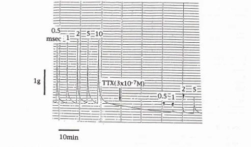
contrast, stimulation of any distal sites including the epididymal tail and vas deferens caused marked elevation of the intraluminalpressure only at the epididynral tail (Figure 5).
Electri-cal stimulation of any segments prepared from the vas deferens caued contraction in the longitudinal
direc-tion
in
vitro.
This
contraction was completelyan-tagonized by the addition
of
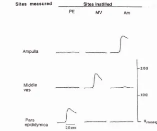
tetrodotoxin, indicating that the muscle contraction by the electrical stimula-tion to be nerve-transmitted (Figure 6). Stretching theportion
of
the vas deferens or the epididymal tail by instilling saline into its lumen caused marked elevation of the intraluminal pressure only at the site stretched(Figure 7). A major portion of the dye instilled into the
Med J Indones
lumen of the epididymal tail was rapidly transported to the ampulla and emitted into the posterior urethra by
the electrical stimulation of any site of the epididymal tail and vas deferens.tt
Th"
above findings appear to shed a light on the mechanism of rapid transport of thespermatozoa stored
in
the epididymal
tail to
the posterior urethra as follows. When sympathetic signalsreach the epididymal
tail, its
intraluminal pressureelevates and pushes out spermatozoa to the ampulla through the shortened and straightened vas deferens''
and the
full
stretch of the ampulla by the transportof
seminalfluid
inducesits
contraction and leads toSEED, The above results indicate that direct electrical stimulation
of
the canine vas deferens causes SEED regardless of the site stimulated.On the transportation of seminal fluid through the vas
deferens,
it
has been generally believed thatcontrac-tion
which starts at the epididymaltail
is
gradually transmittedto
the distal direction and thefluid
istransported gradually to the ampulla and emitted into
the
thraby
f the ampullawit
ls
amplit
sdeferens.l3-study
ect electricalstimulation of the vas deferens caused emission of the
content of the epididymal tail to the posterior urethra
regardless
of
the site stimulated, indicatinga
new concept of mechanism of sperm transport as described above.If
seminal fluid were transported by the abovetraditional hypothesis, muscle contraction at the
stimu-lated site should interfere the flow from the epididymal tail.
Application of the new method to generate semi-nal emission to men : direct electrical stimulation of the vas deferens
Before the above method was applied to nren,
it
wasvalidated
in
dogs whose bilateral hypogastric neryeswere transected. Stimulation
of
any siteof
the vas deferens caused SEED in all dogs receiving the tran-sectionl,
6 and 12 months before the stimulation. Inthe application to men, the middle vas was stimulated directly by penetrating two Pole needles (TOP Corp., Tokyo) through the scrotal skin to the manually
stabi-lized middle vas. Stimulus parameters
for
men were15-20V,2msec and 10H2. for 5 minutes.
All
stinrula-tions of patients with emission loss generated SEED,although pressing
of
the ampulla was necessary forI
L
o--"nl,/idC e va5
[image:6.595.143.429.82.361.2]Pars epididymica
Figure 5' Intralunùnal pressure ofthe epididytuis and vas deferens b1'direct electrical sritlulatio,l (2ntsec, gv, lo Hz; nerve
sti,,tula-tion) of the seninal tracr.
T:
f€,sris, E:
head and bodl' 6711t, epididl,ttris, ET:
epidittl.trtal tail, MV:
ttriddle tas, Attt:
atttltulla.1g
1Omin
Figure 6' Itthibitiott of nuscle contractiott of the atnpulla of the canine vas de.ferens b), rerrodotoïi,t (TTX). stitttulus parattterers for
electrical stintulatiott of the_t'as v:ere 0.5-5 nsec, 80V antl 20 Hz for 5 ,rrorà. The arrou, itrdicates additiott of rcrrodotoxin at
the co,'tcentration of 3 .t l0-' M.
Siteg measured Siles stlmulated
T,E C€ MV
[image:6.595.44.537.410.699.2]Tfi(3x10-20
Kihara et al. Med J IndonesSites
measured Sltes instllledPE MV Am
Ampulla
Middle
VAS
[image:7.595.131.444.84.344.2]Pars epididymica
Figure 7. httralwninal pressure ofthe epididl'tnis and vas deferens Iunten. PE
:
pars epididltnica of the vas, MV:
niddLe vas, AM:
ommHg
b), stretchitrq the wall of the duct bf instillittg saline inro the
anpulLa.
- 200
L
a
slight painat
the stimulating siteat
the scrotum during stimulating in some patients.ll
Previous
trials
for
artifical
seminal emission;sub-arachnoid
injection
of
anticholine esterase, directelectrical stimulation
of
the
superior hypogastricnerve, transrectal electrical stimulation
of
the pelvicplexus or prostatic plexus and vibration
of
the penis have depended upon the existence of the spinal reflex archor at
least thepelvic
plexus.In
contrast, the present method, which stimulates the effective organitself, is applicable to emission loss patients lacking the
reflex arch or the pelvic plexus. The current study has
further demonstrated that there are no specific sites
of
the vas deferens and epididymal
tail for
electrical stimulation to cause SEED. The vas deferens itself can be easily identified and held over the scrotal skin using(he fingers to stabihze
it.
CONCLUSION
The current studies showed the elaborate mechanisnrs
of
the
retroperitoneal and intrapelvic sympathetic nervous system to preserve SEED against various in-juriesof
the peripheral sympathetic nervous systemand introduced such an easy, safe and repeatable
method
of
artificial
seminal emission
from
theejaculatory duct that was based on a new concept of
mechanism of sperm transport and applicable to those
with emission loss.
REFERENCES
1. Langley JM, Anderson HK. Innervation of the pelvic and
adjoining viscera. J Physiol 1896;19:372-84.
2. Leamonth JR. Contribution to neurophysiology of urinary bladder in man. Brain 1931;45:141-76.
3. Semans
IH,
Langworthy OR. Observation on the neurophysiology of sexual function in the rnale cat. I Urol1938;40:836-46.
4. Kimura Y, Miyata K, Adachi K, Kisaki N. Peripheral nerves
controlling the intemal urethral orifice during ejaculation. Urol Int 191 5;30:218 -27.
5. Kihara K, Sato K, Ando M, Sato T. Oshima H. Ability of
each lumbar splanchnic nerve and disability ofthoracic ones
to
generate seminal emissionin
the dog.J
Urol 1992;147:26O-3.6. Ando M, Kihara K, Sato K, Sato T, Oshima H. Regulation of the bladder neck closure by lumbar splanchnic nerves at
ejaculation in the dog. Neurourol Urodynam 1993;12:91-8. 7. Kihara K, Sato K, Ando M, Morita T, Oshirna H. Control of
8. Kihara K, Sato K, Ando M, Sato T, Oshima H. Lumbosacral sympathetic trunk as a compensatory pathway for seminal emission after bilateral hypogastric nerve transections in the dog. J Urol 1991;145:640-3.
9. Sato K, Kihara K, Ando M, Sato T, Oshima H. Seminal emission by electrical stimulation of the spermatic nerve and epididymis. Int J Androl 199l;14:461-7.
10. Kihara K, Sato K, Ando M, Sato S, Oshima fI. A mechanism
of retrograde ejaculation after bilateral hypogastric nerve
,
transectionsinthedog. IUtol 1992;148:1307-9.11. Kihara K, Sato K, Ando M, Ushiyama T, Azuma H, Oshima H. A new method to generate canine seminal emission and its application to men : Direct electrical stimulation of the vas deferens. I Androl in press.
12. Mitsuya H, Asai I, Ushida T, Hosoe K. Application of X-ray cinematography in urology : Mechanism of ejaculation. J Urol 1960;83:86-92.
13. Baumgarten HG, Holstein AF, Rosengren E_ Arrangement, ulttastructute and adrenergfc innervation of smooth mus-culature of the ductuli efferentes, ductus epididymidis and ductus deferens of man. Z Zellforcf,- l97l:l2Oz37-79. 14. Narita H, The study of sperm transport through the human
genital tract. Ipn J Urol 74:1735-48.
15. Ventura WP, Freund M, Davis J, Pannuti C. Influence of
norepinephrine on the motility of the human vas deferens :