Trehalose preincubation increases mesenchymal (CD271
+) stem cells post-
cryopreservation viability
Keywords: cryopreservation, mesenchymal (CD271+) stem cells, trehalose preincubation
pISSN: 0853-1773 • eISSN: 2252-8083 • http://dx.doi.org/10.13181/mji.v25i3.1273 • Med J Indones. 2016;25:129–35
• Received 23 Aug 2015 • Accepted 13 Jun 2016 Corresponding author: Indra Kusuma, [email protected]
Copyright @ 2016 Authors. This is an open access article distributed under the terms of the Creative Commons Attribution-NonCommercial 4.0 International License (http://creativecommons.org/licenses/by-nc/4.0/), which permits unrestricted non-commercial use, distribution, and reproduction in any medium, provided the original author and source are properly cited.
Indra Kusuma,1,2 Restu S. Hadi,1 Bambang Kiranadi,2 Arief Boediono2
1 Faculty of Medicine, Universitas YARSI, Jakarta, Indonesia
2 Department of Anatomy, Physiology, and Pharmacology, Faculty of Veterinary Medicine, Bogor Agricultural University,
Bogor, Indonesia
B a s i c M e d i c a l Re s e a rc h
ABSTRAK
Latar belakang: Dimetil sulfoksida (Me2SO) adalah
krioprotektan yang umum digunakan dalam kriopreservasi sel. Me2SO diketahui menyebabkan perubahan epigenetik yang
mempengaruhi perkembangan sel punca dan diferensiasi sel. Oleh karena itu diperlukan upaya untuk mengembangkan tehnik kriopreservasi yang dapat melindungi fungsi sel dan menghindarkan efek samping Me2SO. Trehalosa diketahui
dapat melindungi organisme dalam kondisi ekstrem seperti dehidrasi dan suhu dingin. Penelitian ini bertujuan melihat efek proteksi pre-inkubasi trehalosa dalam prosedur kriopreservasi.
Metode: Penelitian ini bersifat ekperimental. Sel punca
mesenkim (CD271+) dari biorepositori Universitas YARSI
digunakan dalam eksperimen. Pre-inkubasi trehalosa dilakukan
selama 1 jam, internalisasi trehalosa kemudian dikonfirmasi
dengan pemeriksaan FTIR-ATR. Viabilitas sel dalam kultur dibandingkan antara kelompok yang terdiri dari (1) kriopreservasi tanpa pre-inkubasi trehalosa, (2) kriopreservasi dengan pre-inkubasi trehalosa dan (3) tidak mengalami kriopreservasi (kontrol) setelah 24 jam dibekukan. Absorbansi dari setiap kelompok diperoleh pada panjang gelombang 450 nm. Analisis statistik dilakukan menggunakan student t test.
Hasil: Viabilitas sel punca mesenkim (CD271+) pada kelompok
yang mendapat pre-inkubasi trehalosa lebih tinggi (p<0,05) dari kelompok yang tidak mendapat pre-inkubasi trehalosa. Viabilitas yang lebih baik pada kelompok yang mendapat pre-inkubasi trehalosa dibandingkan kelompok kontrol mengindikasikan adanya proteksi terhadap tripsinisasi. Sel punca mesenkim (CD271+) yang diinkubasi dengan medium yang mengandung 100 mM trehalosa menghasilkan efisiensi
internalisasi trehalosa sebanyak 15%.
Kesimpulan: Hasil menunjukkan efek proteksi prosedur
pre-inkubasi trehalosa dalam kriopreservasi. Penelitian selanjutnya diarahkan untuk menerangkan mekanisme internalisasi trehalosa ke dalam sitoplasma dan mekanisme proteksi yang berperan dalam kriopreservasi.
ABSTRACT
Background: Dimethyl sulfoxide (Me2SO) is a common cryoprotective agent widely used in cell preservation system. Me2SO is currently known to cause epigenetic changes which are critical in stem cells development and cellular differentiation. Therefore, it is imperative to develop cryopreservation techniques that protect cellular functions and avert Me2SO adverse effect. Trehalose was able to protect organism in extreme condition such as dehydration and cold. This study aimed to verify the protective effect of trehalose preincubation procedure in cryopreservation.
Methods: The study was conducted using experimental design. Thawed mesenchymal (CD271+) stem cells from YARSI biorepository were used for the experiment. Trehalose preincubation was performed for 1 hour, internalized
trehalose was confirmed by FTIR-ATR measurement.
Three groups consisted of (1) cryopreserved without trehalose preincubation, (2) cryopreserved with trehalose preincubation, and (3) did not undergo cryopreservation were evaluated after 24 hours in LN2 for viability in culture. The absorbance from each group was measured at 450 nm. The analysis performed using paired student t test.
Results: Viability of thawed mesenchymal (CD271+) stem cells that undergo trehalose preincubation prior
cryopreservation was significantly higher (p<0.05) compared
to group without trehalose preincubation. Higher viability observed between group with trehalose preincubation compared with controlled group suggests protection to trypsinization. Mesenchymal (CD271+) stem cells incubated for 1 hour in 100 mM trehalose supplemented medium
results in 15% trehalose loading efficiency.
Conclusion: These findings confirm the protective effect of
trehalose preincubation in cryopreservation. Future research
Development of regenerative medicine using stem cells as an approach to replace damaged and worn out cells and tissues warrants a storage solution that protects stem cells unique ability to differentiate into many types of cells of the body. Current method of cell cryopreservation incorporated the use of dimethyl sulfoxide (Me2SO) as an intracellular cryomedium. This
practice is currently under criticsm as described by Diaferia et al1 in their review. Me
2SO is known
to cause adverse effects and toxicity to patient, unexpected changes in cell fate, affects epigenetic control by acting on DNA methyltransferases, and loss of pluripotency in human embryonal stem cells.1 Despite the adverse effect of Me
2SO, it is
still commonly used as cell protectant thus we used Me2SO-based cryopreservation in this study.
Therefore, it is necessary to develop alternative procedure that can be adapted to cryopreservation
workflow and avert the potential adverse effect of
using Me2SO as an intracellular cryopreservant.
Among other such as sucrose utilized by embryologist for ovum and sperm preservation, trehalose (378.33 g/mol) is a glucose disaccharides that has been recognized to be able to protect cells,2
organism3 and stabilize intracellular protein in
extreme condition such as dehydration and cold.4
Cellular toxicity of trehalose was also compared in this study to give a picture of the extent of toxicity posed in trehalose preincubation to mesenchymal stem cells along with other disaccharides such as sucrose and known toxic chemicals such as Me2SO
and hydrogen peroxide which is a potent oxydator and caused damage to organic compound. Preincubation was known to lead to trehalose internalization into the cytoplasm. Several methods of trehalose internalization requires cell poration using adenosine triphosphate (ATP) and the use of liposomes is being investigated.5–7
Mesenchymal stem cell is a heterogeneous population recently discovered to contain not only multipotent stem cell but also non-tumorigenic pluripotent stem cell subpopulation known as the multi-lineage differentiating stress enduring (MUSE) cells.8,9 The ability to preserve functional
properties of such unique subpopulation will greatly contribute to the development of regenerative medicine.
Trehalosa as a cryoprotectant has been evaluated in several types of cells such as mesenchymal stem
cells-derived from bone marrow, erythrocytes, platelets, and umbilical cord blood stem cells.10–12
Suggested method of trehalose internalization
was by fluid phase endocytosis after 1–24 hours
of trehalose pre-treatment via clathrin-mediated endocytosis.12 However, this was suggested
as cell-type specific because the endocytosis
mechanism involved.13 This study aimed to verify
the protective effect of trehalose-preincubation in Me2SO-based cryopreservation of mesenchymal
(CD271+) stem cells derived from peripheral
blood mononuclear cells which were a potential source of MUSE cells.
METHODS
Research design
The present study conducted using experimental design from December 2014 to March 2016 at
YARSI University Cell Culture Facility, Jakarta,
Indonesia. Ethical approval received from Komisi Etik Lembaga Penelitian Universitas YARSI (No. 016/KEP-UY/BIA/II/2016).
Cell culture
Mesenchymal stem cells were derived from plastic-adherent mononuclear cells of peripheral blood (PBMC) obtained from Laboratorium Terpadu Universitas YARSI Biorepository. Purified using CD271 magnetic sorting magnetic-activated cell sorting (MACS); Miltenyi, expanded and cultured
in Dulbeco’s modified eagle’s medium (DMEM)
low glucose (Gibco) supplemented with 10%
fetal bovine serum (FBS) heat inactivated (Gibco).
1% penicillin-streptomycin and fungizone (Gibco) added to prevent bacterial and fungal contamination. Mesenchymal stem cells used for experiments were from passage four to six in exponential phase. All experiment described below were performed in triplicate.
Toxicity of trehalose, sucrose, Me2SO and
hydrogen peroxide were compared using CD271+
mesenchymal stem cells. The study was conducted in a 96 microwell plate with as much as 20,000 viable cell cultured. Incubation of the investigated chemicals at 0 mM, 12.5 mM, 25 mM, 50 mM, and 100 mM of trehalose, 0 mM, 18.75 mM, 37.5 mM, 75 mM, and 150 mM of sucrose, 0%, 0.06%, 0.12%, 0.25%, 0.5%, and 1% of Me2SO and 0 µM,
31.25 µM, 62.5 µM, 125 µM, 250 µM, 500 µM hydrogen peroxide for one hour and continued
with 30 minutes water-soluble tetrazolium-1 (WST-1) proliferation assay (Roche) incubation to assess cell viability. Measurement conducted using microplate reader at 450 nm. Long-term toxicity was assessed in seven-days-culture, viability was measured at three different time point (day in vitro/DIV two, four, and seven) also using WST-1 proliferation assays in 96 microwell plate. Osmolarity measurement of medium supplemented by trehalose and sucrose were
conducted at Faculty of Veterinary Medicine, Bogor Agricultural University, Embryology Laboratory.
Trehalose measurement by FTIR-ATR
Mesenchymal (CD271+) stem cells cultured
at 80% confluency using tissue flask 25 sqcm
were incubated with 100 mM trehalose mixed in culture medium for one hour. Cells were harvested by trypsinization using trypsin / ethylenediaminetetraacetic acid (EDTA) 0.05% for
five minutes and washed by centrifugation twice.
Resulting pellet were counted using tryphan blue exclusion method and adjusted for 1,000,000 viable cell resuspended in 50 µl phosphate
buffer saline (PBS), Gibco for FTIR measurement.
Intracellular trehalose detection performed using
fourier transform infrared spectroscopy (FTIR) – attenuated total reflectance (ATR).
Cell pellet were dried using deoxyribonucleic acid (DNA) concentrator (MiVac) at 37°C for 45 minutes. Infrared spectra were generated by putting dried cell pellet on top ATR germanium
crystal. 32 scan performed by FTIR at 1,100–900
wavenumber/cm, peak at 995–991 wavenumber/ cm as reported by Sakurai et al14 was considered
as trehalose. Trehalose concentration was determined using simple Beer’s law principle by
generating trehalosa standard curve in FTIR-ATR
(Nicolet-Bruker). Omnic software was used to measure peak height and area under peak curve to generate a standard curve of known trehalose
concentration with a linear fitting.
Cryopreservation
Trehalose-incubated mesenchymal (CD271+) stem
cells described previously were cryopreserved using standard slow-freeze technique and cryoprotectant (70% culture medium, 20% Me2SO,
and 10% fetal bovine serum). Three groups were established consist of (1) cryopreserved without
trehalose preincubation-slow freeze 721 (SF721)
(group 1), (2) cryopreserved with
trehalose-preincubation-SF721-Tre (group 2), and (3) did
not undergo cryopreservation (control). Cells were stored in -80°C overnight and transferred to liquid nitrogen (LN2) the next day. After 24 hours in LN2 cells were thawed and cultured in 96
microwell plate at 10,000 viable cells per well for functional evaluation using WST-1 proliferation assay. Absorbance of attached and viable cell after 60 minutes plating were measured at 450 nm.
Data analysis
Blank absorbances were subtracted from the original data. Analysis was performed using Excell
(version 2013; Microsoft). Statistical significance
between group 1 and control group and between group 2 and control group were both determined using paired student t test. Phosphate buffer saline spectra were subtracted from all spectra obtained to construct the trehalose standard curve.
RESULTS
Toxicity studies
Toxicity profile of Me2SO, trehalose, and sucrose
hydrogen peroxides were illustrated in Figure
1. These data suggest that 100 mM of trehalose was at comparable toxicity to 1% Me2SO which
was currently accepted as a safe level for cell exposure. A minimum of 150 mM of sucrose was also comparable but the osmolality limited it used. The level of toxicity posed by these dissacharides is comparable with a very low level of hydrogen peroxide concentration.
Further evaluation of the disaccharides were
conducted using 50 mM, 25 mM, and 12.5 mM of trehalose and sucrose. Compared to control (0mM) group, within each time points both disaccharides reduced cells viability in a dose-dependent manner. However, observation of each dose group from three different time points during seven days of culture showed that the
cells continued to have significant proliferation
capacity despite the presence of disaccharides
in the medium (Figure 2). Disaccharides toxicity
was considered related to cellular hydration
status. Figure 3 shows the changes in culture
Figure 1. Toxicity profile of Me2SO (A), trehalose (B), sucrose (C), and hydrogen peroxide (D) to mesenchymal (CD271+) stem cells. Toxicity of 100 mM of trehalose is relative comparable to 1% Me2SO, 160 mM sucrose and 500 µM of hydrogen peroxide. No
significant difference (p>0.05) between trehalose (100 mM) and Me2SO (1%)
extracellular environment and triggered water
efflux from intracellular compartment that
caused dehydration and cell death.
Intracelullar trehalose measurement
Internalization of trehalose into cytoplasm
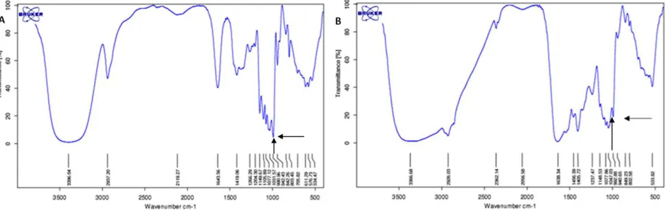
was confirmed by FTIR measurement. Figure
4 shows a spectrum recorded from washed and dried cell pellet with characteristic peak
at the fingerprint region (below 1,500/cm) for
Figure 2. Growth curve of mesenchymal (CD271+) stem cells with trehalose (left) and sucrose (right) as suplementation in culture medium. * p<0.05
trehalose at 991–992 /cm. Absorbance level of trehalose derived from infrared spectra were used to construct trehalose standard curve for
intracellular trehalose quantitation. (Figure
5) the standard curve allowed quantitation of trehalose within the cytoplasm which gives a
resultof 15% loading efficiency. 15.07 mM of
trehalose were estimated within the cytoplasm of approximately 1,000,000 viable cells that
were measured by FTIR-ATR.
http://mji.ui.ac.id
Growth curve with trehalose Growth curve with sucrose
Cryopreservation study
Trehalose-incubated cells showed a greater
(p<0.05) viability and attachment after 24
hours in liquid nitrogen compared with non-trehalose-incubated cells and control group
which was not cryopreserved. These findings confirmed that intracellular trehalose were able
to protect mesenchymal (CD271+) stem cells
in cryopreservation. Figure 6 shows significant
viability retained by trehalose-incubated cells compared to non-cryopreserved cells (control group). These data suggested a certain protection against stress evoked by trypsinization process.
DISCUSSION
Our data showed that extracellular trehalose could cause a toxic effect on cells by inducing osmolality imbalance. Lynch et al15 reported
approximately 60% of cells death recorded after cells loaded with over 200 mM of internal trehalose
Figure 4. A) Spectrum recorded from 250 mM trehalose solution shows a characterisitic peak of trehalose at 990.96 /cm wave-number (arrow); B) Spectrum of dried lysate cell derived from 100 mM trehalose preincubated cell culture shows trehalose peak at 992.65 /cm wavenumber (arrow). The difference in wavenumber attributed to water content from the trehalose solution
Figure 5. Trehalose standard curve with linear fitting
Figure 6. Viability of attached cells in culture after
thaw-ing. SF721: Cryopreserved without trehalose preincubation; SF721-Tre: Cryopreserved with trehalose preincubation; SF721: slow-freeze with 70% culture mediun, 20% FBS and
10% Me2SO
concentration were resuspended in phosphate buffer saline of physiologically osmolality (300 mOsm). Our toxicity data showed a low dose
Medical Journal of Indonesia
0
Disacharides Concentration in medium (mM) Changes of osmolarity by Disacharides
Trehalose Sucrose
Figure 3. Changes in medium osmolarity after disacharides suplementation
Trehalose concentration (M)
Ar
ea under peak [994.73 (SD 1.76)]
Trehalose Sucrose
Disacharides concentration in medium (mM)
Experiment group
R
atio of osmolarity (100%)
A
Disacharides Concentration in medium (mM) Changes of osmolarity by Disacharides
of extracellular trehalose (<50 mM) caused
reduction in viability at day seven of culture
(Figure 2). However, an increase in viability was
clearly observed compared to previous time point in the same dose group. This suggests that trehalose toxicity did not affect all of the cells population and the mesenchymal stem cells population still retained the proliferative capacity.
Zhou et al2 used 50 mM trehalose incubated
for four hours results in 13 mM intracellular trehalose concentration which quite similar with our data (15.07 mM). Zhang et al16 also have a
similar result of 14.57 mM while Oliver et al12
reported at 19 mM. The use of ATP as a poration agent greatly increased the loading kinetics such as intracellular concentration of 50 mM reached after 90 minutes incubation.6 These data were
derived from different cell and different method of quantitation such as anthrone reaction and HPLC measurement. As far as we know our study
is the first that used FTIR-ATR for intracellular trehalose quantification.
Our study suggests that trehalose-preincubation which results a 15 mM of intracellular trehalose concentration alongside the standard slow-freezing, Me2SO-based cryomedium were
sufficient to protect mesenchymal (CD271+)
stem cells from cryoinjury. Although to achieve this we used 100 mM of trehalose with one hour
preincubation protocol which caused significant
reduction in cell viability. This condition warrants a further investigation in trehalose internalization
mechanism. Furthermore, our data suggest that intracellular trehalose was benefit in preventing
trypsinization-induced cell injury. Trypsin, although widely used in passaging procedure, is in fact a protease that can cause stress and cell death. Trehalose was known to help retain cellular integrity and prevent protein denaturation.4
Therefore, intracellular trehalose may prevent cell injury during trypsinization resulting in a higher viability compared to the control group.
Different trehalose loading kinetics reported by comparing different study that has been conducted on platelets, erythrocytes, and stem cells using trehalose incubation method, suggest
there is a cell-type specific of internalization
method. Although Oliver et al12 reveals
bone-marrow derived mesenchymal stem cells used
clathrin-mediated endocytosis for trehalose internalization, this not necessarily the same for other cell type. Elucidating the mechanism of trehalose internalization of different cell type can shed a new understanding of cell membrane regulation.
In conclusion, this study concluded that trehalose-preincubation procedure was able to protect mesenchymal (CD271+) stem cells
from cryoinjury results from slow-freeze
cryopreservation procedure. Further study
should be directed to elucidate factors that regulate trehalose internalization kinetics in different cell types. Research could also be directed to verify trehalose cryoprotection in different type of cryopreservation such as
vitrification.
Acknowledgment
Funding was provided by LPDP scholarship
scheme. We thank Anna Roswiem and Triayu
Septiani for assistance in operating FTIR-ATR
measurement, and Intan Razari for her assistance in using MiVac DNA concentrator.
Conflict of interest
Funding was provided by Lembaga Pengelola
Dana Pendidikan (LPDP) scholarship scheme.
REFERENCES
1. Diaferia GR, Dessi SS, DeBlasio P, Biunno I. Is stem cell chromosomes stability affected by cryopreservation conditions? Cytotechnology. 2008;58(1):11–6.
2. Zhou XL, Zhu H, Zhang SZ, Zhu FM, Chen GM, Yan LX. Freeze-drying of human platelets: influence of
intracellular trehalose and extracellular protectants. Cryo Letters. 2006;27(1):43–50.
3. Streeter JG. Effect of trehalose on survival of Bradyrhizobium japonicum during desiccation. J Appl
Microbiol. 2003;95(3):484–91.
4. Jain NK, Roy I. Effect of trehalose on protein structure.
Protein Sci. 2009;18(1):24–36.
5. Emanuele E, Bertona M, Sanchis-Gomar F,
Pareja-Galeano H, Lucia A. Protective effect of trehalose-loaded liposomes against UVB-induced photodamage in human keratinocytes. Biomed Rep. 2014;2(5):755–9.
6. Elliott GD, Liu XH, Cusick JL, Menze M, Vincent J, Witt
T, et al. Trehalose uptake through P2X7 purinergic channels provides dehydration protection. Cryobiology. 2006;52(1):114–27.
7. Lynch AL, Chen R, Slater NK. pH-responsive polymers for trehalose loading and desiccation protection of human red blood cells. Biomaterials. 2011;32(19):4443–9.
8. Tsuchiyama K, Wakao S, Kuroda Y, Ogura F, Nojima M, Sawaya N, et al. Functional melanocytes are
readily reprogrammable from multilineage-differentiating stress-enduring (muse) cells, distinct
stem cells in human fibroblasts. J Invest Dermatol.
2013;133(10):2425–35.
9. Simerman AA, Perone MJ, Gimeno ML, Dumesic DA,
Chazenbalk GD. A mystery unraveled: nontumorigenic pluripotent stem cells in human adult tissues. Expert Opin Biol Ther. 2014;14(7):917–29.
10. Lynch AL, Slater NK. Influence of intracellular trehalose
concentration and pre-freeze cell volume on the cryosurvival of rapidly frozen human erythrocytes. Cryobiology. 2011;63(1):26–31.
11. Rodrigues J, Paraguassú-Braga FH, Carvalho L, Abdelhay E, Bouzas LF, Porto LC. Evaluation of trehalose and sucrose as
cryoprotectants for hematopoietic stem cells of umbilical cord blood. Cryobiology. 2008;56(2):144–51.
12. Oliver AE, Jamil K, Crowe JH, Tablin F. Loading human mesenchymal stem cells with trehalose by fluid-phase
endocytosis. Cell Preserv Tech. 2004;2(1):35–49. 13. McMahon HT, Boucrot E. Molecular mechanism
and physiological functions of clathrin-mediated endocytosis. Nat Rev Mol Cell Biol. 2011;12(8):517–33.
14. Sakurai M, Furuki T, Akao K, Tanaka D, Nakahara Y, Kikawada T, et al. Vitrification is essential for
anhydrobiosis in an African chironomid, Polypedilum vanderplanki. Proc Nat Acad Sci. 2008;105(13):5093–8. 15. Lynch AL, Slater NK. Mediated trehalose un-loading
for reduced erythrocyte osmotic fragility and phosphatidylserine translocation. Cryo Letters. 2011;32(5):415–24.
16. Zhang S, Qian H, Wang Z, Fan J, Zhou Q, Chen G, et al.
Preliminary study on the freeze-drying of human bone
marrow-derived mesenchymal stem cells. J Zheijang