EISSN: 2086-4094 DOI: 10.4308/hjb.18.3.135
Cloning and Expression Analysis of a Giant Gourami
Vasa
-Like cDNA
ALIMUDDIN1∗∗∗∗∗, IRMA ANDRIYANI1, MUHAMMAD ZAIRIN JUNIOR1, HARTON ARFAH1,
ANNA OCTAVERA1, ODANG CARMAN1, GORO YOSHIZAKI2
1Department of Aquaculture, Faculty of Fisheries and Marine Sciences, Bogor Agricultural University, Bogor 16680, Indonesia
2Department of Marine Biosciences, Tokyo University of Marine Science and Technology, Tokyo 108-8477, Japan
Received August 10, 2010/Accepted September 30, 2011
Molecular marker is useful in the development of testicular cells transplantation for detecting donor-derived germ cells in the recipient gonad. In this study, a giant gourami (Osphronemus goramy) vasa-like gene (GgVLG) was cloned and characterized for use as a molecular marker for germ cells in this species. Nucleotide sequence analysis revealed that GgVLG comprises 2,340 bps with an open reading frame of 1,962 bps encoding 653 amino acids. The deduced amino acid sequence contained 17 arginine-glycine or arginine-glycine-glycine motifs and eight conserved motifs belonging to the DEAD-box protein family. The GgVLG sequence showed high similarity to Drosophila vasa, common carp vasa homolog and tilapia vasa homolog for 66.2, 85.9, and 90.7%, respectively. In adult tissues, the GgVLG transcripts were specifically detected in ovary and testis. In situ hybridization analysis showed that GgVLG mRNA was detected in oocytes of the ovary and spermatogonia of the testis. There was no signal detected in the spermatocytes, spermatids and other gonadal somatic cells. Thus, consensus sequences, specific localization of GgVLG mRNA in the germ cells, amino acid sequence similarity and phylogenic analysis all suggest that GgVLG is the giant gourami vasa-like gene. Further, GgVLG can be used as a molecular marker for giant gourami germ cells.
Key words: germ cell transplantation, ovary, spermatogonia, testis, giant gourami, vasa
___________________________________________________________________________
_________________
∗ ∗∗
∗∗Corresponding author. Phone/Fax: +62-251-8622940,
E-mail: [email protected], [email protected] INTRODUCTION
Giant gourami (Osphronemus goramy) is an important freshwater cultured fish species in Java and Sumatera. The Directorate General of Aquaculture has programmed to increase significantly production of this species (Department of Marine Affairs and Fisheries, 2005). One of the obstacles to meet the production target is seed availability from hatchery. Breeders cultivate fry for 2-3 years to acquire first sexual maturity broodstock. Further, induced maturation and artificial spawning to control seed production of giant gourami remain to be developed. Currently, fry is produced by natural spawning in pond. This seed production system involves maintenance of giant gourami broodstock, which requires considerable space, cost, and labor. Consequently, the need therefore exists to establish a novel method for seed production of giant gourami.
A technique for fish germ cell transplantation using primordial germ cells (PGCs) or spermatogonia (SG) as donor germ cells had recently been developed (Okutsu et al. 2006). Donor germ cells are microinjected into the peritoneal cavities of newly hatched embryos. They subsequently migrate toward and colonize the genital ridges of the recipient embryos. Furthermore, donor-derived germ cells proliferate and differentiate into mature eggs and sperm in the allogeneic (Takeuchi et al. 2003;
Okutsu et al. 2006) and xenogeneic recipient gonads
(Takeuchi et al. 2004; Okutsu et al. 2007); the resulting gametes produce live fry through fertilization. Thus, if the giant gourami germ cell could be transplanted into well-controlled reproduction and smaller fish species such as Nile tilapia, then giant gourami gametes might more easily and rapidly be produced in surrogate Nile tilapia kept in aquaria. Hence, the maintenance of giant gourami broodstock in pond would no longer be required.
MATERIALS AND METHODS
Fish. Ten immature giant gourami fish, Osphronemus goramy were obtained from National Center for Development of Freshwater Aquaculture, Sukabumi. Body weight of fish was 1.03 + 0.17 kg (mean + standard deviation).
RNA Isolation and Synthesis of cDNA. Testes were excised from male giant gourami with gonadosomatic indexes (GSIs) of 0.0089%. The testes were homogenized and used for total RNA extraction using Isogen reagent (Nippon Gene, Tokyo, Japan). First-strand cDNA was synthesized using the Ready-To-Go You-Prime First-Strand Beads Kit (GE Healthcare UK Ltd., England) with an oligo (dT) primer (5’-GTA ATA CGA CTC ACT ATA GGG CAC GCG TGG TCG ACG GCC CGG GCT GGT TTT TTT TTT TTT TTT TTT-3’) according to the manufacturer’s instructions.
Cloning of the Vasa cDNA Fragment by Reverse Transcription Polymerase Chain Reaction (RT-PCR). RT-PCR was performed with degenerate primers as reported by Nagasawa et al. (2009). Primer mix-vasa-Fw: TYCTDCARCAGYTGATGG-3’ and mix-vasa-Rv: 5’-TCAAACTTSCK-GGCYTCMA-3’ were designed using the highly conserved regions of vasa homologs from eight fish species with the following GenBank accession numbers: butterfly fish (Pantodon buchholzi): AF479823, gilthead sea bream (Sparus aurata): AF520608, medaka (Oryzias latipes): AB063484, Nile tilapia (Oreochromis niloticus): AB032467, rainbow fish (Melanotaenia fluviatilis): AF479824, rainbow trout (Oncorhynchus mykiss): AB032566, shiro-uo (Leucopsarion petersii): AB098252, tetra (Hyphessobryon ecuadoriensis): AF479821, and zebrafish (Danio rerio): NM_131057).
The PCR reaction was conducted at 94 oC for 3 min,
followed by 35 cycles of 30 s at 94 oC, 20 s at 56 oC and
30 s at 72 oC, followed by a final extension step of 72 oC for
3 min. The cDNA was amplified using Takara Ex Taq (Takara Bio Inc., Shiga, Japan). PCR products were electrophoresed on a 2.0% agarose gel, and the cDNA fragments that showed the predicted molecular weight were isolated using an UltraClean-15 DNA Purification Kit (MO BIO Laboratories, Inc., CA, USA). The purified cDNA fragments were subcloned into a pGEM T-Easy plasmid vector (Promega, WI, USA), and sequenced using a BigDye Terminator v3.1 Cycle Sequencing Kit (Applied Biosystems, CA, USA). Sequence determination was performed on ABI PRISM 3100-Avant Genetic Analyzer (Applied Biosystems).
Cloning of Full-length Vasa cDNA. The 3’ and 5’ rapid amplification of cDNA ends (RACE) were performed to isolate a full-length cDNA sequence. After determining the DNA sequence of a partial vasa cDNA fragment, two vasa-specific primers (Fw-3’-RACE1: 5’- TGA GAC TGT TGG ATG TGA TCG GAA GA -3’, Fw-3’-RACE2: 5’- TAA GCT GAG GTA CCT GGT GCT AGA -3’) was synthesized for use as the forward primer for 3’-RACE, and adapter primers (AP1: 5’-CCA TCC TAA TAC GAC TCA CTA TAG GGC-3’, AP2: 5’-CTA TAG GGC ACG CGT GGT-3’) were
used as the reverse primers for 3’-RACE. PCR reactions were performed according to the method described previously (Yoshizaki et al. 2000). 5’-RACE was carried out using a GeneRacer Kit with SuperScript III RT (Invitrogen, CA, USA) according to the manufacturer’s instructions. Two primers for giant gourami vasa cDNA (Rv-5’-RACE1: 5’-GCT GCC ACT CCG TCT GCC ATC A-3’, Rv-5’-RACE2: 5’-GCA GCC GTT TTG CCC GAT CC-3’) were synthesized for use as reverse primers for 5’-RACE. The cDNA was amplified using Takara LA Taq (Takara Bio Inc., Shiga, Japan). We estimated the molecular weight and pI of giant gourami vasa homolog using a Compute pI/Mw tool (http://au.expasy.org/tools/ pi_tool.html). Moreover, its similarity and identity were calculated by LALIGN (http://www.ch.embnet.org/softwareLALIGN_ form.html) using vasa homolog sequences from other species. The LALIGN programs compare two sequences and look for local sequence similarities.
Phylogenetic Analysis. A homology search of the deduced amino acid sequence of the obtained cDNA was carried out using the National Center for Biotechnology Information website (http://www.ncbi.nlm.nih.gov/ BLASTP). The deduced amino acid sequences were aligned using Genetyx version 7, and phylogenic tree were constructed by the unweighted pair-group method with arithmetic mean (UPGMA) method.
RT-PCR Analysis. Total RNA extraction and cDNA synthesis were performed using various organs (gill, fin, muscle, liver, intestine, testis, and ovary) of immature giant gourami as described above. The PCR reaction was conducted with giant gourami vasa-specific primers. The forward primer was located between nucleotides 912 and 937 (Fw-PCR: 5’- GTT CCT GCT CCC AAT TCT GCA GCA G -3’), while the reverse primer was located between nucleotides 2,296 and 2,319 (Rv-PCR: 5’-ACG TTC TGT CTG TCA GAC ACA TTG-3’). The PCR reaction was
performed at 94 oC for 3 min, followed by 35 cycles of 30 s
at 94 oC, 30 s at 60 oC and 1.30 min at 72 oC, followed by a
final extension step of 72 oC for 3 min. The cDNA was
amplified using Takara Ex Taq (Takara Bio Inc., Shiga,
Japan). β-actin gene expression also was analyzed as an
internal control for equal loading of RNA with a set of primers; forward (5’-GAC AAC GGM TCY GGY-3’) and reverse (5’-TAG AAG GTG TGR TGC-3’). PCR products were electrophoresed on a 0.7% agarose gel.
In Situ Hybridization Analysis with Digoxigenin
(DIG)-labeled RNA Probes. In situ hybridization was performed using a method developed by Yoshizaki’s laboratory. A 1.4 kb cDNA vasa fragment (nucleotides 912– 2,319 of vasa) was subcloned into the pGEM T-easy vector. Sense and antisense RNA probes were transcribed in vitro using DIG-labeled uridine triphosphate (UTP) (Roche, Mannheim, Germany) and T7 RNA polymerase (Promega). For the in situ hybridization (ISH) of tissue sections, tissue samples from the central region of the gonads were fixed
at 4 oC for 16 h in Bouin’s solution. After dehydration in
increasing concentrations of ethanol, a portion of each
sample was embedded in paraffin wax and cut into 5-µm
were then mounted on Matsunami Adhesive Slides (MAS; Matsunami Glass Ind., Ltd., Osaka, Japan), dewaxed, and dehydrated by immersion in a xylene-ethanol series. The sections were stained with hematoxylin-eosin (HE) or processed for ISH with DIG-labeled RNA probes. The sections were then permeabilized, acetylated, and
incubated with a hybridization mixture of 1 µg/ml RNA
probe, 50% formamide, 29 saline-sodium citrate (SSC) (pH
4.5), 50 µg/ml transfer RNA (tRNA), 50 µg/ml heparin, 1%
sodium dodecyl sulfate (SDS), and 10% dextran sulfate.
After hybridization at 65 oC for 16 h, the sections were
washed as follows: twice in 59 SSC/50% formamide at
65 oC for 30 min, twice in 29 SSC/50% formamide at 65 oC
for 30 min, and once in 19 SSC/25% formamide: 19 Tris buffered saline containing 0.1% Tween-20 (TBST) at room temperature (RT) for 30 min. The sections were then placed in NTE buffer [500 mM NaCl, 10 mM Tris–HCl pH 8.0, 1 mM
ethylenediamine tetraacetic acid (EDTA)] at 37 oC for 5 min
before being washed twice in 0.59 SSC at 65 oC for 20 min
and then twice 19 TBST at RT for 5 min. Hybridized DIG-labeled probes were visualized using a tyramide signal amplification (TSA) Plus 2,4-dinitrophenyl (DNP) alkaline phosphatase (AP)-System (PerkinElmer, CA, USA) as the indirect immunodetection method. Nonspecific binding was blocked in freshly prepared TNB buffer (100 mM Tris– HCl pH 7.5, 0.5% blocking reagent) for 30 min at RT in moist chambers. The sections were incubated for 1 h at RT with anti-DIG-POD Fab fragments (diluted 1:500 in the TNB buffer) (Roche), before being washed twice in TNT wash buffer (100 mM Tris–HCl pH 7.5, 150 mM NaCl, 0.05% Tween-20) for 5 min. DNP-tyramide was diluted (1:50 in the diluent provided) and the sections were incubated for 10 min at RT. The DNP-tyramide signal amplification procedure was performed twice in order to increase the signal-to-noise ratio in ISH. After two washes of 5 min each, anti-DNP-AP (diluted 1:100 in the TNB buffer) was
applied to the sections for 1 h at RT. Finally, each of the sections was rinsed twice in wash buffer for 5 min. The sections were then incubated in a NTMT solution (100 mM NaCl, 100 mM Tris–HCl pH 9.5, 50 mM MgCl2, 0.1% Tween-20, mM, 1 mM Levamisole) containing 0.0035% of nitroblue tetrazolium (NBT; Roche) and 0.0018% of 5-bromo-4-chloro-3-indolyl phosphate (BCIP; Roche) at RT in the dark. After the color reaction had occurred, the slides were mounted using Entellan neu (Merck KGaA, Darmstadt, Germany) and sections were counterstained using Nuclear Fast Red (NFR) (Vector Laboratories, CA, USA) for 16 h. The resulting sections were observed under a BX-50 microscope (Olympus, Tokyo, Japan).
RESULTS
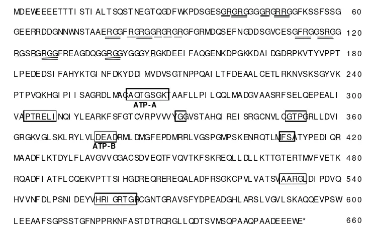
Cloning of Vasa cDNA and Phylogenetic Analysis. The full-length giant gourami vasa cDNA had an open reading frame of 1,962 bp that began with the first start codon, ATG, at position 100, and ended with a stop codon, TAG, at position 2061 (GenBank accession number: GQ422440). The open reading frame encoded 653 amino acids and the predicted sequence had a molecular mass of 70.9 kDa and a pI of 5.55. The deduced amino acid sequence showed 66.2% similarity and 48.0% identity with the Drosophila vasa (Hay et al. 1988; Lasko & Ashburner 1988), 85.9% similarity and 69.2% identity with the common carp vasa homolog (GenBank accession no.: AF479820), and 90.7% similarity and 77.0% identity with the tilapia vasa homolog (Kobayashi et al. 2000). The obtained amino acid sequence contained eight consensus sequences for the DEAD protein family (Figure 1, boxed) (Linder et al. 1989). The amino acid region between the N-terminus and amino acid position 150 contained 17 arginine-glycine repeats (Figure 1, single underline), and there were eight arginine-glycine-glycine repeats (Figure 1, double
MDEWEEEETT T I STI AL T SQST NEGT QGDF WKPDSGESGRGRGGGGRGRRGGFKSSFSSG
GEERRDDGNNWNSTAAERGGF RGRGGRGRGRGFGRMDQSEF NGDDSGVCESGFRGGSRGG
RGSRGRGGFREAGDQGGRGGYGGGYRGKDEEI FAQGENKDPGKKDAI DGDRPKVT YVPPT
L PEDEDSI FAHYKTGI NF DKYDDI MVDVSGT NPPQAI LT F DEAAL CETL RKNVSKSGYVK
PT PVQKHGI PI I SAGRDL MACAQT GSGKT AAF LL PI L QQL MADGVAASRF SELQEPEAL I
VAPTREL I NQI YL EARKF SF GT CVRPVVVYGGVST AHQI REI SRGCNVL CGT PGRL LDVI
GRGKVGL SKL RYL VL DEADRML DMGF EPDMRRLVGSPGMPSKENRQT LMF SATYPEDI QR
MAADF L KT DYL F L AVGVVGGACSDVEQTF VQVTKF SKREQL L DL L KT TGT ERTMVF VET K
RQADF I AT FL CQEKVPT T SI HGDREQREREQALADF RSGKCPVL VAT SVAARGL DI PDVQ
HVVNF DL PSNI DEYVHRI GRT GRCGNT GRAVSFYDPEADGHL ARSL VGVL SKAQQEVPSW
L EEAAF SGPSST GFNPPRKNF AST DT RQRGL L QDT SVMSQPAAQPAADEEEWE*
ATP - A
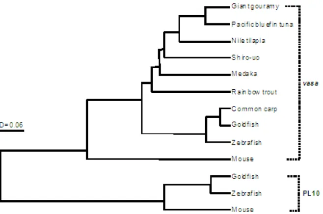
Figure 2. Phylogenic tree of the amino acid sequences of vasa and PL10 constructed using the UPGMA method. The length of horizontal lines indicates genetic distances. The GenBank accession numbers of the aligned amino acid and nucleic acid sequences were as follows: vasa (Pacific bluefin tuna: EU253482, common carp: AF479820, goldfish: AY773078, medaka: AB063484, mouse: AK014844, rainbow trout: AB032566, shiro-uo: AB098252, tilapia: AB032467, zebrafish: NM_131057) and PL10 (goldfish: AY842133, mouse: J04847, zebrafish: BC059794).
M T O G I L S F N M 1.5 1.0 0.3 0.1 Figure 3. RT-PCR analysis of various tissues using vasa-specific primers. cDNAs from various tissues (testis, ovary, gill, intestine, liver, muscle, fin) were used for RT-PCR. β-actin was used as an internal control for RT-PCR amplification. Lane NC was a negative control containing no cDNA template. M represents molecular weight marker (2-log ladder DNA marker, BioLabs, Inc., New England).
underline). The phylogenic trees of the vasa genes and PL10 amino acid sequences belonging to the DEAD protein family constructed using UPGMA method are shown in Figure 2. The sequence obtained in this study belonged to the vasa family and showed a strong association with the other vertebrate vasa homolog examined.
Tissue Distribution of Vasa mRNA by RT-PCR. Tissue distribution patterns of vasa mRNA was analyzed in 2-year-old male (pubertal testes) and female (previtellogenic ovary) tissues (GSI was 0.0089 and 0.23%, respectively). While high levels of transcripts were detected in the gonads of both males and females, none was detected in other tissues (Figure 3).
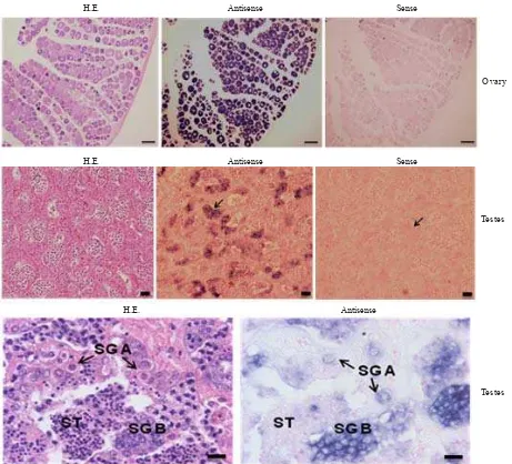
Localization of Vasa mRNA-positive Cells in Gonads by ISH. In pubertal testes (GSI 0.0089%), vasa-positive signals were detected in SG (Figure 4, middle); both at SG types A and B (Figure, bottom). Vasa-positive signals in previtellogenic ovaries (GSI 0.022%) were detected in the oocytes (Figure, top-center). Conversely, no hybridization signals were observed in any of the cells when the sense probes were applied (Figure, top- and middle-right). We
did not observe vasa positive signals in the gonadal
somatic cells of both males and females. No vasa mRNA was also detected in spermatocytes and spermatids (Figure, bottom). Thus, vasa mRNA was predominantly localized in meiotic cells such as SG.
DISCUSSION
It has been demonstrated that vasa is a member of the DEAD protein family that possesses ATP-dependent RNA helicase activity (Hay et al. 1988). The deduced amino acid sequence of the clone isolated in this study contained eight consensus sequences for the DEAD protein family
(Linder et al. 1989), including the ATP-A motif
(AXXXXGKT) and the ATP-B motif (DEAD) (Pause & Sonenberg 1992). In addition, a glycine-rich region in the N-terminal region of giant gourami vasa was observed to contain 17 glycine repeats and 8 arginine-glycineglycine repeats, which is similar to that observed in the vasa orthologs of other species (Raz 2000). This glycine-rich region with several repeated motifs is believed to be a characteristic of single-stranded nucleic acid binding proteins, such as RNA helicase (Liang et al. 1994), and these findings strongly suggest that the cDNA clone isolated in this study encoded a DEAD protein possessing ATP-dependent RNA helicase activity. The phylogenic tree of vasa and the PL10 family revealed that the sequence obtained in this study belongs to a clade containing vasa homologs.
H.E. Antisense Sense
Ovary
H.E. Antisense Sense
Testes
H.E. Antisense
Testes
Figure 4. Vasa expression in ovary and testis. Sequential sections stained with hematoxylin-eosin (H.E.) (left) and hybridized with an antisense (middle) and sense (right) vasa probes. No unspecific staining is observed with the sense probe. The signal of vasa
mRNA is specifically expressed in oocytes (top-center) and spermatogonia types A (SG A) and B (SG B). The scale bars represent 100 μm (ovary) and 20 μm (testis).
cells were detected in the testes and ovaries of adult giant gourami. In pubertal male fish, which contained mainly type SG, vasa-positive signals were specifically detected in SG. In previtellogenic ovaries, vasa mRNA was specifically localized in OC. Furthermore, no vasa mRNA was observed in SC, ST or any of the gonadal somatic cells. Taken together with nucleotide sequence and the spatial expression patterns, we concluded that the clone identified in this study is the giant gourami ortholog of the Drosophila vasa gene and designated it the giant gourami vasa-like gene (GgVLG). Similar expression pattern of vasa gene has also been reported in fish species including rainbow trout (Yoshizaki et al. 2000), tilapia (Kobayashi et al. 2000), and Pacific bluefin tuna (Nagasawa et al. 2009).
Development of SG transplantation technology to generate a surrogate parent fish capable of producing giant gourami gametes is ongoing in our laboratory. The use of highly concentrated SG population, especially SG type A as donor cells is expected to facilitate high colonization efficiency (Nagasawa et al. 2009). The results from the
section HE and ISH showed that, the number of SG was lower in the testes of 2-year-old giant gourami. Thus, in order to increase the successful rate of transplantation in giant gourami using SG, it is desirable to determine the size of giant gourami containing high number of SG. In such observation, the GgVLG cDNA sequence will be a valuable tool for SG quantification. Further, the GgVLG sequence can also be used as a marker to identify donor cells colonized in recipient gonad using ISH and PCR methods.
ACKNOWLEDGEMENT
REFERENCES
Cardinali M, Gioacchini G, Candiani S, Pestarino M, Yoshizaki G, Carnevali O. 2004. Hormonal regulation of vasa-like messenger RNA expression in the ovary of the marine teleost
Sparus aurata. Biol Reprod 70:737-743. http://dx.doi.org/
10.1095/biolreprod.103.021428
Department of Marine Affairs and Fisheries. 2005. Revitalization of Aquaculture 2006-2009. p 275.
Hay B, Jan LY, Jan YN. 1988. A protein component of Drosophila polar granules is encoded by vasa and has extensive sequence similarity to ATP-dependent helicase. Cell 55:577-587. http:/ /dx.doi.org/10.1016/0092-8674(88)90216-4
Kobayashi T, Kajiura-Kobayashi H, Nagahama Y. 2000. Differential expression of vasa homologue gene in the germ cells during oogenesis and spermatogenesis in a teleost fish, tilapia, Oreochromis niloticus. Mech Dev 99:139-142. http:/ /dx.doi.org/10.1016/S0925-4773(00)00464-0
Lasko PF, Ashburner M. 1988. The product of the Drosophila gene vasa is very similar to eukaryotic initiation factor-4A.
Nature 335:611-617. http://dx.doi.org/10.1038/335611a0
Liang L, Diehl-Jones W, Lasko P. 1994. Localization of vasa
protein to the Drosophila pole plasm is independent of its RNA binding and helicase activities. Development 120:1201-1211.
Linder P, Lasko PF, Ashburner M, Leroy P, Nielsen PJ, Nishi K, Schnier J, Slonimski PP. 1989. Birth of the D-E-A-D box.
Nature 337:121-122. http://dx.doi.org/10.1038/337121a0
Miyake A, Saito T, Kashiwagi N, Ando D, Yamamoto A, Suzuki T, Nakatsuji N, Nakatsuji T. 2006. Cloning and pattern of expression of the shiro-uo vasa gene during embryogenesis and its roles in PGC development. Int J Dev Biol 50:619-625. http://dx.doi.org/10.1387/ijdb.062172am
Nagasawa K, Takeuchi Y, Miwa M, Higuchi K, Morita T, Mitsuboshi T, Miyaki K, Kadomura K, Yoshizaki G. 2009. cDNA cloning and expression analysis of a vasa-like gene in Pacific bluefin tuna Thunnus orientalis. Fish Sci 75:71-79. http://dx.doi.org/10.1007/s12562-008-0021-9
Okutsu T, Shikina S, Kanno M, Takeuchi Y, Yoshizaki G. 2007. Production of trout offspring from triploid salmon parents.
Science 317:1517. http://dx.doi.org/10.1126/science.1145626
Okutsu T, Suzuki K, Takeuchi Y, Takeuchi T, Yoshizaki G. 2006. Testicular germ cells can colonize sexually undifferentiated embryonic gonad and produce functional eggs in fish. Proc
Natl Acad Sci USA 103:2725-2729. http://dx.doi.org/10.1073/
pnas.0509218103
Olsen LC, Aasland R, Fjose A. 1997. A vasa-like gene in zebrafish identifies putative primordial germ cells. Mech Dev 66:95-105. http://dx.doi.org/10.1016/S0925-4773(97)00099-3 Pause A, Sonenberg N. 1992. Mutational analysis of a DEAD box
RNA helicase: the mammalian translation initiation factor eIF-4A. EMBO J 11:2643-2654.
Raz E. 2000. The function and regulation of vasa-like genes in germ-cell development. Genome Biol 1: reviews 1017:1-6. Shinomiya A, Tanaka M, Kobayashi T, Nagahama Y, Hamaguchi
S. 2000. The vasa-like gene, olvas, identifies the migration path of primordial germ cells during embryonic body formation stage in the medaka, Oryzias latipes. Dev Growth
Differ 42:317-326.
http://dx.doi.org/10.1046/j.1440-169x.2000.00521.x
Takeuchi Y, Yoshizaki G, Takeuchi T. 2003. Generation of live fry from intraperitoneally transplanted primordial germ cells in rainbow trout. Biol Reprod 69:1142-1149. http://dx.doi.org/ 10.1095/biolreprod.103.017624
Takeuchi Y, Yoshizaki G, Takeuchi T. 2004. Biotechnology: surrogate broodstock produces salmonids. Nature 430:629-630. http://dx.doi.org/10.1038/430629a
Xu H, Gui J, Hong Y. 2005. Differential expression of vasa RNA and protein during spermatogenesis and oogenesis in the gibel carp (Carassius auratus gibelio), a bisexually and gynogenetically reproducing vertebrate. Dev Dyn 233:872-882. http://dx.doi.org/10.1002/dvdy.20410
Ye D, Lv D, Song P, Peng M, Chen Y, Guo M, Yang Q, Hu Y. 2007. Cloning and characterization of a rice field eel vasa-like gene cDNA and its expression in gonads during natural sex transformation. Biochem Genet 45:211-224. http://dx.doi.org/ 10.1007/s10528-006-9066-6
Yoon C, Kawakami K, Hopkins N. 1997. Zebrafish vasa homologue RNA is localized to the cleavage planes of 2- and 4-cell-stage embryos and is expressed in the primordial germ cells.
Development 124:3157-3165.
Yoshizaki G, Sakatani S, Tominaga H, Takeuchi T. 2000. Cloning and characterization of a vasa-like gene in rainbow trout and its expression in the germ cell lineage. Mol Reprod Dev
![KONSTUKTIVISME S2 [Compatibility Mode]](data:image/gif;base64,R0lGODlhAQABAIAAAP///wAAACH5BAEAAAAALAAAAAABAAEAAAICRAEAOw==)