Distal radius morphometry of Indonesian population
Syaiful A. Hadi, Wijiono
Department of Orthopaedic & Traumatology, Faculty of Medicine, Universitas Indonesia, Jakarta, Indonesia
Abstrak
Latar belakang: Morfometri tulang radius distal (radial inclination, palmar tilt, radial height, dan ulnar variance) merupakan parameter penting dalam evaluasi dan tatalaksana fraktur radius distal dimana keselarasan anatomi harus dikoreksi. Pada saat ini, pengobatan cedera radius distal di Indonesia masih didasarkan pada morfometri populasi barat atau dari sisi kontralateral. Tujuan dari penelitian ini adalah untuk menentukan morfometri radius distal pada populasi Indonesia dan untuk mengetahui apakah terdapat perbedaan nilai antara jenis kelamin laki-laki dan perempuan serta antara sisi kanan dan kiri.
Metode: Morfometri radius distal diukur dari 400 foto polos pergelangan tangan kanan dan kiri proyeksi AP dan lateral dari sampel yang memenuhi syarat yang didapat dari RS KESDAM Moh. Ridwan Meuraksa Jakarta pada bulan Juni sampai September 2010. Parameter radial inclination, palmar tilt, radial height, dan ulnar variance diukur. Data dicatat dalam tabel dan dikelompokkan antara jenis kelamin pria dan wanita, sisi kanan dan kiri. Analisis statistik dilakukan dengan uji Mann-Whitney.
Hasil: Dilakukan pengukuran terhadap 400 foto polos (300 pria dan 100 wanita) dengan usia rerata 25,5 tahun (rentang usia 18-48 tahun). Rerata radial inclination 23,99 ± 3,75°, palmar tilt 13,76 ± 4,36°, radial height 11,31 ± 1,66 mm, dan ulnar variance -0,45 ± 2,03 mm. Terdapat perbedaan yang bermakna antara radial inclination, palmar tilt, radial height, dan ulnar variance sisi kanan dan kiri. Terdapat pula perbedaan yang bermakna antara pria dan wanita.
Kesimpulan: Pada penelitian ini diperoleh data morfometri radius distal pada populasi Indonesia yang merupakan data yang berharga dalam evaluasi dan tatalaksana fraktur radius distal. Penggunaan sisi kontralateral sebagai referensi, perlu ditinjau ulang. (Med J Indones. 2013;22:173-7. doi: 10.13181/mji.v22i3.587)
Abstract
Background: Distal radius morphometry (radial inclination, palmar tilt, radial height, and ulnar variance) is an important parameter in the evaluation and treatment of distal radius fractures in which anatomical alignment must be corrected. Currently, treatment of distal radius fractures in Indonesia is still based on morphometry of western population or from the contralateral side. The aim of this study is to determine distal radius morphometry of Indonesian population and to compare between right and left side, male and female gender.
Methods: Distal radius morphometry was measured from 400 plain X-ray of right and left wrist AP and lateral projection. Samples were taken consecutively in Moh. Ridwan Meuraksa Army Hospital, Jakarta, from June to September 2010. Radial inclination, palmar tilt, radial height, and ulnar variance was measured. Data were recorded using tables and grouped between male and female, right and left side, statistical analysis was performed using Mann-Whitney test.
Results: From 400 plain X-ray evaluated, there were 300 males and 100 females with the mean age of 25.5 years old (18-48). The mean of radial inclination was 23.99 ± 3.75°, palmar tilt 13.76 ± 4.36°, radial height 11.31 ± 1.66 mm, and ulnar
variance -0.45 ± 2.03 mm. There were statistically signiicant differences between right and left side of radial inclination, palmar tilt, radial height, and ulnar variance. There was also statistically signiicant difference between male and female.
Conclusion: Distal radius morphometry of Indonesian population may provide valuable data for the treatment of distal radius fractures. The use of contralateral side as reference should be reconsidered. (Med J Indones. 2013;22:173-7. doi: 10.13181/mji.v22i3.587)
Keywords: morphometry, palmar tilt, radial height, radial inclination, ulnar variance
Interaction between distal radius morphometry with wrist biomechanics has been studied in various literatures. Four important parameters of the distal radius are radial inclination, palmar tilt, radial height, and ulnar variance. In 1987, Short, et al1 conducted experiments on cadavers and expressed the importance of anatomic correction of palmar tilt. Decrease in palmar tilt resulted in an increased burden of ulna. Load pressure distribution on the joint surface of distal radius and ulna will also be more concentrated with increasing dorsal angulation. Changes due to the reduced value of radial inclination and radial height has also been reported by several studies.1
Negative ulnar variance is one of the predisposing factors of Kienbock’s disease. In 1984, in a biomechanical study on cadavers, Palmer and Werner concluded that axial force through the wrist was borne by radiocarpal joint 82% and ulnocarpal joint 18%. Positive ulnar variance of 2.5 mm resulted in increased loading on ulnocarpal joint up to 42%. Conversely negative ulnar variance will decrease compression loads borne by ulnocarpal joint up to 4.3%.1,2
treatment of injuries involving the wrist joint. Fractures of the wrist involving distal radius have the prevalence of 8-15% fractures of the upper extremities.3,4 Thus, an understanding of the normal value of distal radius morphometry become very important in the management of distal radius fractures in order to maintain anatomical alignment.5
So far in daily practice we use normal value of distal radius morphometry from western literature as a reference for the evaluation of acceptable value of post reduction distal radius morphometry.5 The aim of this study is to determine distal radius morphometry of Indonesian population. This study also sought to compare the morphometry of right and left distal radius of the sampled population and also between male and female subjects, because some researchers do not agree to use the existing population data as reference values, instead they use the morphometry of the contralateral side as reference.6
This study provides a preliminary data for further analytical or experimental research. The result may also become the reference for the evaluation and treatment of distal radius fracture and injury in Indonesian population.
METHODS
In this cross-sectional study, we performed evaluation of primary data from health survey of Indonesian Army Personnel at KESDAM TK II Ridwan Meuraksa Hospital, Jakarta from June-September 2010. The preference for the use of Indonesian army population as samples is based on the consideration of the uniformity of posture so that the inluence of morphological variation can be minimized as well as the ease in obtaining samples. Samples was estimated using proportion estimation with absolute precission:
With this formula, the minimum sample required was 193 (left and right), for the anticipation of dropouts we took 200 subjects as sample. True anteroposterior (AP) and lateral X-ray was taken after subjects signed the consent for research.
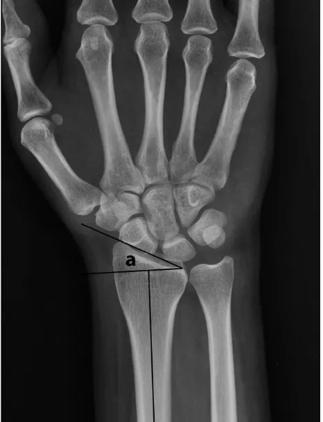
Radial inclination is the angle formed by horizontal line perpendicular to the axis of radius at the level of lunate fossa with tangential line connecting the styloid process of radius and ulnar side of distal radius evaluated from true AP projection of the wrist1,6 (Figure 1).
�ൌα;Ǥ ሺͳ; −ሻ
Figure 1.
Figure 2.
The measurement of radial inclination. (a) is the ra-dial inclination, the angle formed by horizontal line perpendicular to the axis of radius at the level of lu-nate fossa with tangential line connecting the styloid process of radius and ulnar side of distal radius
The measurement of palmar tilt. (b) is the palmar tilt, the angle formed by horizontal line perpendicular to the axis of radius at the level of radial styloid process with tangential line connecting dorsal and volar lip of distal radius
Figure 3.
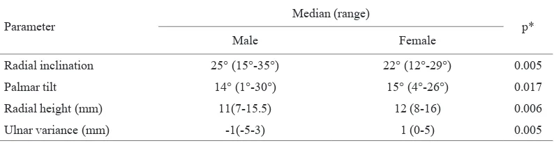
Table 1. Distribution of radial inclination, palmar tilt, radial height, and ulnar variance from 200 subjects (combined right and left side)
Figure 4.
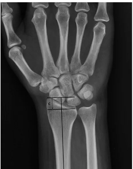
The measurement of radial height. (c) is the radial height, the distance measured from horizontal line perpendicular to the axis of radius at the level of lu-nate fossa or the lowest part of distal radius to the horizontal line perpendicular to the axis of radius at the level of radial styloid process
The measurement of ulnar variances. (d) is the ulnar variance, the distance measured from horizontal line perpendicular to the axis of ulna at the level of the base of ulnar styloid process to the horizontal line perpendicular to the axis of radius at the level of lu-nate fossa or the most proximal part of distal radius Radial height (length) is the distance measured from
horizontal line perpendicular to the axis of radius at the level of lunate fossa or the lowest part of distal radius to the horizontal line perpendicular to the axis of radius at the level of radial styloid process evaluated from true AP projection of the wrist1,6 (Figure 3).
Ulnar variance is the distance measured from horizontal line perpendicular to the axis of ulna at the level of the base of ulnar styloid process to the horizontal line perpendicular to the axis of radius at the level of lunate fossa or the most proximal part of distal radius evaluated from true AP projection of the wrist.1,6 Positive ulnar variance is when the horizontal line at the base of ulnar styloid process is more distal than the horizontal line at the base of lunate fossa (Figure 4). Conversely, negative ulnar variance is when the horizontal line at the base of ulnar styloid process is more proximal than the horizontal line at the base of lunate fossa.
Measurement was done by the researchers, data taken was recorded in tables and grouped between left and right side, male and female gender. Results were compared between left and right side, male and female gender, and also compared to OTA reference value.
Because data distribution was not normal, statistical analysis was done using Mann-Whitney test in SPSS statistical software for Windows version 17.
RESULTS
Two hundred subjects (150 male and 50 female) were involved in this study with mean age 25.50 years old (range 18-48). There was no dropout.
The mean of combined right and left side of radial inclination was 23.99 ± 3.75°, palmar tilt 13.76 ± 4.36°, radial height 11.31 ± 1.66 mm, and ulnar variance -0.45 ± 2.03 mm (Table 1). There were statistically signiicant differences
Parameter Mean ± SD
Radial inclination 23.99 ± 3.75°
Palmar tilt 13.76 ± 4.36°
Radial height 11.31 ± 1.66 mm
between radial inclination, palmar tilt, radial height, and ulnar variance between right and left side (Table 2). There were also statistically signiicant differences between radial inclination, palmar tilt, radial height, and ulnar variance between male and female subjects (Table 3). Male subjects tend to have negative ulnar variance and female subjects tend to have positive ulnar variance (Table 3).
DISCUSSION
The relationship between morphometry and biomechanic of distal radius has been studied in many literatures. Miyake, et al7 in dissection of twelve cadavers has reported the relationship between the concentration of loading force transmitted through the wrist and palmar tilt of distal radius. According to this study, in neutral position (palmar tilt 0°) load was concentrated on volar region of radiolunate articulation, meanwhile in dorsal angulation more than 30° load was concentrated on dorsal region of radiolunate articulation. It was concluded that up to 30° dorsal angulation at radiolunate surface, transmission of load through the wrist and distal radius was not signiicantly distorted.7 Pogue, et al8 reported similar results in their study. Short, et al1 reported that more than 30° dorsal angulation at radiolunate surface resulted in increased load on ulna up to more than 50%.
Adams found that shortening of the radius may result in signiicant distortion of TFCC and may cause major
Parameter
Median (range)
p*
Right side Left side
Radial inclination 24° (12°-35°) 23° (15°-30°) 0.005
Palmar tilt 14° (2°-25°) 15° (1°-30°) 0.006
Radial height (mm) 11(7-15) 11(7-16) 0.008
Ulnar variance (mm) -1(-5-5) 0 (-5-5) 0.023
*Statistical analysis: Mann-Whitney Table 2.
Table 3.
Distribution of radial inclination, palmar tilt, radial height, and ulnar variance between right and left side
Distribution of radial inclination, palmar tilt, radial height, and ulnar variance between male and female subjects
Parameter
Median (range)
p*
Male Female
Radial inclination 25° (15°-35°) 22° (12°-29°) 0.005
Palmar tilt 14° (1°-30°) 15° (4°-26°) 0.017
Radial height (mm) 11(7-15.5) 12 (8-16) 0.006
Ulnar variance (mm) -1(-5-3) 1 (0-5) 0.005
*Statistical analysis: Mann-Whitney
changes in kinematics of the wrist, while the effect on radial inclination and dorsal angulation is not so signiicant.9 Palmer and Werner2 in a biomechanics study on cadaver found that axial force on distal radius was transmitted 82% through radiocarpal joint and 18% through ulnocarpal joint. Positive ulnar variance of 2.5 mm resulted in increased loading on ulnocarpal joint up to 42%. Meanwhile, negative ulnar variance decreased compression loads on ulnocarpal joint up to 4.3%.1,2 This was in accordance with the research performed by De Smet, et al10 which found that negative ulnar variance was associated with Kienbock’s disease (avascular necrosis of the lunate), avascular necrosis of scaphoid, and scapholunate dissosiation. Conversely positive ulnar variance would result in excessive loading on ulnar compartment that may cause triangular ibrocartilage complex (TFCC) degeneration and perforation as well as degenerative changes on carpal bone cartilage.10
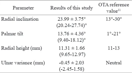
Table 4. Comparison between distal radius morphometry in this study (combined right and left side) and refer-ence value from The Orthopaedic Trauma Associaton (Gartland et al, 1951)
Parameter Results of this study OTA reference value11 Radial inclination 23.99 ± 3.75°
(20.24-27.74)°
13°-30° Palmar tilt 13.76 ± 4.36°
(9.40-18.12)°
1°-21° Radial height (mm) 11.31 ± 1.66
(9.65-12.97)
11-13 Ulnar variance (mm) -0.45 ± 2.03
(-2.45-1.58)
Neutral Altissimi, et al12 in a long-term evaluation of 297 cases of conservative distal radius fracture management found that clinically unsatisfactory result was obtained if radial inclination was less than 5°, palmar tilt more than 15°, and ulnar variance more than 5 mm. Tzabo13 stated that midcarpal instability may occur due to incongruity of distal radioulnar joint. Downing & Karantana14 also stated that irregularity of the distal radioulnar joint is a predisposing factor for degenerative changes in the wrist. Hollevoet, et al6 suggested using the contralateral side (healthy side) as an individual reference parameter which was more similar compared to values obtained from population data.6
From the above facts, it is obvious that it is very important to restore the alignment of distal radius to the normal value. Until now most of clinicians in Indonesia are still using parameters from western literature as reference. It is not wise because the results obtained here was quite different from western literature (Table 4). We also agree on the statement proposed by Hollevoet, et al6 which advised to use the healthy contralateral side for comparison, because in this study we also found that the data was similar between the right and left side (Table 2) which although showed statistically signiicant difference, not clinically signiicant because the difference was small.6
From this study, it was known that the mean value of distal radius morphometry in Indonesian Population are; radial inclination 23.99 ± 3.75°, palmar tilt 13.76 ± 4.36°, radial height 11.31 ± 1.66 mm, and ulnar variance -0.45 ± 2.03 mm. There were statistically significant differences of those values between male and female. Mean radial inclination was greater in males, but the mean of palmar tilt, radial height, and ulnar variance was greater in female. Ulnar variance tends to be negative in male and positive in female.
The mean of radial inclination and radial height was higher on the right side, and the mean of palmar tilt and ulnar variance was higher on the left side, ulnar variance tends to be negative on the right side and neutral on the left side. Although these differences were found to be statistically signiicant, they were not clinically signiicant because the differences were small. Therefore the authors suggested to use morphometry data of the healthy side as individual reference value for the treatment of distal radius injury because it was more suitable compared to population data.
Acknowledgments
I would like to express my gratitude to my colleague at Orthopaedic Department Cipto Mangunkusumo Hospital, secretariat staff at our department, and my family in which patience and encouragement who have made this research possible. I hereby afirm that there is no conlict of interest in this research.
REFERENCES
1. Short WH, Palmer AK, Werner FW, Murphy DJ. A biomechanical study of distal radius fractures. J Hand Surg Am. 1987;12(4):529-34.
2. Palmer AK, Werner FW. Biomechanics of the distal radioulnar joint. Clin Orthop Relat Res. 1984;187:26-35. 3. Austin L, Veillette C. Distal radius fracture. In:
Orthopedia-collaborative orthopaedic knowledgebase. 2009;13:26 4. Nana AD, Joshi A, Lichtman DM. Plating of the distal
radius. J Am Acad Orthop Surg. 2005;13(3):159-71. 5. Sandjaja G. Gambaran nilai rata-rata aksis sudut radius
distal normal pada pengunjung di RSCM [thesis]. Mount Pleasant (MI): Universitas Indonesia; 1993. Indonesian. 6. Hollevoet N, van Maele G, van Seymortier P, Verdonk
R. Comparison of palmar tilt, radial inclination, and ulnar variance in right and left wrists. J Hand Surg Br. 2000;25(5):431-3.
7. Miyake T, Hashizume HI, Inoue H, Shi Q, Nagayama N. Malunited colles fracture, analysis of stress distribution. J Hand Surg Br. 1994;19(6):737-42.
8. Pogue DJ, Viegas SF, Patterson RM, et al. Effect of distal radius fracture malunion on wrist joint mechanics. J Hand Surg Am. 1990;15(5):721-7.
9. Adams BD. Effects of radial deformity on distal radioulnar joint mechanics. J Hand Surg Am. 1993;18(3):492-8. 10. De Smet L. Ulnar variance: facts and iction review article.
Acta Orthop Belg. 1994;60(1):1-9.
11. Gartland JJ, Werley CW. Evaluation of healed colles’ fractures. J Bone Joint Surg Am. 1951;33A(4):895-907. 12. Altissimi M, Attenucci R, Fiacca C, Mancini GB. Long
term results of conservative treatment of fractures of the distal radius. Clin Orthop Relat Res. 1986;206:202-10. 13. Szabo RM. Distal radioulnar joint instability. J Bone Joint
Surg Am. 2006;88(4):884-94.