Abstrak
Terapi radiasi dapat mengakibalkan terjadi proses keganasan di kemudian hari. Dilaporkan satu kasus ôsteosarkoma sehtnder pada klavihtla akibat radiasi I 6 tahun sebelumnya.
Abstract
Radiotherapy can induce a malignancy at a latter time. A case ofsecondary osteosarcoma in the clavicula coused by radiotherapy 16 years earlier is reporled.
Keywords: Osteosarcoma, radiotherapy Vol 9, No 4, Oclober
-
December 2000Post-irradiation
osteosarcoma
:
A
case
report
Enol
U. Hutagalung*, Achmad Basuki*, R. Susworo/
The carcinogenic
effect
of
radiation is a well-known
fact.
Radiotherapy
is used in the
management
of
cancer,
and
at
a latter
time
may contribute to
thedevelopment
of
a
secondarymalignancy
which differ
from
theprimary
one.The frequency
of
malignancy attributed to radiation
isvery low
precededby
along
latentperiod,
so that theincidence
is
usually published
as casereports.l
Herewe
present
a
case
of
osteosarcomaof the
clavicle,
secondary
to
radiotherapy administered
for
a
malig-nancyin
the nasopharyngeal area, 16 years before.CASE REPORT
A
5O-years-oldwoman
washospitalized
for
alump
onthe
left
clavicular bone since
7
months.
Previoushistorv of
trauma andinfection
were denied.She became aware
of
the
lump
atthe medial clavicle
when
it
wasthumb-sized,
which
continued to
enlarge.Throughout this
period
shecontinued
to
lose weight,
Sixteen years
earlier she
underwent
radiotherapy* Subdivision
of
Orthopaedics, Deparlmentof
Surgery, Universily of Indonesia Faculty of Medicine and Cipto Mangunkusumo General Hospital, Jakarta, Indonesia Deparlment of Radiologt, University of Indonesia Facultyof
Medicine and Cipto Mangunkusumo General Hospital, Jakana, Indonesia
P os t-irradia t io n os I eos arcoma
28t
which
amounted
to
5500 cGy
for
a
nasopharyngeal cancer.Physical examination revealed
a lump
measuring
8 x
5
x
3
cm
at the upper
left
hemithorax, which
wasbluish
redin
color
and showedbleeding areas.
It
had a granular surface, hardin
consistency, andfixed
from
the base.
X-ray
and CT-scan
of
the
thorax
showed
a lump
onthe
left
clavicular bone which
has
intruded
into
thethoracic
cavity.
It
was
diagnosed
as a
malignant
tumor
of
the clavicle, and a biopsy was
performed
(PA No.:9406585),
and
diagnosed
as
fibroblastic
osteosarcoma.Due to previous history.of
radiotherapy16 years
earlier,
in
which the middle portion
of
theleft
clavicle,
wherethe lesion was
located, waswithin
the
field
of
radiation, the patient was
diagnosed
ashaving
secondarypost-inadiation
osteosarcoma.DISCUSSION
282
Hutagalung et alHistologically,
primary
osteosarcoma
are mostly
osteoblastic
type,
whereas
in
post-irradiation
osteo-sarcomas
the
fibrolastic
type
is
more
commonly
osbserved.3'6
Cahan and
Arlena'7 described
the criteria
of
post-inadiation
osteosarcoma asfollows
:l.
the irradiated primary tumor
has
no
osteoblasticactivity.
2.
the
secondary
malignancy develop
in
the
areawithin
thefield
of radiation.
3.
the presenceof
arelatively
long
latentperiod
4.
the
presence
of
osteosarcoma
is
confirmed
by
pathological examination
Wiklund8
reported
that
the
incidence
of
post-irradiation
sarcomas
was more frequent
in women
who
have hadprimary
malignancies
of
the breast andgenital
organs, sothat the
sitesfrequently found
with
secondary malignanciesin
women were the
shoulders andhip.i
The
secondary sarcoma
is
attributed
to
geneticmutation
as a consequenceof
irradiation.2,aThe
incidence
of
secondary osteosarcomas
isextremely
low,
about 0.05
-
0.2Yoin patients who
survived 5
years
after irradiation, receiving
between4000
-
7000
cGy.2 Frassicaenoted an inùdence
of
0.035
-
lYo
of
all
malignant
cases
that had
beenirradiated,
whereasHatfieldlo
reported an incidenceof
0.2%o
in
patients
with
breast cancerwho survived
l0
yearsafter irradiation.
The presence
ofa
latentperiod
is arequisite for
a caseto be classified
as
post-irradiation
secondary
malig-nancy.
The latent period reported
in
the
literature
varied
between4
-52
years.r's'e'l'he
latentperiod
will
be shorter
in
the presenceof
a genetic predisposition
such
as
retinoblastoma,
and
v.Recklinghausen's
neurofibromatosis.s'rl The
samecondition
àpplies
tochildren
who are
in
the growing period when
cell
proliferation
is more active.roHatfielde'ro
was
of
the opinion that
post-inadiation
sarcomas
should
be distinguished from the
doubleprimary phenomenon. This
phenomenon explains thehigher probability
of
the
samepatient
to
develop
aMed J Indones
secondary
malignant
processwhich is different
from
the primary one, and
will
manifest after
a
shorter latentperiod
compared to the latentperiod
consequentto
inadiation.
The
average
latent period
for
developing
post-inadiation
secondarymalignancies
isabout
12 years, whereasthat
of
doubleprimary
is
lessthan 5 years.lo
The
-prognosis
of
post-inadiation
osteosarcoma
ispoore which
isattributed
to the facts that :l.
it
isusually
recognized at an advanced stage2. patients
areof old
ageand
afflicted
with
multiple
medical
problems
3.
noeffective
adjuvant therapy is available.In
cases
of
secondary
osteosarcoma
reported
by
Dahlin,l2
noneof
the pâtients survived
for'more
than3 years.
The
present case
was
diagnosed
as
post-irradiation
osteosarcoma
as it
fulfilled the criteria
suggestedby
Cahan and
Arlen
a'7namely
:l.
the
primary
tumor being
inadiated
was
anasopharyngeal
carcinoma
which
did not
have any osteoblasicactivity.
2. the
secondary
osteosarcoma
developed
in
themiddle part
of
left
clavicular bone, which
waswithin
thefield of inadiation.
3.
tËelatent
period
of
16 yearswas
quite long,
so
it
could not be considered as a doubleprimary
case.l04.
the
malignancy
found on the left clavicle
was
of
the
fibroblastic type, which was the
histopatho-logic type
frequently
_encountered
in
post-irradiation
osteosarcoma.6Other supporting findings were the location
of
the\ryas
poor
becauseof
the
advanced stagein which
thetumor
was presented;the
tumor
had already
invadedthe
thoracic
cavity and was
considered
to
beYol 9, No 4, October
-
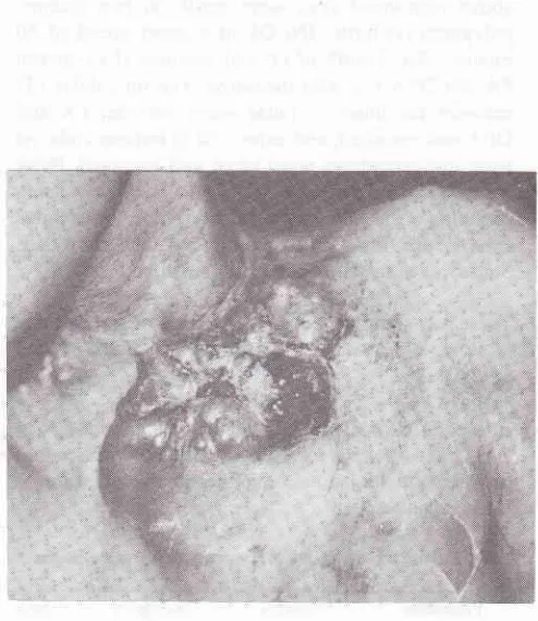
December 2000Figure
l.
The clinical appearence and localization ofthe lumor is unusualfor a primary osleosarcomaREFERENCES
l.
Kim JH, Chu FC, Woodard HQ, Melamed MR, Huvos A, Cantin J. Radiation induced soft tissue and bone sarcoma. Radiology 1978;129:501 - 8.2.
Bechler JR, RobertsonWW,
MeadowsAt,
Womer RB. Osteosarcoma as a second malignant neoplasm in children. J Bone Joint Surg. 1992; 74A: 1079 - 83.3.
Huvos AG, Woodard HQ, Cahan WG, Higinbotham NL,Stewart
FW,
ButterA,
et al.
Post-radiation osteogenic sarcoma of bone and soft tissue.A
çlinic pathologic studyof66 patients. Cancer 1985; 55:1244
- 55.
4.
Arlen
M,
HiginbothamNL,
HuvosAG,
Marcove RC,Miller
T,
ShahIC.
Radiation induced sarcomaof bone.
Cancer l97l;
28: 1087 -99.5.
Tillotson C, Rosenberg A, Gebhardt M, RosenthalDI.
post radiation multicentric osteosarcoma. Cancer 1988; 62: 67 -71.6.
NewtonWA,
MeadowsAT,
ShimadaH,
Bumin
GR,Vawter GF. Bone sarcomas as second malignant neoplasms following chlidhood cancer. Cancer
l99l;
67 : I 93 - 2Cl.Pos t-irradiation osteos arc oma 283
Figure 2. X-ray appearance ofthe case, show a mass in the lefl
iclavicula, which already inJiltrated to left intra thoracic space
7.
Cahan WG, Woodard HQ, HiginbothamNL,
Stewart FW,Coley
BL.
Sarcoma arisingin
irradiated bone. Cancer 1948;l:3
- 29.8.
Wiklund TA, Blomquist CP, Ratly J, Elomaa I, Rissanen p. Post-radiation sarcoma. Analysisof
a nationwidé cancer registry material. Cancer l99l ; 68: 524 - 3l .9. Frassica
FJ. Sim FH, Frassica DA, Wold LE. Survival andmanagement considerations in post radiation osteosarcoma and Paget's osteosarcoma. Clin Orthop 1990;270: 120-7 .
10. Hatfield
PM,
Schulz
MD.
Post
irradiation
sarcoma including 5 cases after x-ray therapy of breast carcinoma, Radiology 1970 96: 593 - 602.ll.
Meadows
AT, Strong
LC,
Li
FD,
D'Angio
GJ,Schweisguth
O,
FreemanAl,
etal.
Bone sarcoma as asecond rnalignant neoplasms
in
children:
influenceof
radiation and genetic predisposition. Cancer 1980; 46: 2603 - 06.