Vol. 20, No. 3, August 2011 Zinc and IL-2 in seropositive leprosy 201
Zinc supplementation could modulate T cell to maintain interleukin-2 level in
seropositive contact of leprosy patients
Mohammad Z. Rahfi ludin,1 Bambang Wirjatmadi,2 Indropo Agusni,3 Yoes P. Dahlan4
1 Department of Nutrition and Health, Public Health Faculty, Diponegoro University, Semarang, Indonesia 2 Department of Nutrition and Health, Public Health Faculty, Airlangga University, Surabaya, Indonesia 3Department of Dermatology and Venerology, Medical Faculty, Airlangga University, Surabaya, Indonesia 4Department of Parasitology, Medical Faculty, Airlangga University, Surabaya, Indonesia
Abstrak
Latar belakang: WHO mencatat jumlah penderita kusta di Indonesia menduduki peringkat ketiga di dunia setelah India dan Brazil. Jumlah penderita kusta baru cenderung meningkat kemungkinan karena yang seropositif kusta telah berubah menjadi manifest klinis. Penelitian ini bertujuan untuk mengetahui pengaruh suplementasi seng terhadap kadar interleukin 2 (IL-2) pada narakontak kusta yang seropositif dengan status seng marjinal.
Metode: Dua puluh dua orang berusia 20 – 40 tahun ikut dalam penelitian ini. Kelompok yang disuplementasi seng menerima dosis 40 mg seng/hari selama 3 bulan. Seropositif kusta ditentukan berdasarkan kadar IgM anti Phenolic Glycolipid–1, dan kadar IL-2 pada supernatan kultur sel limfosit diukur dengan metode Elisa.
Hasil: Kadar IL-2 pada kelompok yang menerima seng relatif tidak berubah (p= 0,721), sedangkan pada kelompok plasebo terjadi penurunan bermakna kadar IL-2 (p= 0,025) pada akhir penelitian.
Kesimpulan: Terdapat perbedaan bermakna perubahan kadar IL-2 di antara ke dua kelompok (p= 0,037). (Med J Indones 2011; 20:201-4)
Abstract
Background: WHO classifi ed the number of leprosy cases in Indonesia as number three in the world after India and Brazil. The number of new leprosy patients tends to increase since there is a possibility that seropositive leprosy is turning into manifest leprosy. The aim of this study was to analyze the infl uence of zinc supplementation on interleukin-2 (IL-2) level of seropositive contact of leprosy patients with marginal zinc defi ciency.
Methods: Twenty two subjects aged 20-40 years were recruited for this study. The zinc-supplemented group received 40 mg elemental Zn/d orally for 3 months. Seropositive leprosy was determined by examining IgM anti Phenolic Glycolipid–1 level and concentration of IL-2 in lymphocyte cell culture supernatant fl uid were measured by Elisa method.
Results: The IL-2 concentration in the subject in the zinc group was relatively not changed (p= 0.721), whereas that in placebo group tended to be signifi cantly lower (p= 0.025) at the end of the study.
Conclusion: There was a signifi cant change of IL-2 level between both groups (p= 0.037). (Med J Indones 2011; 20:201-4)
Key word: IgM anti phenolic glycolipid-1, seropositive leprosy
Correspondence email to: [email protected]
Leprosy is still a major public health problem in Indonesia as well as in the world. The number of leprosy cases in Indonesia could be classifi ed as number three in the world after India and Brazil.1 The number
of new leprosy patients tends to increase since there is a possibility that seropositive leprosy is turning into manifest leprosy. Seropositive leprosy is a condition when someone is infected by M. Leprae, however, they have not shown any clinical symptoms. More than half of healthy inhabitants in endemic leprosy area are anti-micro bacterial antibody positive. Therefore seropositive leprosy should get more attention in the effort to eliminate leprosy. Detection of seropositive leprosy subjects can be done by using various methods; one of them is by examining Immunoglobulin M (IgM) anti Phenolic Glycolipid (PGL)-1 level.2
Cellular immune response level will determine leprosy spectrum. For leprosy pausibasiler type (PB), the cellular immunity is still good; however for leprosy multibasiler type (MB), the cellular immunity is decreased.3 Some researches showed that the immune
response was infl uenced by micro nutrients such as zinc.4 Zinc serum level, which may determine leprosy
spectrum, plays a role in the cellular immune response. Some research results showed gradual decrease in zinc serum level on the transition of leper type PB to MB. The lowest level is found in MB type.5
Rahfi ludin et al.
202 Med J Indones
defi ciency. IFN-γ and IL-2 production is improved when zinc level is maintained to be suffi cient by giving zinc supplement.6
Seropositive contacts of leprosy patients have a tendency to change into leprosy multibasiler type (MB). Cellular immunity of leprosy MB is decreased or even reaches anergy, and it turns into humoral immunity (Th2), which makes the patient’s condition worse. Therefore, this study aimed to analyze the infl uence of zinc supplementation on interleukin 2 (IL-2) level of seropositive contact of leprosy patients with marginal zinc defi ciency. Zinc supplementation is expected to increase IL-2, and in turn will maintain the function of cellular immunity in seropositive leprosy.
METHODS
This study was conducted on people who live in the same house or near the leper population in Brondong subdistric, Lamongan regency, East Java, Indonesia in 2010. The amount of the research sample was predetermined, i.e. 22 subjects who were randomly chosen from those who met the inclusion criteria. The ethical clearance for this study was obtained from Research and Public Service Institute, Airlangga University no. 071/PANEC/LPPM/2009.
Inclusion criteria
The inclusion criteria were individuals of 20 to 40 years old (both male and female) whom were proven to be leprosy serology positive (IgM anti PGL-1 level is 600 – 1500 unit/ml) and had marginal zinc status (zinc plasma level 10.7 - 13.0 μmol/L). In addition, they did not clinically show any leprosy symptoms and did not consume anti-leprosy medicine. They also did not have any disease, which might infl uence their zinc status, did not suffer from tuberculosis and did not consume any anti-tuberculosis drugs; they did not consume any anti-immunosuppressant in the last three months before blood samples were taken; their body mass indexes were more than 18 kg/m2 and were willing to join the study by signing an informed consent form.
Zinc supplementation
Those 22 subjects were randomly assigned in pairs to receive either zinc-supplement (treatment group) or placebo (placebo group). Each day for 3 months, subjects in the treatment group received 1 capsule of zinc sulfate (40 mg elemental zinc), while subjects in the placebo group received placebo capsules in the same manner. The obedience of consuming supplement was monitored by the researchers and local health staff.
Procedures
Plasma zinc levels were measured by using atomic absorption spectrophotometer in Balai Besar Laboratorium Kesehatan Surabaya. Determination of seropositive leprosy was done by examining IgM anti PGl-1 level by enzyme-linked immunosorbent assay using polyclonal rabbit anti human IgM/HRP secondary antibody (Dako ®). The assay was developed
in the Institute Tropical Disease Laboratory, Airlangga University, Surabaya. In addition, daily intake was measured using recall method and food frequency questionnaire, and IL-2 measurements on peripheral blood mononuclear cells in cultures were done.
Peripheral blood mononuclear cell culture and IL2 measurements
Peripheral blood mononuclear cells (3 x 10⁻⁷g/μl per
well) were stimulated with a mitogen (Dharmendra lepromins) in culture for 2 days in 10% CO2 at 37 0C.
Supernatant was collected from lepromin-stimulated cell culture, and the IL-2 level was measured by using commercial enzyme-linked immunosorbent assay kits (Quantikine®catalog number D2050, R&D System, Inc Minneapolis, USA) in Pathology laboratory ofDr. Soetomo hospital, Surabaya. Concentrations of IL-2 were expressed as pg/mL supernatant.
Data collection and analysis
Data collected were characteristics of the sample, daily intake, and anti PGL-1 IgM. In addition, data of plasma zinc and IL-2 levels before and after supplementation were also collected. All data were noted and tabulated. Normality of the data was determined by the Kolmogorov-Smirnov test. Statistical analyses were done using SPSS 16 statistical software, to compare plasma zinc and IL-2 level between treatment and placebo group by independent t test. Further, paired t test was used to compare plasma zinc as well as IL-2 levels of before and after supplementation. Fisher’s exact was used to compare the two sexes between groups.
RESULTS
Vol. 20, No. 3, August 2011 Zinc and IL-2 in seropositive leprosy 203
(37.7%) experienced marginal zinc defi ciency (zinc plasma level 10.7 - 13.0 μmol/L).
The comparative result showed that there was no signifi cant difference of sex, body mass index and IgM PGL-1 level between both groups (table 1). Further, there was no difference in daily intake between the two groups (Data not shown).
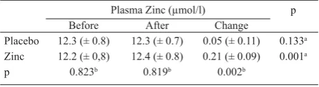
On placebo group, the average of zinc plasma level was relatively the same at the beginning and the end of the study (p= 0.133). However, there was a major increase in zinc plasma level on treatment group (p= 0.001). There was no signifi cant difference between the treatment and placebo group in baseline plasma zinc level (p= 0.823) and 3 months after treatment (p = 0.819). However, there was signifi cant difference in the change of zinc plasma level between both groups (p = 0.002) (Table 2).
On placebo group, IL-2 level after 3 months was signifi cantly lower than baseline (p= 0.025). However, on treatment group, IL-2 level was relatively stable (p= 0.721). There was a signifi cant difference between the treatment and placebo group in IL-2 concentration at baseline (p= 0.032), but after 3 months of treatment there was no signifi cant difference (p= 0.750). Conversely, there was signifi cant change of IL-2 level between the two groups (p= 0.037) (Table 3).
DISCUSSION
Screening results showed that 51 % of inhabitants in Brondong sub district, Lamongan regency, East Java, were leprosy seropositive based on IgM anti PGL–1 examination. This fi nding is higher than that in South Korea that was 12.1 % and in the Philippines that was 13.9%.7 However, this
fi nding is relatively the same compared to that in Semarang, Central Java, namely 53%.8 Indonesia has a higher prevalence of seropositive
leprosy; this fact corresponds with the number of leprosy cases in Indonesia that could be classifi ed as number three in the world after India and Brazil.1
Zinc supplementation on leprosy seropositive subjects for three months was proven to be able to prevent the decrease in IL – 2 level, since zinc can give infl uence on T-cell signal transduction that started from the proximal (cell membrane) level to the distal level (nucleolus transcription).
On proximal level, this fact can be explained since zinc has an effect on lymphocyte protein tyrosine kinase (Lck). On the next signal transduction, zinc infl uences protein kinase C and extracellular-signal regulated kinase (Erk). Zinc is needed for homodimerization and heterodimerization of Lck. Zinc capability on homodimerizationandheterodimerizationwas specifi c for Lck. This capability was due to cystein residues, which form two intermolecular zinc binding-sites that are unique and are not present in other Scr kinase family members.9 Protein kinase C also causes nuclear
factor kappa B (NFκB) to be free from its bond in the cytoplasma and then entered the nucleus. Zinc has an effect on the activation of inhibitor kB (IκB) kinase and the expression of mRNA p105 (prekursor NFκB p50). Then, zinc also infl uences the phosphorylation and ubiquitination process on IκB, so that it is degraded, and makes it possible to translocate NFκB from cytosol to the nucleus. Next, NFκB connects with DNA and then, IL-2 mRNA transcription process happens.10
Zinc can cause phosphorylation of Mek and Erk. Dephosphorylation in vitro to examine the effect of
Variable Groups
p Placebo Zinc
Sex (n)
- Male
- Female
3
8
2
9
1.000a
Age (year) 26 (± 6.3)c 31 (± 5.3)c 0.062b
Body Mass Index 21.3 (± 2.5)c 22.1 (± 2.5)c 0.468b
IgM anti PGL-1 (unit/mL) 993 (± 224)c 968 (± 209)c 0.793b
a= Fisher’s Exact test, b= independent t test, c= mean (± SD)
Table 1. Subject characteristics and IgM anti PGL-1 level
Plasma Zinc (μmol/l) p Before After Change
Placebo 12.3 (± 0.8) 12.3 (± 0.7) 0.05 (± 0.11) 0.133a
Zinc 12.2 (± 0,8) 12.4 (± 0.8) 0.21 (± 0.09) 0.001a
p 0.823b 0.819b 0.002b
Table 2. Average of difference in Zinc plasma level before and after the study
a= paired t test, b= independent t test
IL–2 (pg/ml)
p Before After Change
Placebo 36.1 (± 19.1) 20.1 (± 12.5) -15.1 (± 19.1) 0.025a
Zinc 21.0 (± 11.8) 19.4 (± 11.8) -0.8 (± 7.2) 0.721a
p 0.032b 0.750b 0.037b
a= paired t test, b= independent t test
Rahfi ludin et al.
204 Med J Indones
zinc toward the phosphorylation of Mek and Erk showed that zinc can protect those two kinases from dephosphorylation.11
If the T-cell signal transduction runs well, transcription factors such as nuclear factor of activated T–cells (NFAT), NFκB and activator protein-1 (AP–1) that control IL – 2 gene transcription will work well.12
Signifi cant decrease of IL-2 level on placebo group can be explained since Mycobacterium leprae is able to destroy T-cell signal transduction, which causes T-cell proliferation decrease. Mycobacterium leprae defects the activities of jun-NH2-terminal kinase (JNK) and ERK. The defected activities of JNK and ERK will cause the decreased activity of AP–1. Moreover, there are also reduced calcium levels, protein kinase C activity and calcineurin activity. Lower calcium level, activities of protein kinase C and calcineurin can interfere with the transcription factors ofNFAT and NFkB. 13
Mycobacterium leprae can interfere with host immune response by loweringcostimulatory molecules activity of host cell. The expression of B7-1 and CD 28 were significantly decreased in patients with untreated lepromatous leprosy disease. Down regulation of B7-1 and CD 28 in lepromatous leprosy may be responsible for a defective T cell signaling by the B7-1/CD 28 pathway due to M. leprae antigens.14
In conclusion, Zinc supplementation could modulate T cell to maintain IL–2 level production in leprosy seropositive subjects with marginal zinc defi ciency.
Acknowledgments
Our grateful thank to Prof. Shinzo Izumi, MD, PhD, Leprosy Study Group of Tropical Disease Center, Airlangga University, Surabaya, for providing us mitogen Dharmendra lepromins.
REFFERENCES
World Health Organization (WHO). Global leprosy 1.
situation. Wkly Epidemiol Res. 2006; 81: 309-16.
Izumi S. Subclinical infection by Mycobacterium leprae. 2.
Int J Lepr. 1999; 67 (4) (Suppl):S67-71.
Scollard DM, Adams LB, Gillis TP, Krahenbuhl JP, Truman 3.
RW, Williams DL. The continuing challenges of leprosy. Clin Microbiol Rev. 2006; 19: 338-81.
Prasad AS. Zinc: Mechanisms of host defense. J Nutr. 2007; 4.
137: 1345-9.
George J, Bathia VN, Balakrishnan S, Ramu G. Serum zinc/ 5.
copper ratio in subtypes of leprosy and effect of oral zinc therapy on reactional states. Int J Lepr. 1991; 59: 20-4. Prasad AS, Bao B, Beck FWJ, Sarkar FH. Zinc enhances 6.
the expression of interleukin–2 and interleukin–2 receptors in HUT–78 cells by way of NF-κB activation. JLab Clin Med. 2002; 140; 272 – 89.
Cho SN, Kim SH, Cellona RV, Chan GP, Fajardo TT, 7.
Walsh GP, et al. Prevalence of IgM antibodies to phenolic glycolipid I among household contacts and controls in Korea and the Philippines. Lepr Rev. 1992; 62: 12-20. Rahfi ludin MZ, Kartasurya MI, Purwaningsih E. The 8.
different levels of interferron gamma capacity production on several stages of leprosy. Med J Indones. 2007; 16: 224-7. Haase H, Rink L. Functional signifi cance of zinc–related 9.
signaling pathways in immune cells. Annu Rev Nutr. 2009; 29: 133-52.
Prasad AS. Clinical, immunological, anti–infl ammatory and 10.
antioxidant roles of zinc. Exp Gerontol. 2008; 43: 370-7. Ka
11. ltenberg J, Plum LM, Ober-Blöbaum JL, Hönscheid A, Rink L, Haase H. Zinc signals promote IL–2 – dependent proliferation of T cells. Eur J Immunol. 2010; 40: 1496-1503.
Abbas AK, Lichtman AH. Basic immunology functions and 12.
disorders of the immune system. third edition. Philadelphia: Saunders Elsevier; 2011.
Chat
13. tree V, Khanna N, Rao DN. Alterations in T cell signal transduction by M. leprae antigens is associated with downregulation of second messengers PKC, calcium, calcineurin, MAPK and various transcription factors in leprosy patients. Mol Immunol. 2007; 44 (8): 2066-77. A