Effect of equiosmolar solutions of hypertonic sodium lactate
versus mannitol in craniectomy patients with moderate
traumatic brain injury
Abstrak
Latar belakang: Kraniektomi memerlukan relaksasi otak dan pencegahan edema cerebri. Hal tersebut dicapai dengan osmoterapi yang bertujuan menarik cairan dari parenkim otak. Selama ini digunakan manitol 20% namun dapat mengganggu hemodinamik karena merangsang produksi urin. Sodium laktat hipertonik 0,5M (SLH) merupakan zat alternatif untuk menurunkan edema cerebri dan memberi relaksasi. Penelitian ini bertujuan melihat pengaruh SLH terhadap relaksasi otak, kadar glukosa darah, dan hemodinamik pada pasien kraniektomi akibat cedera otak sedang.
Metode: Penelitian ini menggunakan desain uji klinik terkontrol secara random terhadap 42 penderita cedera otak sedang, berusia 18 - 65 tahun, ASA 1 - 3 antara September-November 2012. Pasien dibagi dua kelompok yaitu kelompok SLH 0,5M (n = 21) 2,5 mL/kg dan kelompok manitol 20% ( n = 21) 2,5 mL/kg. Pengukuran tekanan arteri rerata (TAR), tekanan vena sentral (TVS) dan produksi urin dilakukan sebelum dan setelah induksi anestesi, serta 15, 30, 45, dan 60 menit setelah pemberian larutan hiperosmolar. Relaksasi otak dinilai dengan skala 4 angka, sesaat setelah membuka duramater. Kadar glukosa darah diukur sebelum induksi dan 60 menit setelah pemberian larutan hiperosmolar. Uji-t tidak berpasangan digunakan untuk membandingkan hemodinamik, kadar glukosa darah dan uji kai-kuadrat untuk analisis relaksasi otak.
Hasil: Pada menit ke-60, pengukuran hemodinamik kelompok SLH (81,66 ± 7,85mmHg) lebih tinggi dari kelompok MAN (74,33 ± 6,18 mmHg) (p = 0,002). Relaksasi otak pada kedua kelompok tidak berbeda bermakna (p = 0,988) . Glukosa darah meningkat secara bermakna pada kelompok SLH (17,95 ± 11,46 mg/dL; p = 0,001).
Kesimpulan: SLH 0,5M memberikan proil hemodinamik yang lebih stabil, menyebabkan peningkatan glukosa namun relaksasi otak tetap sama dibandingkan mannitol 20%.
Abstract
Background: Brain relaxation and prevention from cerebral edema are essential in craniectomy. Osmotherapy with 20% mannitol are generally used to withdraw
luid from the brain parenchyma, however may cause hemodynamic luctuation, due to increase diuresis. On
the other hand 0.5 M hypertonic sodium lactate (HSL) appeared as an alternative of osmotherapy. This study aimed to observe the effect of hypertonic sodium lactate (HSL) on brain relaxation, blood glucose level and hemodynamic variables in craniectomy due to moderate brain injury.
Methods: A randomized controlled study of 42 cases with moderate brain injury, aged 18 - 65 years, ASA 1 - 3, between September-November 2012, was carried out. The patients were divided into group M (n = 21) that received 2.5 mL/kg 20% mannitol and group HSL that received 2.5 mL/kg 0.5M HSL. Mean arterial pressures (MAP), central venous pressures (CVP) and urine output were measured after induction, and at 15, 30, 45, 60 min after infusion. Brain relaxation was assessed at a four-point scale after opening the duramater. Blood glucose levels were measured before induction and at 60 min after the infusion. Appropriate statistical tests were used for comparison. Unpaired t-test was used to compare hemodynamic and blood glucose level, and chi-square was used to compare brain relaxation.
Results: MAP at 60 minute was signiicantly higher in HSL group than M group (81.66 ± 7.85 vs 74.33 ± 6.18 mmHg; p = 0.002). There was no difference in brain
relaxation (p = 0.988). A signiicant increase in blood
glucose level was observed in group HSL (17.95 ± 11.46 mg/dL; p = 0.001).
Conclusion: Half-molar HSL was as effective as 20% mannitol in producing brain relaxation, with better
hemodynamic stability and gave signiicant increase in
blood glucose level.
Keywords: brain relaxation, hemodynamic, hypertonic sodium lactate, mannitol, traumatic brain injury
pISSN: 0853-1773 • eISSN: 2252-8083 • http://dx.doi.org/10.13181/mji.v23i1.686 • Med J Indones. 2014;23:30-5
Correspondence author: Muhammad R. Ahmad, [email protected]
C l i n i c a l R e s e a r c h
Copyright @ 2014 Authors. This is an open access article distributed under the terms of the Creative Commons Attribution-NonCommercial-ShareAlike 4.0 International License (http://creativecommons.org/licenses/by-nc-sa/4.0/), which permits unrestricted non-commercial use, distribution, and reproduction in any medium, provided the original author and source are properly cited.
Muhammad R. Ahmad, Hanna
Mannitol has been used as the standard and recommended hyperosmotic agent to provide brain relaxation.1-4 Nevertheless, the use of
mannitol is associated with several side effects, such as hypotension, hypovolemia, hyponatremia, and renal failure.5-7 Two studies comparing the
effects of mannitol and hypertonic saline (HTS) on brain relaxation have suggested that HTS is as effective as and better than mannitol in providing brain relaxation.7-8 However, the presence of
non-metabolized chloride anion (Cl-) in HTS may have
a detrimental effect. The equivalent load of sodium (Na+) and Cl- from HTS infusion will decrease the
strong ion difference (SID) and in turn increases the
degree of water dissociation, and inally results in
metabolic acidosis.9
Lactate is now recognized as a key inter-cellular metabolite between glycolysis and oxidative phosphorylation that can be both produced and utilized by the brain under pathological conditions.10
Animal and human studies showed that lactate can prevent the detrimental neurological effect of hypoglycemia, indicating that systemic lactate can be metabolized by the brain.11 Experimental data on
rat hippocampi submitted to an ischemia reperfusion injury found that lactate was a better substrate than glucose.12
Hypertonic sodium lactate (HSL) solution has currently been an interesting alternative both as
a resuscitation luid and osmotherapy. A study comparing the eficacy of HSL and mannitol in
decreasing intracranial pressure (ICP) found that
HSL produced a signiicantly more profound and
more prolonged decrease in ICP than mannitol.13 However, the same study also showed a signiicant
increase in blood glucose in patients receiving HSL. An increase in blood glucose to a certain limit is associated with detrimental effects on neurological outcome.14,15
This study aimed to compare the eficacy of HSL
and mannitol in providing brain relaxation during craniectomy procedure in patients with moderate traumatic brain injury, as well as the effects of HSL and mannitol on hemodynamic parameters and blood glucose.
METHODS
Study approval was obtained from the Hasanuddin University Ethical Committee, letter number
UH12110335. The study population were patients, aged 18 - 65 years, presented with acute to moderate
head injury within the irst 48 hours, with Glasgow coma score (GCS) of 9 - 13 and midline shift 5-10
mm, underwent craniectomy with opening of the duramater. Exclusion criteria was patients with trauma other than head injury, epidural hemorrhage, pregnant women, patients with (i) diabetes mellitus, (ii) history of untreated hypertension, (iii) initially hyponatremia (< 130 mmol/L), (iv) initially hypernatremia (> 155 mmol/L), (v) initially hyperglycemia (> 180 mg/dL), and (vi) initial Central venous pressures (CVP) < 8 mmHg.
Patients were randomly allocated to group M that received 20% mannitol, 2.5 mL.kg-1 and group HSL
that received 0.5M HSL 2.5 mL.kg-1. HSL was
designed to have a similar osmolarity to 20% mannitol (1100 mosm/L for HSL and 1160 mosm/L for 20% mannitol). HSL contains 504 mmol/L Na+ (equivalent
with Na+ in 3% NaCl), 4 mmol/L K+, 1.35 mmol/L
Ca2+, 6.74 mmol/L Cl-, and 504.1 mmol/L lactate.
Patients were premedicated with 50 mg ranitidine and 4 mg ondansetron. Fentanyl 2 mcg.kg-1 and
lidocaine 1.5 mg.kg-1 were given to facilitate
intubation, and anesthesia was induced using propofol 2 mg.kg-1. Vecuronium 0.1 mg.kg-1 was
given as muscle relaxant. Patients were intubated using direct laryngoscopy within 30 seconds. Patients with intubation time exceeding 30 seconds or more than one intubation attempt were excluded.
Anesthesia was maintained using isolurane 1%
vol, fentanyl 1 mcg.kg-1.h-1, and vecuronium 0.05
mg.kg-1 every 30 minutes. Patients received either
infusion of 20% mannitol (group M) or 0.5M hypertonic sodium lactate (group HSL) over 15 minutes, started at the induction of anesthesia.
CVP and mean arterial pressures (MAP) were measured before infusion of osmotic solutions and at 15, 30, 45 and 60 minutes after infusion completed. Blood pressures were measured non-invasively. Blood glucose was measured before infusion and 60 minutes after the end of the infusion. Brain relaxation was assessed by the neurosurgeon immediately after the opening of the duramater, based on a four-point scale (1 = excellent, brain parechyma did not stick to the duramater; 2 = satisfactory, brain parenchyma
stick to the duramater but still relaxed; 3 = irm,
Fluid balance was maintained with normal saline and Ringer’s Lactate with 3:1 ratio at a rate of 1.5
mL/kg/h. PaCO2 was maintained at 35-40 mmHg
using ETCO2. If the brain tension interfered the surgical manipulation, furosemide 20 mg or extra 20% mannitol 0.25 mg.kg-1 was given as rescue therapy. Hypotensive episodes, deined as 20%
decrease in MAP from the baseline or systolic blood pressure < 90 mmHg, were treated with 5-10 mg ephedrine, followed by CVP measurement and 200 mL crystalloid bolus if CVP < 8 mmHg.
Categorical data were compared by chi-square test and Mann-Whitney test. Differences within the groups were evaluated using paired Student t test, and differences between the two groups were evaluated using unpaired Student-t test. A p value
of ≤ 0.05 was considered signiicant. All numerical
data are expressed as means ± SD.
RESULTS
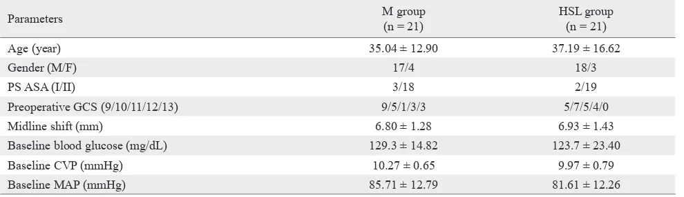
Forty-two patients were included in the study, assigned by random envelope to M (n = 21) or HSL (n = 21) groups. The two groups were similar
with respect to sex, age, GCS, ASA PS, midline
shift, baseline MAP, CVP and blood glucose, as presented in table 1.
Signiicant differences ( p < 0.05) between the groups were found in the CVP at 30, 45, and 60 minutes where the CVP in group M was getting lower and lower and reached its lowest level after 60 minutes (7.87 ± 0.37 mmHg) while in group HSL the CVP tended to be stable with slight decrease, the lowest level after 60 minutes was 9.38 ± 0.37 mmHg (Table 2).
The same pattern was also observed in MAP with 4 hypotensive episodes in group M and one hypotensive
Parameters M group
(n = 21)
HSL group (n = 21)
Age (year) 35.04 ± 12.90 37.19 ± 16.62
Gender (M/F) 17/4 18/3
PS ASA (I/II) 3/18 2/19
Preoperative GCS (9/10/11/12/13) 9/5/1/3/3 5/7/5/4/0
Midline shift (mm) 6.80 ± 1.28 6.93 ± 1.43
Baseline blood glucose (mg/dL) 129.3 ± 14.82 123.7 ± 23.40
Baseline CVP (mmHg) 10.27 ± 0.65 9.97 ± 0.79
Baseline MAP (mmHg) 85.71 ± 12.79 81.61 ± 12.26
Table 1. Baseline demographic data of patients
episodes in group HSL. The baseline MAP in group M was higher than group HL (85.71 ± 12.79 vs 81.61 ± 12.26 mmHg) however at 60 minutes, it was
signiicantly lower in group M (74.33 ± 6.18 vs 81.66 ± 7.85, p = 0.002) (Table 2, Figure 1).
Diuresis was signiicantly higher (p = 1.001) after
30 minutes in group M and increasing about twice of the previous volume every 15 minutes. The volume of water loss through diuresis in group M was 2.35 X the group HSL after 60 minutes (Table 2).
There was no difference in brain relaxation between the two groups, with three patients in group HSL and two patients in group M requiring rescue therapy for tense brain conditions.
Blood glucose level at 60 minutes was signiicantly
higher in group HSL than in group M (141.81 ± 19.09 vs 122.71 ± 17.89, p = 0.027); the blood glucose difference in group HSL between baseline
value and after 60 minutes was signiicant (17.95 ± 11.46, p = 0.001) (Table 2).
DISCUSSION
This study aimed to compare the effect of equiosmolar boluses of HSL and mannitol on clinical brain condition, hemodynamic variables (MAP and CVP) and blood glucose level in patients with moderate traumatic brain injury underwent craniectomy with opening of the duramater. The
main indings were that during craniectomy, (1)
either equiosmolar boluses of 2.5 mL.kg-1 20%
Table 2. Measured parameters during study period in M and HSL group.
Parameters M Group
(n = 21)
HSL Group
(n = 21) p
Baseline CVP (mmHg)* 10.27 ± 0.65 9.97 ± 0.79 0.188
CVP at 15 min (mmHg)* 10.25 ± 0.54 10.20 ± 0.57 0.784
CVP at 30 min (mmHg)* 9.54 ± 0.46 9.93 ± 0.62 0.028
CVP at 45 min (mmHg)* 8.85 ± 0.50 9.51 ± 0.52 0.001
CVP at 60 min (mmHg)* 7.87 ± 0.37 9.38 ± 0.37 0.001
Baseline MAP (mmHg)* 85.71 ± 12.79 81.61 ± 12.26 0.296
MAP at 15 min (mmHg)* 84.14 ± 10.44 80.71 ± 12.07 0.331
MAP at 30 min (mmHg)* 79.95 ± 9.99 79.47 ± 8.62 0.870
MAP at 45 min (mmHg)* 75.80 ± 8.26 81.66 ± 10.43 0.051
MAP at 60 min (mmHg)* 74.33 ± 6.18 81.66 ± 7.85 0.002
Baseline blood glucose (mg/dL)* 129.30 ± 14.82 123.7 ± 23.40 0.392
Blood glucose at 60 min (mg/dL)* 122.71 ± 17.89 141.81 ± 19.09 0.027
Blood glucose change (mg/dL)* -6.38 ± 7.12 17.95 ± 11.46 0.001
Diuresis at 15 min (mL) 66.67 ± 22.21 59.05 ± 39.48 0.445
Diuresis at 30 min (mL) 147.62 ± 30.31 96.67 ± 52.28 0.001
Diuresis at 45 min (mL) 293.81 ± 62.64 135.95 ± 65.71 0.001
Diuresis at 60 min (mL) 476.19 ± 85.87 203.57 ± 86.21 0.001
Brain relaxation† (number of patients)
excellent/satisfactory/irm/bulging 3/13/4/1 2/15/3/1 0,988 *unpaired t-test; †chi-square test; P ≤ 0.05 was considered signiicant
MAP; (3) 2.5 mL.kg-1 bolus of 0.5M HSL was
associated with a signiicant increase in blood
glucose level, while 20% mannitol did not.
The effect of mannitol and HSL on ICP has been investigated by Ichai, et al13 with HSL was reported
to be more effective in reducing ICP and had more
prolonged effect than mannitol. Rozet, et al7 compared
5 mL.kg-1 boluses of 3% HTS and mannitol, did not ind any difference in brain relaxation between
the two groups. Wu CT, et al8 also compared the
effect of 160 mL 3% HTS and 150 mL of 20% mannitol infusion on brain relaxation, and showed that hypertonic saline did better than 20% mannitol,
85.71
84.14
79.95
75.8
74.3
81.61
80.71
79.47
81.6
81.6
68
70
72
74
76
78
80
82
84
86
88
MAP0
MAP15
MAP30
MAP45
MAP60
Group M
Group HL
mmHg
0
15
30
45
60
Figure 1. Mean arterial pressure (MAP in mmHg) during the study period in patients treated with mannitol (group M) or hypertonic
sodium lactate (group HSL). MAP was signiicantly higher in group HSL at 60 minutes (P = 0.002)
M group
which is not in agreement with our inding (HSL
has a similar sodium concentration to 3% HTS). The discrepancy could be due to differences in the
volume of the hyperosmotic luids administered to
the patients and the populations studied.
The principal mechanism of action of hyperosmotic
luids such as HSL and mannitol is to create osmotic
gradient across the blood-brain-barrier (BBB) due to impermeability of BBB to mannitol and sodium.7-8 but
not to lactate. Therefore, the hyperosmotic property of HSL is derived from the sodium concentration in the solution, while lactate serves as the alternative energy source for the brain tissue, particularly in the post-ischemic condition.12,16 The effectiveness of the hyperosmotic luid depends on the “relection coeficient” of its solute, which determines the
relative impermeability of BBB to the solute. A
relection coeficient of 1 means the BBB is absolutely
impermeable to the solute, and 0 means totally
permeable. The relection coeficient of sodium is 1
and mannitol is 0.9,7-8 therefore hypertonic solutions
containing sodium may theoretically be superior to mannitol in reducing ICP or brain bulk. Unfortunately, the evidences are not yet enough to support this theory.
This study showed that HSL was associated with
signiicantly less diuretic effect compared to
mannitol, as was already reported by other studies.7-8
Rozet, et al7 also reported a more negative luid
balance after mannitol administration and an increase in blood lactate over time, whereas no changes in blood lactate after HTS administration. The increase in lactate concentration may be secondary to effective hypovolemia induced by mannitol. Both 3% HTS and HSL have similar sodium concentration and cause an increase in serum sodium.13 The increase in
serum sodium stimulates the release of antidiuretic hormones, leading to the absorption of free water from the kidney,17 which may explain the lower
diuretic effect of HSL compared to mannitol.8 We
also found that mannitol was associated with a more
signiicant decrease in CVP compared with HSL,
possibly related to the diuretic effect of mannitol. We
observed a signiicant increase in CVP at 15 minutes
after HSL administration, which may be due to the volume expansion of the hyperosmotic solution, but
the increase was not maintained. The same inding was also shown in a study by Gemma et al18 after
administration of 7.5% HTS.
In our study, MAP was more stable after HSL administration than mannitol. Blood pressure is
determined by cardiac output and systemic vascular resistance. The determinants of cardiac output include stroke volume and heart rate; the stroke volume is determined by the preload volume and cardiac contractility. Assuming all other factors were
comparable, the more negative luid balance induced
by mannitol may explain the more pronounced decrease in MAP.
Lactate was thought to increase the blood glucose due to hepatic gluconeogenesis (Cori cycle).13 Patki,
et al19 showed that administration of Ringer Lactate
20 mL.kg-1 will increase the blood glucose level as
much as 14.56 ± 0.51 mg.dL-1, while Ichai, et al13
showed 3.8% increase in blood glucose compared to baseline value after administration of 100 mL HSL.
In our current study, a signiicant increase in blood
glucose level was seen in Group HSL which could be due to larger volume of HSL given to the patient,
the surgical stress response, and the inluence of isolurane used during the procedure.
High level of blood glucose may have detrimental effect to the brain14-15 as was shown in a study by
Lam, et al14 that severe traumatic brain injury patients with post-operative blood glucose ≥ 200 mg.dL-1 had signiicantly worse outcome than those < 200
mg.dL-1. Older theory stated that a high glucose
level is detrimental in ischemic conditions because glucose will be converted to lactic acid, which causes neuronal damage.20 However, more evidence have
supported the newer theory proposed by Schurr, et al21
that the detrimental effect of hyperglycemia during cerebral ischemia is not associated with lactate, but with the corticosterone released, in response to high glucose level. In contrast to post-ischemic conditions when the oxygen delivery has just started to resolve; lactate serves as a ready substrate, which can be directly converted to pyruvate.12 Pyruvate then enters
the tricarboxylic acid cycle to produce ATP, in which one lactate molecule yields 17 ATP.
Beside their hyperosmotic property, hyperosmolar
luids such as HSL, HTS and mannitol can improve the
cerebral perfusion by their effects on blood rheology, shrinkage of erythrocytes, and the decreased CSF production.8 Both HSL and mannitol also has anti-inlammatory properties. mannitol has the free-radical
scavenging property, which can reduce the increased malondialdehyde, catalase, and glutathione peroxidase in traumatic brain injury.22 This effect is not owned by
post-ischemic conditions. mannitol does not have this advantage. Therefore, despite their osmotic properties, both HSL and mannitol have their own advantages
in speciic patient conditions. HSL is recommended
for patients with suspected hypovolemia or hypoxia, while mannitol for cases with high baseline blood glucose, history of diabetes, or in hypernatremic conditions.
Due to limited facilities, observation for patient outcome such as non-invasive measurement of blood pressure, ICP and arteriovenous difference of oxygen and lactate levels, were not performed.
In conclusion, 0.5M HSL was equivalently effective as 20% mannitol to produce brain relaxation.
Administration of 2.5 mL.kg-1 0.5M HSL was
associated with signiicant increase in blood glucose. To our knowledge, this is the irst prospective,
randomized human study comparing the effect of equiosmolar and equivolemic solutions of HSL and mannitol on intraoperative brain relaxation, blood pressure, CVP, and blood glucose level. HSL may be recommended as an alternative to mannitol for providing brain relaxation during craniectomy procedure in cases with moderate traumatic brain injury, unstable hemodynamic and/or when high risk of hypovolemia is expected and not for diabetic cases.
Conlict of interest
The authors declare that this study is free of conlict
of interest.
REFERENCES
1. Morgan GE, Mikhail MS, Murray MJ. Anesthesia for neurosurgery. In:Clinical anesthesiology. 4th ed. New York:
Lange Medical Books/McGraw-Hill Companies; 2006. p.
631-46.
2. Tenenbein P, Kincaid S, Lam AM. Head trauma – anesthetic consideration and management. In: Smith CH, editor. Trauma anesthesia. Cambridge: Cambridge University Press; 2008. p. 172-82.
3. Cottrell JE, Newield P. Anesthesia for Traumatic Brain
injury. In : Textbook of neuroanesthesia. 4th ed. New York: Lippincott Williams and Wilkins; 2007. p. 317-27.
4. Bratton SL, Chestnut RM, Ghajar J, McConnell Hammond
FF, Harris OA, Hartl R, et al. Hyperosmolar therapy. J Neurotrauma. 2007;24(Suppl 1):S14-20.
5. Haddad SH, Arabi YM. Critical care management of severe traumatic brain injury in adults. Scand J Trauma Resusc Emerg Med. 2012;20:2.
6. Roberts I, Schierhout G, Wakai A. Mannitol for acute
traumatic brain injury. Cochrane Database Syst Rev 2003:CD001049.
7. Rozet I, Tontisirin N, Muangman S, Vavilala MS, Souter MJ, Lee LA, et al. Effect of equiosmolar solutions of mannitol versus hypertonic saline on intraoperative brain relaxation and electrolyte balance. Anesthesiology. 2007;107(5):697-704.
8. Wu CT, Chen LC, Kuo CP. Ju DT, Borel CO, Cherng CH, et al. A comparison of 3% hypertonic saline and mannitol for brain relaxation during elective supratentorial brain tumor surgery. Anesthe Analg. 2010;110(3):903-7.
9. Mustafa I, Leverve X. Metabolic and hemodynamic effects of hypertonic solutions: sodium-lactate versus sodium chloride infusion in postoperative patients. Shock. 2002;18(4):306-10.
10. Leverve X, Mustafa I. Lactate: a key metabolite in the intercellular metabolic interplay. Crit Care. 2002;6(4):284-5. 11. Rice CA, Zsoldon R, Chen T, Wilson MS, Ales Sandri B,
Hamm RJ, et al. Lactate administration attenuates cognitive
deicits following traumatic brain injury. Brain Res.
2002;928(1-2):156-9.
12. Cater HL, Chandratheva A, Benham CD, Morrison B, Sundstorm LE. Lactate and glucose as energy substrates during, and after, oxygen deprivation in rat hippocampal acute and cultured slices. J Neurochem. 2003;87:1381-90.
13. Ichai C, Armando G, Orban JC, Berthier F,
Sama-long C, et al. Sodium lactate versus mannitol in the treatment of intracranial hypertensive episodes in severe traumatic brain-injured patients. Intensive Care Med. 2009;35(3):471-9.
14. Lam AM, Winn HR, Cullen B, Sundling N. Hyperglycemia and neurological outcome in patients with head injury. J Neurosurg. 1991;75(4):545-51.
15. Rovlias A, Kotsou S. The inluence of hyperglycemia on
neurological outcome in patients with severe head injury. Neurosurgery 2000.46(2);335-42.
16. Gladden LB. Lactate metabolism: a new paradigm for the
third millenium. J Physiol. 2004;558(1): 5-30.
17. Tyagi R, Donaldson K, Loftus CM, Jallo J. Hypertonic saline: a clinical review. Neurosurg Rev. 2007;30(4):277-90.
18. Gemma M, Cozzi S, Tommasino C, Mungo M, Calvi
MR, Cipriani A, et al. 7.5% hypertonic saline versus 20% mannitol during elective neurosurgical supratentorial procedures. J Neurosurg Anesth. 1997;9(4):329-34.
19. Patki A, Shelgaonkar VC. Effect of 6% hydroxyethyl starch-450 and low molecular weight dextran on blood sugar levels during surgery under subarachnoid block: A prospective randomised study. Indian J Anaesth. 2010;54(5):448-52.
20. Rehncrona S, Rosen I, Siesjo BK. Brain lactic acidosis and ischemic cell damage: biochemistry and neurophysiology. J Cereb Blood Flow Metab. 1981;1(3):297-311.
21. Schurr A, Payne RS. Hyperglycemia and neuronal damage in cerebral ischemia and beyond. Crit Care & Shock. 2003;6(4):184-90.