ROLE OF LEUKOCYTE PROFILES IN WOUND HEALING
BETWEEN H-PLASTY AND LINEAR CLOSURE IN
DOMESTIC HOUSE CATS (
Felis catus
)
LIM SU-SZIEN
DEPARTMENT OF CLINIC, REPRODUCTION AND PATHOLOGY FACULTY OF VETERINARY MEDICINE
BOGOR AGRICULTURAL UNIVERSITY BOGOR
STATEMENT ON UNDERGRADUATE THESIS AND
INFORMATION SOURCES AND COPYRIGHT
I hereby state that this undergraduate thesis entitled “Role of Leukocytes
Profile in Wound Healing between H-Plasty and Linear Closure in Domestic House Cats (Felis catus)” is my own work with the assistance of my supervisors and has not been submitted in any form to other institutes. Sources of information used in this undergraduate thesis quoted from other sources both publicized and unpublicized are written at the end of this thesis.
I hereby present the copyright of my undergraduate thesis to Bogor Agricultural University.
ABSTRACT
LIM SU-SZIEN. Role of Leukocyte Profiles in Wound Healing between H-Plasty and Linear Closure in Domestic House Cats (Felis catus). Supervised by DENI NOVIANA and GUNANTI.
Skin wounds commonly seen in veterinary practice can be repaired with a variety of reconstructive choices such as skin flaps. The research purpose is to learn and understand the healing process between skin flap H-Plasty and linear closure in domestic house cats by observing leukocyte profiles. Six male cats divided into two groups were used of which open wounds of 2x2 cm were made on the skin of lateral thorax. In H-Plasty group, 1 cm parallel incisions were made on each sides of the defect, skin is undermined, advanced over the defect and sutured. In linear closure group, the elliptic defect was sutured across the widest part, continued by dividing each remaining segment in half with subsequent sutures. Blood was drawn of 1 ml from the medial saphenous vein for an alternating 3 days of a 12-day period and the profiles’ results were observed. Both flaps showed insignificant differences (P>0.05) in the profiles of segmented neutrophils, lymphocytes and monocytes whereas significant differences (p<0.05) was seen in the profiles of leukocytes, banded neutrophils and eosinophils when compared between observation time. The profiles’ results were all within reference range, therefore both skin flaps can be used to close wounds.
ABSTRAK
LIM SU-SZIEN. Peran Gambaran Leukosit terhadap Kesembuhan Luka antara H-Plasty dengan Linear Closure pada Kucing Lokal (Felis catus). Dibimbing oleh DENI NOVIANA dan GUNANTI.
Luka pada kulit yang sering terlihat dalam praktik kedokteran hewan dapat diperbaiki dengan berbagai pilihan rekonstruksi tersedia seperti skin flap. Penelitian ini bertujuan untuk mempelajari dan memahami proses persembuhan antara skin flap H-Plasty dengan linear closure pada kucing lokal dengan mengamati gambaran leukosit. Penelitian ini menggunakan enam ekor kucing jantan yang dibagi menjadi dua kelompok. Luka terbuka berukuran 2x2 cm dibuat pada kulit lateral thorax pada kedua kelompok. Pada kelompok H-Plasty, tambahan sayatan paralel dilakukan pada keempat sisi luka, kulit dipisahkan dari subkutan, kemudian ditarik untuk menutupi luka dan dijahit. Pada kelompok linear closure, penjahitan luka dimulai pada sayatan terlebar dan dilanjutkan dengan penutupan luka pada pembagian setengah tiap segmen tersisa dengan jahitan. Darah sebanyak 1 ml diambil dari vena saphena medialis setiap 3 hari dalam kurun waktu 12 hari untuk dilakukan pengamatan gambaran leukosit. Hasil dari gambaran leukosit kedua flap menunjukkan perbedaan tidak nyata (P>0.05) dalam gambaran neutrofil bersegmen, limfosit dan monosit sedangkan perbedaan nyata (P<0.05) dapat diamati pada gambaran leukosit, neutrofil batang dan eosinophil apabila dibandingkan antar waktu pengamatan. Hasil gambaran kedua flap dalam kisaran normal sehingga dapat disimpulkan keduanya dapat digunakan untuk menutupi luka.
Undergraduate Thesis as a requirement for a degree in Bachelor of Veterinary Medicine
Faculty of Veterinary Medicine
ROLE OF LEUKOCYTE PROFILES IN WOUND HEALING
BETWEEN H-PLASTY AND LINEAR CLOSURE IN
DOMESTIC HOUSE CATS
LIM SU-SZIEN
DEPARTMENT OF CLINIC, REPRODUCTION AND PATHOLOGY FACULTY OF VETERINARY MEDICINE
BOGOR AGRICULTURAL UNIVERSITY BOGOR
FOREWORD
Praise and grace to the Lord for His guidance and blessings during the writing of this undergraduate thesis. The research entitled “Role of Leukocytes Profile in Wound Healing between H-Plasty and Linear Closure in Domestic Cats (Felis catus)” was done from December 2014 until March 2015 in the Department of Clinic, Reproduction and Pathology.
The writer would like to thank everyone who made this undergraduate thesis possible:
1. Prof Drh Deni Noviana, PhD and Dr Drh Hj Gunanti, MS as the supervisors for the assistance and guidance in making this thesis,
2. Drh Erwin, MSc for the guidance during the research and writing of thesis, 3. Ibu Siti Sa’diah, SSi, Apt, MSi as the academic advisor for the assistance
and support these past three years,
4. Father Dr Roland Lim Swee Guan, mother Ng Yoon Land and siblings for the love, spirit and support,
5. The TaiTai-s: Charisha, Kartini and Clarisse for the friendship, laughter and support.
The writer hopes that this thesis can be beneficial for the readers, especially small animal practitioners and the writer herself.
CONTENT
LIST OF TABLES vi
LIST OF FIGURES vi
INTRODUCTION 1
Background 1
Objective 1
Benefit 1
LITERATURE REVIEW 2
Skin 2
Skin Flap 2
Blood 4
Leukocyte 4
Differential Leukocyte 5
MATERIALS AND METHOD 6
Time and Place 6
Animals and Materials 6
Method 6
Animals 6
Preparation 7
Surgical Technique 7
Blood Sampling 7
Data Analysis 7
RESULT AND DISCUSSION 7
CONCLUSION 12
REFERENCES 12
LIST OF TABLES
1 Leukocyte reference range 4
2 Total average of leukocytes 8
3 Total average of segmented neutrophils 9
4 Total average of banded neutrophils 9
5 Total average of lymphocytes 10
6 Total average of monocytes 11
7 Total average of eosinophils 11
LIST OF FIGURES
1 Cutaneous vascular supply in cats 3
2 Paired single advancement flaps 3
INTRODUCTION
Background
The Indonesian Domestic House Cat, usually short-haired, are members of one species, Felis catus. Indoor cats would explore crevices and dangerous household hazards that lead to them getting hurt. Skin wounds commonly seen in veterinary practice can be repaired relatively simply with a combination of undermining, apposition and suturing of a variety of reconstructive choices available including skin stretching, free skin grafts, microvascular free tissue transfer and a variety of skin flaps (Nevill 2010).
A flap is tissue that is transferred from a donor site to a recipient site while maintaining its own blood supply. Flaps can be categorised by type of blood supply, type of tissue to be transferred, and location of donor site which may further be classified based on geometric design or method of transfer (Oliaei and Chu 2013).
Common postoperative complications following flaps are edema, seroma formation, wound margin necrosis, poor wound drainage due to extensive soft tissue dissection at the donor and recipient sites and even flap failures which can predispose to wound infection, may cause discomfort and prevent healing of tissues on to wound beds that leads to inflammatory cell changes which can be observed by blood tests (Shin et al. 2014, Nelissen and White 2014). The haematology profile can be used as a platform to predict, prevent and treat a disease in an animal and help to monitor the progress of medical treatment and health of the animal (Özalp et al. 2013).
Objective
The objective of this study was to learn and understand the healing process between skin flap H-Plasty and linear closure in domestic house cats (Felis catus) by observing the leukocyte profile.
Benefit
2
LITERATURE REVIEW
Skin
The skin is composed of the epidermis, dermis and hypodermis. The outermost layer, epidermis, known to be hard, dry and contains no blood vessels which continuously cast off dead cells. In hair-bearing areas, the epidermis of hairy skin consists of three major layers: stratum cylindricum, stratum spinosum and stratum corneum. The dermis layer, below the epidermis contains nerves, blood vessels, glands and hair roots, is divided into superficial stratum papillare and deep stratum reticulare. The innermost layer, hypodermis is composed primarily of fat with loose collagenous trabeculae and elastic fibers (Dallas 2006, Pavletic 2010).
The integument serves as the body’s first defence line against microorganisms which is injured more frequently than any other tissue and resulting damage, while repairable, leads to permanent scarring in mammals (Seifert et al. 2012). The skin is a sensory receptor for touch, pressure, vibration, pain, heat and cold. Its multiple functions include vitamin D production, storage of water, fat, electrolytes, carbohydrates and proteins; a barrier against chemicals and radiation; and along with subcutaneous fat for insulation (Pavletic 2010). .
Skin Flap
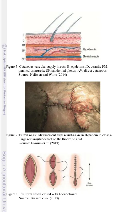
A skin flap is a piece of healthy skin and tissue that is partly detached and moved to cover a nearby wound which is often still attached to its original site at one end and remains connected to at least one source of vascular support from the donor site during its transfer as shown in Figure 1, offering a reconstructive method without the undesirable effect of second intension healing (Koch 2011). Flaps are normally used to cover large open wounds, ulcers, tumour excisions, and congenital abnormalities. Consequently, flap surgery is one of the most important techniques in reconstructive surgery (Wang et al. 2013). Square or rectangular defects may be closed with centripetal, unilateral, or bilateral advancement flap. An H-plasty is used to close large defects that have mobile skin available on two sides of the defect as shown in Figure 2. On the other hand, fusiform, or elliptic defects are closed by first placing a suture across the widest part of the defect which is then continued by dividing each remaining segment in
3
Figure 1 Fusiform defect closed with linear closure Source: Fossum et al. (2013)
Figure 3 Cutaneous vascular supply in cats. E, epidermis; D, dermis; PM, panniculus muscle; SP, subdermal plexus; AV, direct cutaneous Source: Nelissen and White (2014)
Figure 2 Paired single advancement flaps resulting in an H-pattern to close a large rectangular defect on the thorax of a cat
4
Blood
Blood is a connective tissue made up of formed elements in a fluid matrix which connects all the cells in the body together, highly specialised, circulating within a fluid medium called plasma and is termed serum when depleted of fibrinogen (Bacha and Bacha 2012, Dallas 2006). Blood consists of erythrocytes, thrombocytes and leukocytes.
Erythrocytes are found most abundant, with several million erythrocytes per microliter of blood in mammals and thrombocytes are the next most abundant cell blood type. Total leukocyte count is much lower than erythrocyte and thrombocyte counts, with total leukocyte counts ranging from 5.4 to 15.4 thousand leukocytes per microliter in cats. The proportion of leukocyte types differs by species, with neutrophils found the most in carnivores and lymphocytes found the most in the blood of ruminants and rodents (Harvey 2012).
Leukocyte
5 Differential Leukocyte
Neutrophils are the most predominant leukocyte in circulation and undergo complete replacement relatively 2.5 times each day (Brockus 2006). Neutrophils defence against invading microorganisms, primarily bacteria. To be effective, they must recognise inflammatory signals, leave the blood, migrate through tissue to a site where bacteria are present, and then neutralise the bacteria (Harvey 2012). Neutrophilia is a condition when measured blood neutrophil concentration increases while neutropenia is a term used when measured blood neutrophil concentration decreases (Stockham and Scott 2002).
Eosinophils play an important role in the host defence against helminth infections and are responsible for type 1 hypersensitivity allergic reactions (Harvey 2012). In most species, eosinophils have round granules but domestic cats have rod-shaped ones easily identifed through light microscopy. Eosinophilia is due to increased production and/or release of eosinophils from bone marrow reserves (Harvey 2012). On the other hand, eosinopenia is identified when measured blood eosinophil concentration decreases. Eosinopenia may be observed with chemotherapeutic agents, stress or steroid-like compounds (Poitout-Belissent and McCartney 2010).
Basophils normally are the least common leukocyte observed in normal feline peripheral blood smears (Bacha and Bacha 2012, Solomon et al. 2015) but contain most of the histamine measured in blood (Harvey 2012). The granules in
cat’s basophil are small lavender-grey, by Romanowsky staining, compared to the metachromatic staining of other species. Basophilia is a term where measured blood basophil concentration increases or more than 200 basophils per microliter (Brockus 2006). Although basophilia is rarely observed, when occurred, it can be associated with allergic, parasitic, and neoplastic states. Basopenia is not known to be clinically significant (Stockham and Scott 2002).
Monocytes are the largest leukocytes and its nucleus’ shape is highly variable (Bacha and Bacha 2012). In mammals, monocytes’ three major functions are phagocytosis, antigen presentation to T lymphocytes, and immunomodulation associated with cytokines production involved in the inflammation regulation and haematopoiesis (Koh and DiPietro 2011). Monocytosis, more than 850 monocytes per microliter is a nonspecific finding in cats associated with purulent inflammation, tissue destruction, neutrophilia, trauma, necrosis, malignancy, immune-mediated injury, haemorrhage, haemolysis, and pyogranulomatous inflammation. Monocytopenia is difficult to document because there can be relatively few blood monocytes in healthy domestic mammals (Stockham and Scott 2002).
6
MATERIALS AND METHOD
Time and Place
The study was done from December 2014 until March 2015. The surgery was carried out at the surgical laboratory of the Division of Surgery and Radiology, Department of Clinic, Reproduction and Pathology, Faculty of Veterinary Medicine, Bogor Agricultural University. The blood test was carried out at a commercial laboratory of Apotek YASA.
Animals and Materials
The animals used for this research were six healthy male Indonesian Domestic House Cats which were divided into two groups of three cats each.
The equipment used for this research were cat cages, manual weighing scale (10 kg capacity), surgical instruments consisting of towel clamps, scalpel handle #10, blade #10, scissors, forceps, Mathieu needle holder, needle, hematology analyzer, vacuum whole blood collection tube (Vaculab EDTA K3 Glass 3 ml), disposable syringes (Terumo), gauze and gloves.
Materials used for this research includes pre-anesthetic adjuvant (atropine sulfate 0.25 % 0.04 mg/kg body weight (BW)), anesthetic Ketamine HCl (Ketalar® 100 mg/ml 10 mg/kg BW), Xylazine HCl (Xyla® 0.20 mg/ml 1 mg/kg BW), Claneksi® (Amoxicillin 25 mg/ml-Clavulanic Acid 6.25 mg/ml 62.5 mg/kg BW), Procaine Penicillin-G Meiji (liquid procaine benzylpenicillin 15000 IU/ml), Flagyl® (metrodinazole 20 mg/kg BW), Drontal® (praziquantel 20 mg/kg BW), Silk 3.0 (Silkam®), povidone-iodine, sodium chloride (NaCl) 0.9 % solution, 70 % ethanol solution, antibacterial gauze dressing (Sofra-tulle®), commercial cat food and water.
Method
Animals
7 Preparation
Food was withheld from the cats 8 hours prior to the surgery. The cats were injected with pre-anesthetic adjuvant of atropine sulfate (0.04 mg/kg BW) subcutaneously. After ten minutes, the cats will be anesthetized with a combination of xylazine (1 mg/kg BW) and ketamine (10 mg/kg BW). After the cats were under the influence of general anesthesia and before the surgery, the cats were put in lateral recumbent position and the fur on the lateral thorax was shaved before swabbing the area with povidone-iodine.
Surgical Technique
Open wound of 2x2 cm was made on the lateral thorax. For the H-Plasty group, the surgery began by making parallel incisions on each side of the defect at least half as long as the width of the defect, skin is undermined, advanced over the defect and sutured. For the linear closure group, the elliptic defect was closed by first placing a suture across the widest part of the defect, continued by dividing each remaining segment in half with subsequent sutures. The wound was dressed with sterile gauze impregnated with povidone-iodine. The cats were given antibiotic Penicillin-G topically of 0.5 ml after surgery and Claneksi orally with the dosage of 6.25 mg/kg BW twice a day for five consecutive days. Stitches were removed on the 10th day after the suturing and blood tests were done to observe the phases of wound healing.
Blood Sampling
One ml of blood was drawn from the medial saphenous vein with a 1 ml syringe after the cat was anesthetized and placed in vacuum whole blood collection tube containing anticoagulant ethylenediaminetetraacetic acid (EDTA). The blood collection tubes were sent to a commercial laboratory of Apotek YASA to measure the total leukocyte count and leukocyte differential count.
Data Analysis
The quantitative properties of the leukocytes were evaluated and analyzed using a hematology analyzer and a statistical analysis software package (SPSS Version 20.0).
RESULT AND DISCUSSION
8
wound healing period to understand the systemic changes which might possibly occur. Leukocyte differentials are widely available, simple, inexpensive and appear to have independent prognostic significance beyond traditional risk factors (Vaduganathan et al. 2012). The leukocyte profile observed comprises of leukocytes, segmented neutrophils, banded neutrophils, lymphocytes, monocytes, eosinophils and basophils.
Based on the statistic test result using univariate analysis, leukocyte when compared by the observation time of both groups has a significant difference (P<0.05) between Day 0 and 3 with Day 12 whereas when compared between both groups, they are of insignificant difference (P>0.05). The average is higher in Group II compared to Group I as shown in Table 2 may be due to the fact that the normal physiological state of the cats were already high before the operation. The leukocytes are highest on Day 0 in Group I before the surgery might be due to excitement, fear, forced flight, and excessive muscular activity by environmental stresses (Latimer and Bienzle 2010, Valenciano et al. 2010). In Group I, leukocytes decreased from Day 0 to Day 6, and increased on Day 9 and continued to decrease on Day 12. On the other hand, in Group II, leukocytes increased on Day 3 and decreased from Day 6 to Day 12. They increased on Day 9 and Day 3 in each respective group can be possibly due to inflammation, stress due to handling, or fight or flight response to pain or fright (Stockham and Scott 2002).
Table 2 Total leukocytes (×103/µL) during the wound healing period of skin flaps in cats
9
Based on the statistic test result using univariate analysis, segmented neutrophils when compared by the observation time of both groups have an insignificant difference (P>0.05) between all the days whereas when compared between groups, they are of significant difference (P<0.05) on Day 0 and 9. The average in Group II slightly higher than Group I as displayed in Table 3, may be due to stress in the cats during venipuncture or according to Kim et al. (2008), neutrophil influx is an early inflammatory response that is essential for the clearance of bacteria and cellular debris during cutaneous wounding. Nevertheless, the total is still within reference range. Segmented neutrophils are observed to increase in Group I possibly due to an inflammation (Valenciano et al. 2010) whereas in Group II, they are seen to decrease on Day 3, increase on Day 6, decrease on Day 9 and increase on Day 12 may be due to stress or excitement (Stockham and Scott 2002).
Table 3 Total segmented neutrophils (×103/ µL) during the wound healing period of skin flaps in cats and row followed by the same alphabet (a) and (x,y) respectively indicate an insignificant result at a 5% level of significance test.
10
Banded neutrophils when compared by observation time of both groups, portrayed a significant difference (P<0.05) between Day 0, 3, 6 and 12 with Day 9 but when compared between both groups, they are of insignificant difference (P>0.05). The average in Group II, slightly higher than Group I as shown in Table 4, may be due to stress during venipuncture or an acute inflammation because stress mediators act as regulators of inflammation (Stella et al. 2013). The average of banded neutrophils is slightly higher than the reference range is due to the fact that the work of the neutrophils is crucial within the first days after injury because their ability in phagocytosis and protease secretion kills local bacteria and helps to degrade necrotic tissue (Reinke and Sorg 2012). In Group I, banded neutrophils increased on Day 3 till Day 9 and decreased on Day 12 whereas in Group II, they increased on Day 3, decreased on Day 6, increased on Day 9 and decreased on Day 12. The increase is due to inflammation which is the most common cause of a pathologic neutrophilia (Valenciano et al. 2010) and the decrease is the body returning to its physiological state.
Lymphocytes reveal an insignificant difference (P>0.05) when compared by the observation time between all days of both groups and between both groups except on Day 0. The average in Group I is higher than Group II as seen in Table 5 which is resulted from infection due to the bigger wound size between the two skin flaps however, the total is still within reference range. Lymphocytes are observed to decrease on Day 3 till Day 9 and slightly increase on Day 12 in Group I whereas they slightly increased on Day 3 and Day 9 and slightly decreased on Day 6 and Day 12 in Group II. The increment might be due to the fact that the cats used in the research are young because younger animals are prone to lymphocytosis secondary to excitement of fear (Brockus 2006, Stockham and Scott 2002, Valenciano et al. 2010) and the decrement is the body’s effort in getting back to its normal condition.
Table 5 Total lymphocytes (×103/ µL) during the wound healing period of skin flaps in cats
11
Monocytes when compared by observation time of both groups and between both groups except Day 9 displayed an insignificant difference (P>0.05) based on the statistic test result using univariate analysis. The average in Group II is higher than Group I as displayed in Table 6, resulted from an acute inflammation on the wound but the amount is still within reference range. In wound healing, the sequence of events awaiting a complete wound closure and repair can be divided into three overlapping phases: inflammation, proliferation, and matrix formation and remodelling (Koh and DiPietro 2011). In Group I, monocytes are observed to increase on Day 3 till Day 6 and decrease on Day 9 till Day 12 whereas in Group II, they increased on Day 3, insignificantly decreased and increased on Day 6 and Day 9 respectively and decreased on Day 12. Monocytosis occurs in many conditions, including inflammation and tissue destruction which might have cause the increment (Valenciano et al. 2010).
Table 6 Total monocytes (×103/ µL) during the wound healing period of skin flaps in cats and row followed by the same alphabet (a,b) and (x,y) respectively indicate an insignificant result at a 5% level of significance test.
12
Based on the statistic test result using univariate analysis, eosinophils when compared by observation time of both groups manifested a significant difference (P<0.05) between Day 0 with Day 3, 6, 9 and 12. They also showed a significant difference on Day 0 but insignificant difference (P>0.05) on Day 3, 6, 9 and 12 when compared between both groups. The average in Group II is higher than Group I as shown in Table 7, however, it is still within reference range. Eosinophils are observed to increase and stayed stagnant from Day 3 to Day 9 and decreased on Day 12 in Group I whereas they increased and stayed stagnant from Day 3 to Day 6, decreased on Day 9 and increased on Day 12. The increase in eosinophils is due to tissue remodelling of the wound (Young and Meadows 2012) and the decrement is the body working its way to its physiological condition.
Basophils release mediators for inflammation and allergy (Worm et al. 2013). They were not observed during the blood sampling; basopenia is not a recognised clinical problem (Brockus 2006, Valenciano et al. 2010). Therefore, the data collected of zero basophil to be found is within reference range. No basophil observed does not indicate that the animal is not having an inflammation or allergy nonetheless; changes in other leukocyte profiles should be observed to ensure the presence or absence of an inflammation (Mochizuki et al. 2014).
CONCLUSION
The leukocyte profiles observed from both skin flaps “H-Plasty” and linear closure are all within reference range therefore they can be used to repair wounds in domestic house cats (Felis catus).
REFERENCES
Bacha JW, Bacha LM. 2012. Color Atlas of Veterinary Histology. 3rd ed. UK: Wiley-Blackwell.
Brockus CW. 2006. Consultations in Feline Internal Medicine. In August JR, editor. US: Elsevier Saunders.
Dallas SE. 2006. Animal Biology and Care. 2nd ed. UK: Blackwell Publishing Ltd Fossum TW, Dewey CW, Horn CV, Johnson AL, MacPhail CM, Radlinsky MG,
Schulz KS, Willard MD. 2013. Small Animal Surgery. 4th ed. Missouri (US): Mosby Elsevier.
Harvey JW. 2012. Veterinary Hematology: A Diagnostic Guide and Color Atlas. Missouri (US): Elsevier Saunders.
Kim MH, Liu W, Borjesson DL, Curry FRE, Miller LS, Cheung AL, Liu FT, Isseroff RR, Simon SI. 2008. Dynamics of neutrophil infiltration during cutaneous wound healing and infection using fluorescence imaging. J Invest Dermatol. 128:1812-1820.doi:10.1038/sj.jid.5701223.
13
May 12]. On
http://www.dkoch.ch/xp_wysiwyg_media/English_Website/Handout_Advance d_Course_in_Soft_Tissue_Surgery_2011.pdf.
Koh TJ, DiPietro LA. 2011. Inflammation and wound healing: the role of the macrophage. Expert Rev Mol Med. 13(23):1.doi:10.1017/S1462399411001943. Latimer KS, Bienzle D. 2010. Schalm’s Veterinary Hematology. 6th ed. In Weiss
DJ, Wardrop KJ, editor. Iowa (US): Blackwell Publishing Ltd.
Liu Y, Jiao H, Ji X, Liu C, Zhong X, Zhang H, Ding X, Cao X. 2014. A comparative study of four types of free flaps from the ipsilateral extremity for finger reconstruction. PLoS ONE. 9(8):1.doi:10.1371/journal.pone.0104014. Merck Manuals. 2012. Hematologic Reference Ranges. The Merck Veterinary
Manual [Internet]. [Accessed 2015 April 3]. On http://www.merckmanuals.com/vet/appendixes/reference_guides/hematologicr eference_ranges.html.
Mochizuki H, Seki T, Nakahara Y, Tomita A, Takahashi M, Fujino Y, Ohno K, Tsujimoto H. 2014. Chronic myelogenous leukaemia with persistent neutrophilia, eosinophilia and basophilia in a cat. J Feline Med Surg. 16(6):517-21.doi:10.1177/1098612X13505576
Nagatomi R. 2013. Alteration in blood leukocyte profile due to exercise and its implication. J Phys Fitness Sports Med. 2(4):451-455.doi:10.7600/jpfsm.2.451. Nelissen P, White D. 2014. Feline Soft Tissue and General Sugery. In
Langley-Hobbs SJ, Demetriou JL, Ladlow JF, editor. Missouri (US): Elsevier Saunders. Nevill BG. 2010. Bilateral axillary skin fold flaps used for dorsal thoracic skin
wound closure in a dog. J S Afr Vet Assoc. 81(1):58-61.
Oliaei S, Chu EA. 2013. Encyclopedia of Otolaryngology, Head and Neck Surgery. In Kountakis SE, editor. US: Springer Link.
Özalp GR, Temizel EM, Özocak-Batmaz. 2013. Clinical, ultrasonography and haematology of aglepristone-induced mid-gestation pregnancy terminations in rabbits. J S Afr Vet Assoc. 84(1):1.doi:10.4102/jsava.v84i1.998.
Pavletic MM. 2010. Atlas of Small Animal Wound Management and Reconstructive Surgery. 3rd ed. Iowa (US): Wiley-Blackwell.
Poitout-Belissent FM, McCartney JE. 2010. Schalm’s Veterinary Hematology. 6th ed. In Weiss DJ, Wardrop KJ, editor. Iowa (US): Blackwell Publishing Ltd. Reinke JM, Sorg H. 2012. Wound repair and regeneration. Eur Surg Res.
49(1):35-43.doi:10.1159/000339613.
Rodrigues HG, Vinolo MAR, Magdalon J, Vitzel K, Nachbar RT, Pessoa AFM, Santos MF, Hatanaka E, Calder PC, Curi R. 2012. Oral administration of oleic or linoleic acid accelerates the inflammatory phase of wound healing. J Invest Dermatol. 132:208-215.doi:10.1038/jid.2011.265.
Seifert AW, Monaghan JR, Voss SR, Maden M. 2012. Skin regeneration in adult axolotls: a blueprint for scar-free healing in vertebrates. PLoS ONE. 7(4):1.doi:10.1371/journal.pone.0032875.
Shin SJ, Han DH, Song H, Jang YJ, Park DH, Park MC. 2014. Continuous high-pressure negative suction drain: new powerful tool for closed wound management: clinical experience. J Craniofac Surg. 25(4):1427-1431.doi:10.1097/SCS.0000000000000575.
14
Stella J, Croney C, Buffington T. 2013. Effects of stressors on the behaviour and physiology of domestic cats. Appl Anim Behav Sci. 143(2-4):157-163
Stockham SL, Scott MA. 2002. Fundamentals of Veterinary Clinical Pathology. Iowa (US): Blackwell Publishing.
Vaduganathan M, Greene SJ, Butler J, Sabbah HN, Shantsila E, Lip GYH, Gheorghiade M. 2012. The immunological axis in heart failure: importance of the leukocyte differential. Heart Fail Rev. 18:835-45.doi:10.1007/s10741-012-9352-9.
Valenciano AC, Decker LS, Cowell RL. 2010. Schalm’s Veterinary Hematology. 6th ed. In Weiss DJ, Wardrop KJ, editor. Iowa (US): Blackwell Publishing Ltd. Wang X, Yu M, Zhu W, Bao T, Zhu L, Zhao W, Zhao F, Wang H. 2013.
Adenovirus-mediated expression of keratinocyte growth factor promotes secondary flap necrotic wound healing in an extended animal model. Aesth Plast Surg. 37(1):1023-1033.doi:10.1007/s00266-013-0200-7.
Worm M, Patel D, Creticos PS. 2013. Cat peptide antigen desensitisation for treating cat allergic rhinoconjunctivitis. Expert Opin Investig Drugs. 22(10):1347-57.DOI:10.1517/13543784.2013.827661
15
BIOGRAPHY
Su-Szien was born on September 24th, 1993 and raised in Penang, Malaysia. She is the youngest child of three siblings of Lim Swee Guan and Ng Yoon Land.