PARTIAL PURIFICATION AND CHARACTERIZATION OF CHITIN
DEACETYLASE PRODUCED BYBacillus thermoleovorans LW-4-11
Aris Toharisman1and Maggy T. Suhartono2
1)
Indonesian Sugar Research Instutute
Jl. Pahlawan 25 Pasuruan 67126, East Java-INDONESIA Email: [email protected]
2)
Research Center for Biotechnology IPB, Jln. Puspa, Kotak Pos 1 Darmaga Campus, Bogor Agricultural University, Bogor 16180 – INDONESIA
ABSTRACT
Purifikasi Parsial dan Karakterisasi Enzim Kitin Deasetilase yang Dihasilkan
Bacillus thermoleovorans LW-4-11. Kitin deasetilase yang dihasilkan dari bakteri
Bacillus thermoleovorans LW-4-11 telah dimurnikan menggunakan amonium sulfat 70% diikuti dengan perlakuan panas (suhu 70oC selama 1 jam). Kemurnian enzim yang dipisahkan dari medium fermentasi meningkat 4,28 kali dengan aktivitas spesifik sekitar 4,37 mU/mg. Enzim memiliki suhu dan pH optimum masing-masing 80oC dan 6,0 dalam substratO-hydroxyethylated chitin(glycol chitin). Kitin deasetilase ini relatif tahan panas dengan waktu-paruh sekitar 30 menit pada suhu 80oC. Enzim dihambat oleh ion Li+, Zn2+, Mn2+, Co2+and Ni+pada konsentrasi 1 mM, tetapi diaktifkan oleh EDTA 1 mM.
Kata kunci: Purifikasi parsial, karakterisasi, kitin deasetilase,B. thermoleovoransLW-4-11
INTRODUCTION
Chitin, a homopolymer of-(1-4)-linkedN-acetyl-D-glucosamine, is one of the most
abundant, easily obtained, and renewable natural polymers, second only to cellulose. It is
commonly found in fungi, marine vertebrates, and insects (Sandford 1989; Patil et al.
2000). Chitin is an insoluble material and its industrial use is still limited. Chitosan, a
partially deacetylated form of chitin, is water soluble and has a large number and a wide
variety of important applications (Muzzarelli 1996; Somashekar and Joseph 1996; Shahidi
et al. 1999; Tsigoset al. 2000).
Chitosan is produced by the thermochemical deacetylation of chitin which leads to
heterogeneous end-product owing to the severity of the treatment (Chang et al. 1997;
Kolodziejska et al. 2000). The enzymatic conversion of chitin to chitosan using chitin
been identified and characterized from several extracts of fungi (Gaoet al. 1995; Deising
and Siegrist 1995; Alfonso et al 1995; Tokuyasu et al. 1996; Tsigos and Bouriotis 1995;
Christodoulidouet al. 1999). However, CDA’s from bacteria have been rarely reported.
This paper describes the isolation, characterization, and partial purification of
thermostable CDA fromB. thermoleovorans LW-4-11.
MATERIALS AND METHODS
Materials
Chitin, glycol chitosan and glucosamine were purchased from Sigma Chemicals.
Glycol chitin was prepared from glycol chitosan with reacetylation using acetic anhydride
(Trudel and Asselin 1990). Reagents for protein determination and sodium dodecyl
sulphate-polyacrylamide gel electrophoresis (SDS-PAGE) analysis were purchased from
Bio-Rad. All other chemicals were the highest grade available.
Microorganims and Cultivation
B. thermoleovorans LW-4-11, a chitinase producing bacterium, was used for the
production of CDA. The strain was isolated from Langoan hot spring water in North
Sulawesi by the method of Srinivasan (2000) on minimal medium containing of (g l-1):
Bacto yeast extract, 10; (NH4)2SO4, 4: KH2PO4, 0,15 g; and 100 mg chitin (100 mesh).
For enzyme production, bacterium was cultured on Thermus medium (Takayanagi et al,
1991) containing (NH4)2 SO4, 0,7%; K2HPO4, 0,1%; NaCl, 0,1%; MgSO4 7H2O, 0,01%,
Bacto yeast extract, 0,2%, Bacto trypton, 0,1% and colloidal chitin, 1% and incubated at
70oC for 3 days.
Preparation of Enzyme
The bacterial cells were separated from culture broth by centrifugation at 10 000 x g
for 15 minutes. The supernatant was brought to 70% saturation with ammonium sulphate
at 4 oC and allowed to settle down overnight. The precipitate was recovered by
centrifugation (10 000 x g, 15 minutes) and the pellet formed was solubilized in 20 mM
Tris-HCl buffer, pH 7,0. The solution was dialyzed overnight against the same buffer at
Chitin Deacetylase Assay
Chitin deacetylase activity was estimated using glycol chitin as a substrate (Tokuyasu
et al. 1996). The assay mixture contained 0,15% glycol chitin dissolved in 20 mM
tetraborate/HCl buffer (pH 8,5). Reaction was initiated by the addition of 200 l (crude
extract) or 50l (ammonium sulphate precipitated) enzyme solution to 123l of reaction
mixtures, incubated at 70oC for 30 min. The reaction was terminated by the addition of
200 l of 33% (v/v) acetic acid. The control was prepared by adding the enzyme solution
after inactivation. Upon termination of the reaction, the concentration of glucosamine
residues produced by the deacetylation reaction was estimated by oxidation using NaNO2,
followed by a spectrophotometric method using indole/HCl (Dische and Borenfreund
1950). Protein content was determined using the dye-binding method with bovine serum
albumin fraction V as a standard (Bradford 1976).
Effect of pH, temperature and additives
The pH and temperature optima of the enzyme as well as the effect of metal ion
concentration on the activity were measured after incubation for 30 min at various pHs,
temperatures (60-100oC) and metal ion concentration, respectively. The pH was adjusted
by using the following buffers: glycine-HCl (pH 3,0-5,0), NaH2PO4 – Na2HPO4 (pH 6,
0-8,0), borate (pH 8,0-9,0), and glycine-NaOH (pH 10-12). Metal ions analysed included
LiCl, MnCl2, ZnCl2, CoCl2, MgCl2, and NiCl2,. The effect of EDTA upon the enzyme
activity was also examined at a concentration of 0,1 mM.
The thermostability of enzyme was measured by the residual CDA activities after
incubation of enzyme solution at optimum temperature with the corresponding buffers.
Samples were withdrawn every 15 minutes and the activities were measured.
RESULTS
Isolation and Identification of Microorganism
Microorganisms isolated from hot water collected at different locations in Langoan,
North Sulawesi were screened on agar plates containing 1% of colloidal chitin at 70oC for
the supernatants were collected for measurement of CDA activity. One strain showed the
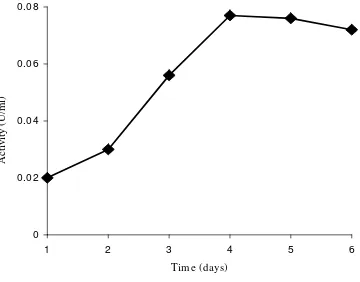
highest activity and used throughout the study. It produced CDA extracellularly at the
maximum level after 4th days cultivation (Fig 1). According to 16S rRNA sequence, the
[image:4.595.119.479.186.468.2]strain was identified asB. thermoleovorans (Suhartono 2002).
Fig 1. Time course of CDA production byB. thermoleovoransLW-4-11
Enzyme Preparation
The result of partial purification procedure are summarized in Table 1. The enzyme
from 4 days cell-free medium of LW-4-11 was precipitated by the addition of ammonium
sulphate up to 80% saturation. This step resulted in 1.50-fold increase in specific activity.
The heat treatment of enzyme increased the purity of enzyme to be 4,28 fold.
0 0.02 0.04 0.06 0.08
1 2 3 4 5 6
Time (days)
A
ct
iv
it
y
(U
/m
Table 1. Partial Purification Steps of CDA Produced byB. thermoleovorans LW-4-11
Steps Total
Activity (mU)
Protein (mg)
Specific Activity (mU/mg)
Purification (Fold)
Recovery (%)
Free-Cell Medium 21,4 2,10 10,2 1,00 100,0
Ammonium Sulfate ppt (70%)
18,5 1,21 15,3 1,50 86,4
Heat Treatment (70oC, 1 h)
7,9 0,18 43,7 4,28 36,7
Characterization of Enzyme
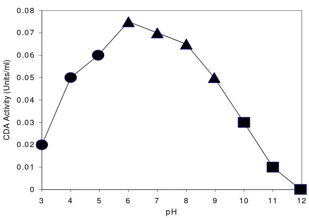
The effect of pH, temperature and addition of additives on CDA activity was studied
by using glycol chitin as a substrate under the standard assay conditions. The dialyzed
CDA exhibited maximum activity at pH 6,0 (Fig 2). The optimum temperature of enzyme
was found to be 80 oC (Fig 3). To examine the thermostability of the enzyme, CDA
solution in 50 mM sodium tetraborate buffer (pH 7.0) was allowed to stand during 5 h at
various temperatures, and the residual activity was measured. The enzyme was stable up to
80 oC with a loss of more than 80% (data not shown). However about 50% of the original
CDA activity was lost after incubation at 90 oC for 40 minutes and at 100 oC after 5
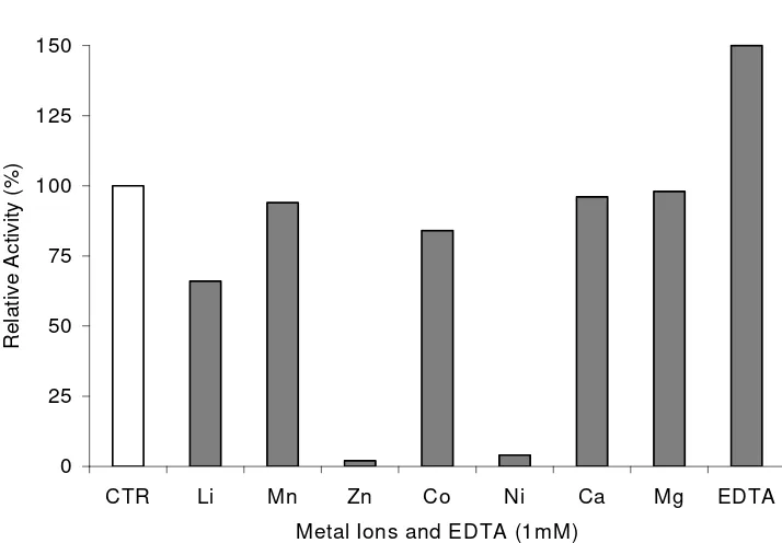
minutes, respectively (Fig. 3). The CDA was activated by EDTA. It was inhibited by Li+,
Mn+, Zn+, Co+and Ni+tested as chlorides at 1 mM concentration (Fig. 4).
DISCUSSION
Chitosan is usually produced from chitin by thermochemical deacetylation. This
process results in a polymer product having a broad distribution of molecular mass and a
heterogeneous extent of deacetylation. However, for many potentially important
applications, uniform material with specific physical and chemical properties is required
degradation of sugar chain and produces no alkaline wastes (Tokuyasu et al. 1996). Chitin
deacetylases (EC 3.5.1.41) which represent a class of hydrolytic enzymes were mainly
found in fungi. It was first demonstrated in extracts of the fungusMucor rouxii(Davis and
Garcia 1984). Chitin deacetylase activity also had been found in other Zygomycetes
(Trudel and Asselin 1990; Alfonsoet al. 1995), in Colletotrichum lindemuthianum(Tsigos
et al. 2000), in Uromyces viciae-fabae (Deising and Siegrist 1995), in Saccharomyces
cerevisiae (Mishra et al. 1997; Christodoulidou et al. 1999) and in cucumber leaves
[image:6.595.124.440.285.508.2]infected withColletotrichum lagenarium(Siegrist and Kauss 1990).
Figure 2. The optimal pH of chitin deacetylase fromB. thermoleovorans LW-4-11;, 0,2 M citrate buffer; ▲, 0,2 M phosphate buffer; and , 0,2 M glycine-NaOH buffer
CDA from other microorganisms mainly bacteria was rarely reported. We have
isolated and characterized CDA from acidophillic Bacillus sp. isolated from Kamojang
Crater West Java (Natsir 2000; Rahayu 2000) and B. stearothermophilus from Langoan,
North Sulawesi (Toharisman et al. 2001). In this study, we tried to isolate another
thermophile from hot spring water and sediment from North Sulawesi having different
characteristics.Bacillus thermoleovorans LW-4-11 was able to grow at 70oC and had the 0
0.01 0.02 0.03 0.04 0.05 0.06 0.07 0.08
3 4 5 6 7 8 9 10 11 12
pH
C
D
A
A
c
ti
v
it
y
(U
n
it
s
/m
high CDA activity. It produced CDA extracellularly having optimum temperature of 80
o
C. It might be one of the highest optimum temperatures for CDA as all optimum
temperature of CDAs reported so far was 50 oC (Gao et al. 1995; Deising and Siegrist
1995; Alfonso et al. 1995; Tokuyasu et al. 1996; Tsigos and Bouriotis 1995;
[image:7.595.140.470.224.467.2]Christodoulidouet al. 1999).
Figure 3. Enzyme stability at;, 70oC;, 80oC;▲, 90 oC; and, 100oC
The activity of the enzyme was retained by over 50% when it was kept for 45 minutes
at 70oC. The half-lifes of the activity (T ½) at 80 and 90oC were about 30 min and 20 min,
respectively. When the enzyme was incubated at 70oC for 1 h, heat-labile proteins were
denatured. This strategy was applied in partial purification processes described in this
paper and could purify enzyme of 4,28 fold with overall yield of 36,7%. Similar
procedures were conducted for CDA purification fromM. rouxii at 50 oC for 10 minutes
(Martinouet al, 1993) and fromBacillus sp. at 50oC for 24 h (Rahayu 2000).
Other characteristics of the enzyme were also performed. The optimum pH of the
0 10 20 30 40 50 60 70 80 90 100
0 15 30 45 60
Time (min)
R
e
la
ti
v
e
A
c
ti
v
it
y
(%
optimum pH of CDAs reported so far was between 4,5 and 8,5 (Tsigos et al. 2000). The
effect of cations were those similar to those of CDA of C. lindemuthianum (Tsigos and
Bouriotis 1995; Tokuyasuet al.1996) andAspergillus nidulans (Alfonsoet al. 1995). The
enzyme was activated by EDTA, whereas and inhibited by metal ions such as Zn2+, Mn2+
[image:8.595.155.512.233.481.2]and Cu2+(1 mM).
Figure 4. Effect of various cations and EDTA (1 mM) on CDA activity
REFERENCES
Alfonso, C., O.M. Nuero, F. Santamaria, & F. Reyes. 1995. Purification of a heat-stable
chitin deacetylase from aspergillus nidulans and its role in cell wall degradation.
Curr. Microbiol.30 (1): 49-54.
Bradford, M.M. 1976. A rapid and sensitive method for quantitation of microgram
quantities of protein utilizing the principle of protein-dye bindingh. Anal. Biochem.
72: 248-254.
0 25 50 75 100 125 150
CTR Li Mn Zn Co Ni Ca Mg EDTA
Metal Ions and EDTA (1mM)
R
e
la
ti
v
e
A
c
ti
v
it
y
(%
Chang, K.L.B., G. Tsai, J. Lee , & W.R. Fu. 1997. Heterogeneous N-deacetylation of
chitin in alkaline solution. Carbohydr. Res.303: 327-332.
Christodoulidou, A., P. Briza, A. Ellinger, & V. Bouriotis. 1999. Yeast ascospore wall
assembly requires two chitin deacetylase isozymes.FEBS Lett. 29 (2):275-279.
Davis, L.L., & S.B. Garcia. 1984. Chitosan synthesis by the tandem action of chitin
synthetase and chitin deacetylase fromMucor rouxii. Biochemistry. 23: 1065-1073.
Deising & J. Siegrist. 1995. Chitin Deacetylase Activity of the rust Uromyces
viciae-fabaeis controlled by fungal morphogenesis. FEMS Microbiol. Lett.127: 207-212.
Dische, Z., & E. Borenfreund. 1950. A Spectrophotometric methods for the
microdetermination of hexosamines.J. Biol. Chem. 184: 517-522.
Gao, X.D., T. Katsumoto, & K. Onodera. 1995. Purification and characterization of chitin
deacetylase fromAbsidia coerulea.J. Biochem. 117 (2):257-63.
Kafetzopoulos, D., A. Martinou, V. Bouriotis. 1993. Bioconversion of chitin to chitosan:
purification and characterization of chitin deacetylase from Mucor rouxii. Proc.
Natl. Acad. Sci. USA. 90: 2564-2568.
Kolodziejska, I., A. Wojtasz-Pajak, G. Ogonowska, & Z.E. Sikorski. 2000. Deacetylation
of chitin in a two-stage chemical and enzymatic process. Bulletin of the Sea Fisheries
Institute. 2 (150): 15-24.
Natsir, H.D. 2000. Biochemical Characteristics of chitinase enzyme fromBacillusSp. of
Kamojang Crater, Indonesia. Master Thesis, Bogor Agricultural University.
Martinou, A., D. Kafetzopoulos, & V. Bouriotis. 1993. Isolation of chitin deacetylase from
Mucor rouxiiby immunoaffinity chromatography.J. Chromatogr.644: 35-41.
Mishra, C., S.E. Semino, K.J. McCreath, H. de la Vega, B.J. Jones, C.A. Specht, & P.W.
Robbins. 1997. Cloning and expression of two chitin deacetylase genes of
Saccharomyces cerevisiae.Yeast. 13(4): 327-336.
Muzzarelli, R.A.A. 1996. Chitosan-based dietary foods. Carbohydr. Polym.29:
309-316.
Rahayu, S. 2000. Biochemical characteristics of thermostable chitinase and chitin
deacetylase enzymes from the Indonesian Bacillus K29-14. Master Thesis, Bogor
Agricultural University.
Sandford, P.A. 1989. Chitosan: Commercial Uses and Potential Applications. In.
Skjak-Braeket al(Editors). Chitin and Chitosan. Elsevier Applied Science, London.
Shahidi, F.J., K.V. Arachchi, & J. You-Jin. 1999. Food applications of chitin and
chitosans. Trends Food Sci. & Technol.10: 37-51.
Siegrist, J., & H. Kauss. 1990. Chitin deacetylase in cucumber leaves infected by
Collelotrichum lagenarium. Physiol. Mol. Plant Pathol.36:267-275.
Somashekar, D., & R. Joseph. 1996. Chitosanases-properties and applications: a review.
Biores. Technol. 55: 35-45.
Suhartono, M.T. 2002. Personal Communication.
Srinivasan, V.R. 2000. Biotransformation of chitin to chitosan. US Patent 5,739,015.
Takayanagi, T., K. Ajisaka, Y. Takiguchi, & K. Shimahara. 1991. Isolation and
characterization of thermostable chitinases fromBacillus licheniformisX-7u. Biochim
Biophys Acta. 1078: 404-410.
Toharisman, A., E. Chasanah, E.Y. Purwani, J.F.L Jayanti, V. Welan, M.T. Suhartono,
J.K. Hwang, & Y.R Pyun. 2001. Screening of thermophilic microorganisms
producing thermostable chitin deacetylase. Indonesian Biotechnology Conference; An
International Seminar and Symposium. Yogyakarta, 2000.
Tokuyasu, K., M. Ohnishi-Kameyama, & K. Hayashi. 1996. Purification and
characterization of extracellular chitin deacetylase from Colletotrichum
lindemuthianum.Biosci. Biotechnol. Biochem. 60 (10): 1598-1603.
Trudel, J., & A. Asselin. 1990. Detection of chitin deacetylase activity after polyacrylamide
gel electrophoresis.Anal. Biochem.189(2):249-253.
Tsigos, I., & V. Bouriotis. 1995. Purification and characterization of chitin deacetylase
fromColletotrichm lindemuthianum.J. Biol. Chem.270: 26280-26291.
Tsigos, I., A. Martinou, D. Kafetzopoulos, & V. Bouriotis. 2000. Chitin deacetylases: new,