Investigation on Association and Expression of
ESR2
as a Candidate Gene for Boar
Sperm Quality and Fertility
A Gunawan1, MU Cinar1, MJ Uddin1,2, K Kaewmala1, D Tesfaye1, C Phatsara1,3, E Tholen1, C Looft1and K Schellander1 1Institute of Animal Science, Animal Breeding and Husbandry Group, University of Bonn, Bonn, Germany;2Department of Medicine, Faculty of Veterinary Science, Bangladesh Agricultural University, Mymensing, Bangladesh;3Department of Animal and Aquatic Sciences, Faculty of Agriculture, Chiang Mai University, Chiang Mai, Thailand
Contents
ESR2 is involved in oestrogen-related apoptosis in cell cycle spermatogenesis but their effects have not yet confirmed in pig. Therefore, this study was aimed to investigate the association ofESR2 polymorphism with sperm quality and boar fertility traits and to analyse theESR2mRNA and protein expressions in boar reproductive tissues. DNA samples from 203 Pietrain (PI) and 100 Pietrain·Hampshire (PIHA) pigs with records of
sperm quality [sperm concentration (SCON), motility (MOT), semen volume (VOL), plasma droplet rate (PDR) and abnor-mal spermatozoa rate (ASR)] and fertility [non-return rate (NRR) and number of piglet born alive (NBA)] traits were available. A SNP in coding region ofESR2g.35547A>G in exon 5 was associated with MOT and PDR in the PI and with SCON, VOL, MOT and PDR in PIHA population. For mRNA and protein expression study, a total of six boars were divided into two groups with group I (G-I) and group II (G-II) where G-I characterized for relatively a better sperm quality according to the mean of two groups. mRNA expression was higher in brain and testis than that in all parts of epididymis. Both qRT-PCR and western blot analysis revealed that the
ESR2gene expression and protein expression were significantly higher in testis collected from G-II compared with that of G-I boars. Moreover, ESR2 protein localization in germ cell, Leydig and Sertoli cells, epithelial cells and spermatozoa was remarkable, which indicated the important role of ESR2 in spermatogenesis process. These results might shed new light on the roles ofESR2 in spermatogenesis as candidate for boar fertility, but still the lack of association across populations should be considered.
Introduction
Oestrogens are classically known to play a major role in female reproduction, but there is now compelling evidence that they may also be involved in the regulation of male reproductive functions. Oestrogens are inti-mately involved in male fertility, and their function is mediated through binding with the oestrogen receptors 1 and 2 (ESR1 andESR2) (Couse and Korach 1999). It has been speculated thatESR2 could act as a negative regulatory partner for ESR1(Weihua et al. 2000). The
lack of ESR1 leads to reduced epididymal sperm
content, reduced sperm motility and fertilizing ability (Couse and Korach 1999), while the overexpression of ESR2results in germ cell cycle arrest or apoptosis and infertility (Selva et al. 2004). Aschim et al. (2005) reported a significant association of ESR2 polymor-phism with infertility in human with less sperm concen-tration, testicular cancer and cryptorchidism. TheESR2 mRNA was highly expressed in the epididymis of adult mouse, rat, dog, cat and monkey (Hess et al. 1997;
Saunders et al. 1997; Nie et al. 2002; Zhou et al. 2002). Rago et al. (2007) and Saunder et al. (2001) localize ESR2 protein in spermatozoa within the testis in human and primates, respectively. However, theESR2gene has scarcely been investigated as a candidate gene for sperm quality and fertility traits in pigs. In pig,ESR2is located at the telometric end on the q-arm of SSC1 (Munoz et al. 2004). In this region, QTL for total sperm per ejaculate and sperm motility in boars (Xing et al. 2008) and QTL for number of nipples and age at puberty in sows are reported (Cassady et al. 2001).
Before reaching sexual maturity, a number of germ cells undergo physiological apoptotic death, which has been shown to be controlled by a large number of genes, including theESR2(Delbes et al. 2004). When germ cell development is complete, the mature spermatids are released from the Sertoli cells into the tubule lumen and proceed through the excurrent duct system, known as the rete testis, until they enter the epididymis via the efferent ducts. In spermatogenesis, functional male gametes are produced through complex processes in the testis, epididymis and other male reproductive tract (Frungieri et al. 2006). Failure in any of these events leads to disturbances in male fertility.ESR2is expressed in the cellular type during normal spermatogenesis but its function is still unknown. For the better understand-ing of ESR2 functions in spermatogenesis in pig, the expression and localization ofESR2at different parts of reproductive tract including non-reproductive tissues are important. Considering together, it could be spec-ulated that ESR2 might be a functional as well as a positional candidate gene for male reproduction traits in pigs. But to the author’s knowledge, no study was devoted to unravel its association with sperm quality and boar fertility traits, and the functions of ESR2 in boar spermatogenesis within the reproductive tracts by mRNA and protein expression are poorly understood. Therefore, the aims of this research were to study the
association of ESR2 with boar sperm quality and
fertility traits and to investigate the ESR2 mRNA and protein expressions in sperm and reproductive tissues from boars with divergent phenotype.
Material and Methods
Animals and traits used in association study
Semen samples from Pietrain (PI, n = 203) and Pie-train ·Hampshire crossbred (PIHA, n = 100) boars
were used for association analysis in this study. These animals were used for AI in commercial pig herds in
north-western Germany. Details of the populations and phenotypes were described previously by Wimmers et al. (2005), Lin et al. (2006) and Kaewmala et al. (2011). In brief, sperm samples of more than 31 000 ejaculates were repeatedly collected from these boars. Whole ejaculates were obtained from purebred Pietrain and crossbred Pietrain·Hampshire boars aged between 2 and 5 years
with an average age of 3.5 years. Sperm quality traits included sperm concentration [SCON (·108ml)], semen
volume per ejaculate [VOL (ml)], sperm motility [MOT (%)], plasma droplets rate [PDR (%)] and abnormal spermatozoa rate [ASR (%)] and were obtained from each ejaculate employing light microscopic evaluation according to the guidelines of the World Health Organi-zation. Semen was collected by the vinyl glove hand method twice⁄week. For each boar, the repeated
mea-surements of sperm quality traits were available. Fertility data [non-return rate data (NRR) at 42 days after insemination (%) and number of piglet born alive (NBA) per litter] of each boar were available as the deviation from the population means within sow breed, parity of sow, farm and season classes as described earlier by Lin et al. (2006) and Kaewmala et al. (2011).
Genotyping of ESR2 SNP
As a single nucleotide polymorphism, the arginine (A)-to-guanine (G) transversion of ESR2at g. 35547A>G in exon 5 reported by Munoz et al. (2007) was further investigated in this study. For PCR amplification, the forward (5¢-cttccttggatttagcc-3¢) and reverse (5¢
-at-gctctccttcttcggtga-3¢) primer pairs were designed covering
exon 5 of porcine ESR2 genomic sequence (GenBank
accession No. ENSSSCG00000005632) using the Primer3 tool (Rozen and Skaletsky 2000). Polymerase chain reactions (PCR) were performed in a 20-ll volume containing 100 ng of porcine genomic DNA, 1· PCR
buffer (with 1.5 134 mMMgCl2), 0.25 mMof each dNTP, 5 pmol of each primer and 0.1 U of Taq DNA polymerase (GeneCraft). The PCR were performed under the follow-ing conditions: initial denaturation at 95C for 5 min
followed by 35 cycles of 30 s at 95C, 30 s at 57C, 30 s at
72C and final elongation of 10 min at 72C. After
checking the PCR products in 1.5% (w⁄v) agarose gels,
genotyping was carried out following the restriction fragment length polymorphism (RFLP) analysis. The digestion of restriction enzyme FatI (New England BioLabs, Ipswich, MA, USA) was carried out in 10ll of reaction mixture of each sample and incubated overnight at 65C. Restriction fragment length
polymor-phisms of 304 boars were detected by electrophoresis in 3% (w⁄v) agarose gels.
Statistical analysis for sperm quality traits
The association of the ESR2 genotypes with sperm
quality and quantity traits was carried out using mixed
model (PROC MIXED) in the SAS software package
(SAS Institute Inc., ver. 9.2, Cary, NC, USA) as described by Kaewmala et al. (2011).
yijkl=l+seasoni+genotypej+agek+ ejaculationl+
ijkl[Model 1]
where yijkl is the sperm quality traits (SCON, VOL,
MOT, PDR and ASR); l is the overall population
mean; seasoniis the fixed effect of thei-th season (i= 1 through 8; four seasons⁄year, in total eight seasons
within 2 years from January 2000 to December 2001); genotypejis the fixed effect of thej-th genotype (j= 1, 2 and 3); agek is the effect of boar age (covariable); ejaculationl is the permanent environmental effect of the l-th boar (random); and ijkl is the residual error. As ejaculation was recorded as repeated measurement, it was considered as random effect in the statistical model.
The association analysis between ESR2 and the
fertility traits was carried out using the following generalized linear model (PROC GLM) inSAS
(Kaew-mala et al. 2011).
yij=l+ genotypei+ yearj+ij[Model 2]
whereyijis the boar fertility trait (NRR and NBA);lis the overall population mean; genotypeiis the fixed effect of thei-th genotype (i= 1, 2 and 3); yearj is the fixed effect of the j-th boar year of birth (j= 1 through 3: boar born before 1996, in 1996–97 and in 1998–99); and ijis the residual error.
The distribution of the genotype was tested for Hardy–Weinberg equilibrium by chi-square (v2) test. Least-square mean values for theESR2genotypes were compared byt-test and p-values, adjusted by the Tukey– Kramer correction.
Selection of animals for mRNA and protein expression The reproductive (testis, head, body and tail of epidid-ymis, vas deferens, bulbourethral gland, vesicular glands and prostate gland) and non-reproductive tissues (brain, muscle and liver) from six breeding boars with divergent phenotypes were collected from the AI station (SuisAG, Sempach, Switzerland) for mRNA and protein study as described earlier by Kaewmala et al. (2011). For differ-ential expression study between reproductive and non-reproductive tissues by reverse transcription PCR (RT-PCR) study, mRNA from all six boars were pooled together according to the tissues. On the other hand, the differential mRNA and protein expression study in different reproductive tissues from two divergent groups of animals was performed by RT-PCR, qRT-PCR and western blot, respectively. For these purposes, the six boars were divided into two groups based on extreme
phenotypes (high⁄low SCON, SMOT and SVOL).
Correlation between sperm quality traits was carried out using correlation analysis (PROC CORR) in SAS.
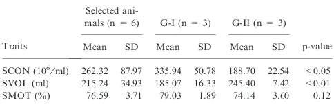
The SCON (average sperm concentration) was highly negatively (r=)0.8) correlated with SVOL (average semen volume), whereas SCON was highly positively (r= 0.7) correlated with SMOT (average sperm motil-ity). Moreover, SVOL was highly negatively (r=)0.8) correlated with SMOT. Therefore, grouping was made on the basis of SCON, SVOL and SMOT (Table 1). The six boars were selected and equally divided into group I (G-I) with high SCON (>262.32·106ml), high SMOT
(>76.59%) and low SVOL (<215.24 ml⁄ejaculation)
differ-ence between the two groups was calculated using PROC T-Test inSAS. There were differences for SCON
(p < 0.05) and for SVOL (p < 0.01) between G-I and G-II, whereas for the SMOT, the difference was not significant (p = 0.12).
Reverse transcription PCR
Total RNA was isolated using TRI Reagent
(Guani-dinium thiocyanate-phenol-chloroform extraction)
(Sigma-Aldrich, Munich, Germany) from different reproductive and non-reproductive tissues of breeding boars mentioned earlier. RNA was purified using RNeasy Mini Kit (Qiagen, Hilden, Germany) according to the manufacturer’s instructions. Total RNA was
treated using on-column RNase-Free DNase set
(Promega, Mannheim, Germany) and quantified spec-trophotometrically (ND8000; Nano Drop, Thermo Scientific, Wilmington, DE, USA). Furthermore, RNA integrity was checked by 2% agarose gel electrophoresis. First-strand cDNA were synthesized from individual RNA using Superscript II enzyme (Invitrogen, Darm-stadt, Germany).
cDNA amplification was performed by using specific forward and reverse primers (forward: 5¢
-ggccccatata-tatccctcc-3¢ and reverse: 5¢-gagttggccacaacacattg-3¢)
derived from porcine ESR2 sequence (GenBank acces-sion AF164957). Amplification was performed with an initial heating at 95C for 5 min followed by 35 cycles of
95C for 45 s, annealing temperature at 58C for 1 min
and 72C for 1 min, on the PCR Thermal Cycler
(BioRad, Munich, Germany). PCR products were electrophoresed on a 1.5% agarose gel and visualized upon staining with ethidium bromide. Glyceraldehyde 3-phosphate dehydrogenase (GAPDH) served as house-keeping gene.
Quantitative real-time PCR
For real-time PCR, total RNA was isolated using TRI reagent (Sigma; Sigma-Aldrich, Munich, Germany) from different reproductive tissues of two divergent groups of animals (G-I and G-II) as described in aforesaid section. cDNA synthesis was carried out as described in the previous section. The same primer pairs used in RT-PCR were also used in qRT-PCR. Ninefold serial dilutions of plasmid DNA were prepared and used as template for the generation of the standard curve. In each run, the 96-well microtitre plate contained each cDNA sample, plasmid standards for the standard curves and no-template control. To ensure the repeatability of the experiments, each plate was run in three replications. qRT-PCR was
set up using 2ll first-strand cDNA template, 7.6 ll deionized H2O, 0.2lM of upstream and downstream
primers and 10ll 1· Power SYBR Green I master mix with ROX as reference dye (BioRad). The thermal cycling conditions were 3 min at 94C followed by 40
cycles of 20 s at 94C and 1 min at 60C. Experiments
were performed using the ABI prism 7000 (Applied Biosystems, Carlsbad, CA, USA) qRT-PCR system. The
housekeeping gene GAPDH (forward: 5¢
-acccagaa-gactgtggatgg-3¢ and reverse: 5¢-acgcctgcttcaccaccttc-3¢)
derived from porcine sequence (GenBank accession No. AF017079) was used for the data normalization. Glyc-eraldehyde 3-phosphate dehydrogenase was quantified twice as technical replication and the average was used for the normalization of target gene ESR2. Final results were reported as the relative abundance level after normalizing with mRNA expression level of the
house-keeping gene. Differences in ESR2 mRNA expression
were analysed with the simplet-test inSASsoftware (SAS
Institute Inc., ver. 9.2). Values of p < 0.05 were consid-ered to indicate statistically significant differences.
Western blotting
The protein was extracted from different reproductive tissues (testis, head, body and tail of epididymis) from the two divergent groups of breeding boars as used in qRT-PCR. However, for western blot study, proteins from three G-I boars were pooled together and proteins from three G-II boars were pooled together according to the tissues. The proteins extracted from tissues were separated by SDS-PAGE (gradient 4–18%). Sub-sequently, the proteins were transferred onto a nitro-cellulose membrane (Amersham Biosciences, Freiburg, Germany). After blocking in blocking buffer (20 mM
Tris pH 7.5, 150 mM NaCl, 0.05% Tween-20 and 1%
polyvinylpyrrolidone) at room temperature for 1 h, the membrane was incubated with the anti-ESR2 antibody purified from goat polyclonal antibody (Cat.nr. Sc6822; Santa Cruz, Heidelberg, Germany) in the blocking medium (diluted 1 : 500) overnight at 4C. Non-specific
binding of antibody was washed off with six changes of 0.1% PBST (10 min to time). The horseradish peroxi-dase–conjugated goat anti-goat IgG (Cat.nr. Sc2020; Santa Cruz) was used as the secondary antibody (diluted 1 : 50 000). The membrane was incubated for 1 h at room temperature with secondary antibody, followed by washing with six changes of 0.1% PBST (10 min to time). The chemiluminescence was detected by using the ECL plus western blotting detection system (Amersham Biosciences) and was visualized by using Kodak BioMax XAR film (Kodak, Stuttgart, Germany). Glyceralde-hyde 3-phosphate dehydrogenase was used as a loading control and for normalization. The membrane was stripped by incubation in 2% SDS, 100 mM Tris–HCl
and 0.1% b-mercaptoethanol for 30 min at 60C and
reprobed with GAPDH antibody (Cat.nr. Sc20357; Santa Cruz).
Protein localization by immunofluorescence
Owing to the limitations of fresh samples from G-I and G-II boars, we collected different fresh reproductive
Table 1. Means, standard deviation (SD), number of boars and ranges of traits selected for mRNA and protein expression study
Traits
Selected
ani-mals (n = 6) G-I (n = 3) G-II (n = 3)
p-value
Mean SD Mean SD Mean SD
SCON (106⁄
ml) 262.32 87.97 335.94 50.78 188.70 22.54 <0.05 SVOL (ml) 215.24 34.93 185.07 16.33 245.40 7.42 <0.01
tissues from a healthy breeding boar after slaughtering for protein localization by immunofluorescence (Kaew-mala et al. 2011). Immunofluorescence staining was performed on 8-lm cryostat sections of snap-frozen tissues. All sections were kept at )80C for further analysis. To block unspecific staining, sections were incubated for 30 min at room temperature with 5%
bovine serum albumin in PBS (50 nM sodium
phos-phate, pH 7.4; 0.9% NaCl). Sections were incubated overnight at 4C with the ESR2 goat polyclonal primary
antibody (Cat.nr. Sc6822; Santa Cruz) diluted at 1 : 50 in PBST followed by six times (10 min to time) washing with PBS. Then, the sections were incubated for 1 h at room temperature with the biotinylated donkey anti-goat IgG-B conjugated with fluorescein isothiocynate (FITC)-reactive water-soluble fluorescent dye (Cat nr. Sc2090; Santa Cruz) (dilution 1 : 200) as a secondary antibody for ESR2 followed by six times (10 min to time) washing with PBS. Finally, the samples were counterstained with vectashield mounting medium (Vec-tor Labora(Vec-tories, Loerrach, Germany) containing 4,6-diamidino-2-phenyl indole (DAPI) and covered with a cover glass slip. The staining was observed by confocal laser scanning microscope (Carl Zeiss, Oberkochen, Germany). In case of negative controls, PBS was used instead of the primary antibody.
Results
Association study
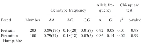
The polymorphism ofESR2at g. 35547A>G in exon 5 was used to genotype the PI and PIHA population by PCR–RFLP. The DNA restriction fragments obtained for the ESR2-FatI polymorphism were 284, 146 and 28 bp for the AA genotype, 284, 174, 146 and 28 bp for the AG genotype and 284 and 174 bp for the GG genotype. The genotype and allele frequencies of porcine ESR2 gene calculated are shown in Table 2. Homozy-gote AA was more frequent, and homozyHomozy-gote GG was rare in both populations. The chi-square test revealed that the locus of ESR2 was in Hardy–Weinberg equi-librium in both populations (Table 2).
The general descriptions of sperm quality and fertility traits are shown in Table 3. Means of VOL, MOT and PDR traits in the crossbred PIHA were higher com-pared with those in the PI populations. The NRR42 was higher in PIHA compared with PI, while the NBA was similar in both PI and PIHA populations.
Association analysis of g.35547A>G with sperm quality and fertility traits revealed significant (p < 0.01) association with MOT and PDR and suggestively
(p = 0.06) with NBA in PI population. SCON, VOL, MOT and PDR were found to be significantly (p < 0.05) associated with the g.35547A>G genotype in the PIHA population (Table 4). This association with MOT and PDR was consistent in PI and PIHA populations. The genotype AA is different from
AG⁄GG genotype. The genotypes AG and GG were
associated with lower MOT and higher PDR than the animals having a AA genotype in the PI population (Table 4). In the PIHA population, the animals AG and GG genotypes were associated with lower SCON and MOT, but higher VOL and PDR than animals having AA genotype. The polymorphism g.35547A>G of
ESR2 showed highly additive effect on MOT
(p < 0.01) in PI population. The results also indicated higher additive effect on SCON and VOL (p < 0.05) in PIHA population (Table 4).
mRNA expression by reverse transcription PCR
ESR2 gene expression was higher in brain and testis, and lower expression was found in the head, body and tail of epididymis. The mRNA expression ofESR2was not detectable in accessorial gland (vas deferens, bulbo-urethral, vesicular and prostate glands), muscle and
liver. The RT-PCR result of GAPDH showed no
remarkable differences among tissues (Fig. 1).
mRNA and protein expression study in tissues from G-I and G-II boars
TheESR2mRNA was expressed in testis, body and tail of epididymis from both the G-I and G-II boars but higher expression was found in testis of G-II than in that of G-I boars by RT-PCR (Fig. 2a). These mRNA expression results of RT-PCR appeared to be consistent
with the results of the qRT-PCR. The ESR2 mRNA
expression was higher in testis of G-II compared with that of G-I boars (p < 0.01), whereas the difference in expression level was not statistically significant in case of head, body and tail of epididymis between G-I and G-II boars (Fig. 2b). ESR2 protein with 56 kDa molecular weight was detected in testis, head, body and tail of
Table 2. Genotype, allele frequencies and the chi-square test of the porcineESR2gene in different pig populations
Breed Number
Pietrain 203 0.89(176) 0.10(20) 0.01(7) 0.92 0.08 0.01 0.98 Pietrain·
Hampshire
100 0.79(77) 0.18(18) 0.03(5) 0.86 0.14 0.02 0.99
Table 3. Means, standard deviation (SD), sample size, ranges of traits in Pietrain (PI) and Pietrain·Hampshire (PIHA)
Population Traits Sample size Mean SD Minimum Maximum
PI SCON (108⁄ml) 20 077 3.03 0.94 1 6
VOL (ml) 21 248 237.03 57.32 25 522
MOT (%) 20 782 84.72 4.37 65 95
PDR (%) 20 805 5.41 3.33 0 15
ASR (%) 21 056 6.53 4.18 0 20
NRR42 (%)a
203 0.50 7.00 )24.07 18.62
NBA (per litter)a
203 0.04 0.54 )1.69 1.27
PIHA SCON (108⁄
ml) 7327 2.95 0.97 1 6
VOL (ml) 7826 297.50 81.62 56 546
MOT (%) 7610 85.46 4.03 70 95
ASR, abnormal spermatozoa rate; MOT, motility; PDR, plasma droplet rate, SCON, sperm concentration; VOL, semen volume.a
epididymis in both G-I and G-II boars (Fig. 2c). The western blot result showed that the ESR2 protein was higher in testis in G-II compared with G-I boars. This protein expression seemed to be consistent with the results of transcription levels.
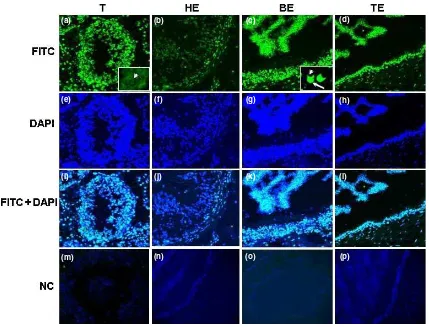
Localization of ESR2 protein in boar reproductive tissues by immunofluorescence
Sections of testis, head, body and tail of epididymis were stained through the same optical panel for the cell-surface ESR2 protein expression (Fig. 3). The Leydig and Sertoli cells in testis and epithelial cells in all parts of epididymis showed signals for ESR2 immunoreactivity
(Fig. 3a–d). Immunoreactive ESR2 protein was
observed as strong staining in germ cell cytoplasm of Sertoli cells and Leydig cells in testis (Fig. 3a). ESR2 protein was expressed in epithelial cells of head (Fig. 3 b), body (Fig. 3c) and tail of epididymis (Fig. 3d). In case of spermatogenesis, ESR2 protein was expressed in spermatogonia, primary spermatocyte and spermatid (arrow head) within seminiferous tubules (Fig. 3a). The ESR2 protein was expressed in the spermatozoa located within the lumen of the body of epididymis. The ESR2 protein was expressed in the acrosomal cap (arrow) of boar spermatozoa (Fig. 3c).
Discussion
Association of SNP with sperm quality and boar fertility traits
This study revealed an association ofESR2with sperm quality and fertility traits in boars. The exonic SNP g.35547A>G was found to be significantly associated with sperm motility and plasma droplet rate and suggestively (p = 0.06) associated with number of piglet born alive in PI populations, whereas it was significantly associated with sperm concentration, semen volume, sperm motility and plasma droplet rate in PIHA populations. Sperm motility and plasma droplet rate are consistently associated in both the PI and PIHA populations. It could be seen in both populations that AA genotype contributed significantly to have higher sperm motility and less plasma droplet rate (Table 4). It is important to note that sperm motility and plasma droplet are significantly negatively correlated in our populations. In case of PIHA, genotype AA significantly contributed to higher sperm concentration and lower semen volume (Table 4), which are also in agreement with our correlation results that SVOL and SCON are significantly negatively correlated. In case of ESR2, association has been described in sows by Munoz et al. (2004, 2007) but they did not find any statistically
Fig. 1. mRNA expression ofESR2
in reproductive and non-reproduc-tive tissues by reverse transcription PCR
Table 4. Association ofESR2genotypes with sperm quality and fertility traits
Population Trait
ESR2genotype (l± SE) Effect (l± SE)
AA AG GG Additive Dominance
Pietrain (PI) No. of observationsg
17 608 3189 589
SCON (108⁄
ml) 2.98 ± 0.04 3.03 ± 0.14 2.89 ± 0.28 0.04 ± 0.14 )0.09 ± 0.20
VOL (ml) 244.37 ± 3.28 247.79 ± 10.15 231.89 ± 19.82 6.23 ± 10.04 )9.66 ± 14.28
MOT (%) 85.38 ± 0.24c
83.76 ± 0.75d
81.55 ± 1.46d
1.91 ± 0.74** )0.29 ± 1.05
PDR (%) 5.04 ± 0.19c
6.99 ± 0.59d
7.13 ± 1.16d
)1.04 ± 0.59 )0.90 ± 0.84
ASR (%) 6.17 ± 0.20 7.09 ± 0.63 8.05 ± 1.23 )0.93 ± 0.62 0.01 ± 0.88
No. of boars 176 20 7
NRR42 (%) 0.58 ± 0.56 )0.59 ± 1.74 )0.87 ± 4.10 4.22 ± 2.60 )3.05 ± 2.60
NBA (per litter) 0.04 ± 0.04e
0.32 ± 0.13f
0.38 ± 0.31f
)0.17 ± 0.16 )0.10 ± 0.20
Pietrain·Hampshire (PIHA) No. of observationsg
6270 1266 323
SCON (108⁄
ml) 3.16 ± 0.07a
2.98 ± 0.15a
2.33 ± 0.33b
0.41 ± 0.17* )0.24 ± 0.22
VOL (ml) 265.34 ± 6.84a
271.85 ± 13.94a
360.03 ± 31.19b
)47.34 ± 15.97* 40.83 ± 21.17
MOT (%) 85.13 ± 0.47a
82.33 ± 0.97b
83.32 ± 2.18b
0.90 ± 0.11 1.89 ± 1.48
PDR (%) 5.43 ± 0.33a
7.58 ± 0.68b
7.54 ± 1.52b
)1.05 ± 0.78 )1.09 ± 1.03
ASR (%) 6.06 ± 0.40 7.27 ± 0.82 5.84 ± 1.83 0.11 ± 0.94 )1.31 ± 1.24
No. of boars 77 18 5
NRR42 (%) 0.46 ± 0.53 1.86 ± 1.11 3.31 ± 2.49 )1.42 ± 1.24 0.02 ± 1.57
NBA (per litter) )0.01 ± 0.06 )0.09 ± 0.14 0.01 ± 0.30 )0.01 ± 0.15 0.12 ± 0.19
ASR, abnormal spermatozoa rate; MOT, motility; PDR, plasma droplet rate, SCON, sperm concentration; VOL, semen volume.g
Repeated measurements;
a,b
p < 0.05;c,d
p < 0.01;e,f
significant association. However, in human, there are two reported polymorphisms ofESR2 at 1082 (G>A) and 1730 (G>A) in exons 5 and 8, respectively, but only the SNP at 1082 (G>A) showed an association with male infertility (Aschim et al. 2005). This finding is almost similar to our study describing that the hetero-zygote AG genotype has negative effect on sperm quality. Aschim et al. (2005) reported a significantly increased frequency of the ESR2 AG genotype among infertile man, compared with fertile control.
Polymorphism in ESR2 at g.35547A>G in exon 5 had effect on sperm quality traits in this study. This SNP was observed in the coding region of the ESR2 gene leading to an amino acid substitution (MetfiVal) in
the hormone-binding domain, which may be critical for its role as transcription factor (Munoz et al. 2007). Moreover, this alteration involves the replacement of the non-polar amino acid, valine, by the polar sulphur-containing amino acid, methionine, which could modify the secondary and tertiary structures of the protein because of the different ability of these amino acids to form hydrogen and disulphide bonds. The potential deleterious effect of the mutation p.Val317Met located in the ligand-binding domain is suggested by its conserved amino acid position among some mammalian species (Rattus norvergicus, GenBank accession number:
Q62986, Mus musculus: O08537 and Bos taurus:
Q9XSB5). In human, it has been reported that the ESR2polymorphism could have a direct effect through changing the nucleotide sequence and thereby the
secondary structure of the ESR2 mRNA, possibly
leading to changes in mRNA synthesis, splicing, matu-ration, transport, translation or degradation (Iida and Akashi 2000). These data support the possible biological relevance of this amino acid change, and we found association with sperm quality traits. However, we could not detect any significant association of these SNPs with fertility traits. We found a suggestive association of g.35547A>GESR2with number piglet born alive in PI population (p = 0.06) but not in PIHA population. Munoz et al. (2004) reported that SNP inESR2are not associated with litter size in Iberian and Chinese– European sows. In addition, sampling additional ani-mals for DNA sequencing may provide detection of other sequence variants in this gene that could be associated with fertility traits. An association study with more individuals should be conducted with this Met⁄Val
substitution.
mRNA and protein expression in boar reproductive tissue TheESR2was highly expressed in brain and testis when compared with other tissues, indicating that this gene might have important functions in these tissues.
Repro-Fig. 2. mRNA and protein expression ofESR2in reproductive tissues (testis, head, body and tail of epididymis). (a)ESR2mRNA expression in
different reproductive tissues from G-I and G-II boars by RT-PCR. (b)ESR2mRNA expression in different reproductive tissues from G-I and
ductive processes were maintained by hormone level initiated in the brain (Balthazart and Ball 1997a; Thompson et al. 1998). Enzyme CYP19 catalyses the oestrogen production in brain, andESR2is reported to
express at the same area at brain where CYP19
(aromatase subfamily 19) is reported to express (Bal-thazart 1998). It has been reported that testosterone must be transformed into an oestrogen by aromatization to activate the male sexual behaviour (Balthazart and Ball 1997b). Two responses that were known to be oestrogen dependent are the control of aromatase synthesis and the activation of reproductive behaviour (Balthazart 1998).
The expression of ESR2 gene in the reproductive tissues of G-II was higher than that in G-I boars. The protein expression coincided with mRNA expression for
ESR2. There were significantly higher ESR2 mRNA
and protein expressions in testis from G-II boars than in the testis from G-I boars. TheESR2tended to be higher in G-II, indicating that higher mRNA and protein expressions might have negative effect on boar sperm quality and fertility.ESR2 is reported to be expressed higher in the semen of infertile males compared with fertile (Bujan et al. 1993). Moreover, the overexpression of ESR2 is reported to cause apoptosis in cell cycle spermatogenesis (Selva et al. 2004). Another study
reported that ESR2 is involved in oestrogen-related apoptosis of germ cells, and as a consequence, there is a blockade of germ cell growth during foetal and neonatal life (Delbes et al. 2004). ESR2 plays a role either in regulating the progression of the first meiotic division or in favouring the entrance of primary spermatocytes to an apoptotic pathway (Selva et al. 2004). One of the mechanisms by whichESR2might regulate cell cycle is through its direct and specific interaction with the cell cycle spindle assembly checkpoint protein,Mad2(Poelzl et al. 2000). In the spermatogenesis process, the over-expressed ESR2 interacted with Mad2 in meiosis I in mouse leading to a cell cycle arrest or apoptosis in metaphase I (Wassmann et al. 2003). However, ESR2 gene has been expressed in diseased as well in normal testis (Makinen et al. 2001). It has been reported in
ESR2 knockout mice (ESR2KO) that ESR2 is
impor-tant for maintaining the testicular function (Oliveira et al. 2004). Adult ESR2 knockout mice study showed that inactivation of ESR2 affected the cellular compo-sition of the testis (Gould et al. 2007), which may indeed have a direct role in the spermatogenesis. Yet, it is important to note thatESR2does not play a role alone in spermatogenesis. ESR2 regulation is most likely of extratesticular origin, and a plausible candidate for the regulation of ESR2 in the testis and epididymis would
(a)
(e)
(i)
(m) (n) (o) (p)
(j) (k) (l)
(f) (g) (h)
(b) (c) (d)
Fig. 3. Localization of ESR2 protein in different parts of the boar reproductive tissues. (a) Immunofluorescence detection of ESR2 in germ cell and in cytoplasm of Sertoli and Leydig cells. Germ cells were stained with ESR2 (arrows), and the nuclei were counterstained with diamidino-2-phenyl indole (DAPI). (b–d) ESR2 protein localization in epithelial cells in the head, body and tail of epididymis. (c) The ESR2 localized in acrosomal cap (arrow) of spermatozoa (arrow head) within the lumen body of epididymis. (e–h) The cell nuclei were counterstained with DAPI.
(i–l) Merged images. (m–p) Negative control. Magnification 40·. T, Testis; HE, Head of epididymis; BE, Body of epididymis; TE, Tail of
be luteinizing hormone (LH) because a correlation betweenESR2andLHR(luteinizing hormone receptor) concentration is reported in the male tract (Derecka et al. 1999; Guo et al. 2001). LH controls epididymal differentiation, function and sperm maturation through ESR2, and lack of LHR function results in serious disorders in the development of puberty and fertility (Zhang et al. 1997).
Localization of protein
The concentration of ESR2 appears to be high in germ cell and in cytoplasm of Sertoli and Leydig cells in testis. A number of studies reported that ESR2 is predominant in germ cells of rodents and humans (van Pelt et al. 1999; Jefferson et al. 2000; Makinen et al. 2001; Zhou et al. 2002). A similar expression pattern of ESR2 described in this study suggests that the locally produced oestrogens in these cells might act through ESR2. Furthermore, there are reports of ESR2 expression in other germ cell types such as spermatogonia (van Pelt et al. 1999; Jefferson et al. 2000; Makinen et al. 2001; Zhou et al. 2002) and elongated spermatids (Rosenfeldt et al. 1998). Our observations are in agreement with the results reported in adult rodents, primate goats, dog and cats (Goyal et al. 1997; Nie et al. 2002; Hess and Carnes 2003). Expression of ESR2 seemed rather homogeneous within and between cell types, and there was a little variation in staining intensity. These data revealed on the protein level were confirmed on the mRNA level, indicating that transcription and translation of ESR2 occur in the positive staining cells. Oestrogen activity of pig testis is unknown, but it has been reported that there is a functional linkage between ESR2 and embryonic growth of pig (Kowalski et al. 2002). In fact, the ESR2 immunostaining pattern in testicular somatic and germ cells as well as in immature and mature gonads suggested that oestrogens might modulate sper-matogenesis and testis development via a differential expression of two oestrogen receptor subtypes (Rago et al. 2007).
In this study, the ESR2 immunostaining was found in the epithelial cells from three epididymal regions. The findings of the present study confirmed that ESR2 is expressed in a specific manner in the epididymis and suggest that oestrogens might modulate the epididymal function. Head, body and tail of the epididymis are reported to be involved in morphological and biochem-ical sperm maturation, in the progression of sperm towards the vas deferens and in its storage (Carpino et al. 2004). We have found ESR2 in the acrosomal cap of pig spermatozoa within the lumen of the body of
epididymis. The acrosomal cap is a cellular site closely related to the exocytotic event preceding the oocyte fertilization, and in human, it has been reported that oestradiol is able to influence capacitation and acroso-mal reaction of spermatozoa (Aquila et al. 2005). The localization of ESR2 in the acrosomal region implies its involvement in the fertilization process (Solakidi et al. 2005). However, it is important to note that there are more other proteins identified in the post-acrosomal region of sperm, such as equatorin and oscilin, which are important for successful fertilization (Ramalho-Santos et al. 2002; Solakidi et al. 2005).
Conclusion
It might suggest that the higher expression of ESR2 might have negative effect on boar sperm quality validated through association study and by profiling of mRNA and protein expression in non-reproductive and reproductive tissues. Therefore, with regard to the association, the transcript and differential expression and protein localization depending on boar sperm quality traits provide experimental evidence for the role of ESR2 in male fertility. However, the results of this study have to be validated in another breed⁄crossbreed
population in order to evaluate its potential in genomic selection.
Acknowledgements
This research was financially supported by FBF (Fo¨rderverein Biotechnologieforschung e. V., Bonn, Germany). We thank Dr A. Niggemeyer, Dr M. Friedrichs, Dr S. Bru¨ning and Dr A. Riesenbeck (GFS Artificial Insemination Station: Die Genossenschaft zur Fo¨rde-rung der Schweinehaltung, Ascheberg, Germany) and Dr A. Hofer, Dr H. Luther and Dr K. Caspari (SuisAG Artificial Insemination Station, Sempach, Switzerland) for their support during the sample collection and the arrangements of the reproduction records. We are also thankful to Prof Dr C. Knorr (Institute of Veterinary Medicine, Georg-August-University, Goettingen, Germany) for his cooperation.
Conflict of interest
None of the authors have any conflict of interest to declare.
Author contributions
AG performed the experiments and wrote the manuscript; MUC partly supervised the work and revised the manuscript; MJU edited the manuscript; KK performed the experiment with AG; DT was responsible for kits and reagents; CP partly supervised the work; ET was responsible for the statistical analysis; CL revised the manuscript; KS was responsible for the whole experiment and supervised the overall work.
References
Aquila S, Gentile M, Middea E, Catalano S, Ando S, 2005: Autocrine regulation of insulin secretion in human ejaculated
spermatozoa. Endocrinology 146, 552–
557.
Aschim EL, Giwercman A, Stahl O, Eber-hard J, Cwikiel M, Nordenskjold A, Haugen TB, Grotmol T, Giwercman YL, 2005: The RsaI polymorphism in
the estrogen receptor-beta gene is associ-ated with male infertility. J Clin
Endocri-nol Metab90,5343–5348.
Balthazart J, 1998: The Japanese quail as a model system for the investigation of steroid-catecholamine interactions
medi-ating appetitive and consummatory
aspects of male sexual behavior. Ann
Rev Sex Res9,96–176.
Balthazart J, Ball GF, 1997a: New insights into the regulation and function of brain
estrogen synthase (aromatase). Trends Neurosci21,243–249.
Balthazart J, Ball GF, 1997b: Neuroendo-crine regulation of appetitive and
con-summatory aspects of male sexual
behavior in Japanese quail. In: Etches R, Harvey S (eds), Perspectives in Avian Endocrinology. Society for Endocrinol-ogy, Bristol, UK, pp. 241–255.
level in seminal plasma in infertile men.
Hum Reprod8,74–77.
Carpino A, Bilinska B, Siciliano L, Maggi-olini M, Rago V, 2004: Immunolocaliza-tion of estrogen receptor beta in the epididymis of mature and immature pigs.
Folia Histochem Cytobiol42,13–17.
Cassady JP, Johnson RK, Pomp D, Rohrer GA, Van Vleck LD, Spiegel EK, Gilson KM, 2001: Identification of quantitative trait loci affecting reproduction in pigs. J Anim Sci79,623–633.
Couse J, Korach K, 1999: Estrogen receptor null mice: what have we learned and where will they lead us?. Endocr Rev20,358–417. Delbes G, Levacher C, Pairault C, Racine C, Duquenne C, Krust A, Habert R, 2004: Estrogen receptor beta-mediated inhibi-tion of male germ cell line development in mice by endogenous estrogens during perinatal life. Endocrinology 145, 3395– 3403.
Derecka K, Zhang FP, Ziecik AJ, Huhtan-iemi I, 1999: Ontogeny of LH receptor gene expression in the pig reproductive tract. J Reprod Fertil115,365–372. Frungieri M, Gonzalez-Calvar S, Parborell
F, Albrecht M, Mayerhofer A, Calandra RS, 2006: Cyclooxygenase-2 and prosta-glandin F2 alpha in syrian hamster Leydig cells: inhibitory role on luteinizing
hor-mone⁄human chorionic gonadotropin
stimulated testosterone production.
Endocrinology147,4476–4485.
Gould ML, Hurst PR, Nicholson HD, 2007: The effects of oestrogen receptors alpha and beta on testicular cell number and steroidogenesis in mice. Reproduction
134,271–279.
Goyal HO, Bartol FF, Wiley AA, Neff CW, 1997: Immunolocalization of receptors for androgen and estrogen in male caprine reproductive tissues: unique distribution of estrogen receptors in efferent ductule epithelium. Biol Reprod56,90–101. Guo W, Okamoto M, Lee YM, Baluda MA,
Park NH, 2001: Enhanced activity of cloned hamster TERT gene promoter in transformed cells. Biochim Biophys Acta
1517,398–409.
Hess R, Carnes K, 2003: The role of oestrogen in testis and male reproductive tract: a review and species comparison.
Anim Reprod1,5–30.
Hess R, Gist D, Bunick D, 1997: Estrogen receptor (alpha and beta) expression in the excurrent ducts of the adult male rat reproductive tract. J Androl18,602–611. Iida K, Akashi H, 2000: A test of transla-tional selection at ‘silent’ sites in the human genome: base composition com-parisons in alternatively spliced genes.
Gene261,93–105.
Jefferson WN, Couse JF, Banks EP, Korach KS, Newbold RR, 2000: Expression of estrogen receptor beta is developmentally regulated in reproductive tissues of male
and female mice. Biol Reprod 62, 310–
317.
Kaewmala K, Uddin MJ, Cinar MU, Gro-ße-Brinkhaus C, Jonas E, Tesfaye D, Phatsara C, Tholen E, Looft C, Schel-lander K, 2011: Investigation on
associa-tion of PLCz and COX2 as a candidate gene for boar sperm quality and fertility.
Reprod Domest Anim, (in press).
doi:10.1111/J.1439-0531.2011.01831.x. Kowalski AA, Graddy LG, Vale-Cruz DS,
Choi I, 2002: Molecular cloning of por-cine estrogen receptor-b complementary DNAs and developmental expression in periimplantation embryos. Biol Reprod
66,760–769.
Lin C, Jennen D, Ponsuksili S, Schellander K, Wimmers K, 2006: Candidate gene markers for sperm quality and fertility of
boar. Anim Reprod Sci92,349–363.
Makinen S, Makela S, Weihua Z, Warner M, Rosenlund B, Salmi S, Hovatta O, Gustafsson J, 2001: Localization of estro-gen receptor alpha and beta in human
testis. Mol Hum Reprod7,497–503.
Munoz G, Ovilo C, Amills M, Rodriguez C, 2004: Mapping of the porcine oestrogen receptor 2 gene and association study with litter size in Iberian pigs. Anim Genet35,
242–244.
Munoz G, Ovio C, Estell J, Silio L, Fernadez A, Rodriguez C, 2007: Association with litter size of new polymorphism on ESR1 and ESR2 genes in a Chinese-European pig line. Genet Sel Evol39,195–206. Nie R, Zhou Q, Jassin E, Saunders P, Hess
R, 2002: Differential expression of oes-trogen receptors alpha and beta in the reproductive tract of adult male dogs and cats. Biol Reprod66,1161–1168. Oliveira C, Mahecha G, Carnes K, Prins G,
Saunders P, Franca L, Hess R, 2004: Differential hormonal regulation of estro-gen receptors ESR alpha and ESR beta and androgen receptor expression in the rat efferent ductules. Reproduction128,
73–86.
van Pelt AM, de Rooij DG, van der Burg B, van der Saag PT, Gustafsson JA, Kuiper GG, 1999: Ontogeny of estrogen receptor-beta expression in rat testis. Endocrinol-ogy140,478–483.
Poelzl G, Kasai Y, Mochizuki N, Shaul PW, Brown M, Mendelsohn ME, 2000: Spe-cific association of estrogen receptor beta with the cell cycle spindle assembly check-point protein, MAD2. Proc Natl Acad Sci
U S A97,2836–2839.
Rago V, Aquilla S, Panza R, Carpino A, 2007: Cytochrome P450arom, androgen and estrogen receptors in pig sperm. Reprod Biol Endocrinol5,23.
Ramalho-Santos J, Schatten G, Moreno R, 2002: Control membrane fusion during spermiogenesis and the acrosome reac-tion. Biol Reprod67,1043–1051. Rosenfeldt H, Lee DJ, Grinnell F, 1998:
Increased c-fos mRNA expression by human fibroblasts contracting stressed
collagen matrices. Mol Cell Biol 18,
2659–2667.
Rozen S, Skaletsky H, 2000: Primer3 on the WWW for general users and for biologist
programmers. Methods Mol Biol 132,
365–386.
Saunder P, Sharpe RM, Williams K, Mac-pherson S, Urquart H, Irvine DS, Millar MR, 2001: Differential expression of oes-trogen receptor alpha and beta proteins in
the testes and male reproductive system of human and non-human primates. Mol
Hum Reprod7,227–236.
Saunders P, Fisher J, Sharpe R, Millar M, 1997: Expression of oestrogen receptor beta (ESR2) occurs in multiple cell types, including some germ cells, in the rat testis. J Endocrinol156,13–17.
Selva DM, Tirado OM, Toran N, Suarez-Quian CA, Reventos J, Munell F, 2004: Estrogen receptor beta expression and apoptosis of spermatocytes of mice over-expressing a rat androgen-binding protein
transgene. Biol Reprod71,1461–1468.
Solakidi S, Psarra AM, Nikolaropoulos S, Sekeris CE, 2005: Estrogen receptors alpha and beta (ERalpha and ERbeta) and androgen receptor (AR) in human sperm: localization of ERbeta and AR in mitochondria of the midpiece. Hum
Reprod20,3481–3487.
Thompson RR, Goodson JL, Ruscio MG, Adkins-Regan E, 1998: Role of the arc-histriatal nucleus taeniae in the sexual behavior of male Japanese quail (Coturnix japonica): a comparison of function with the medial nucleus of the amygdala in
mammals. Brain Behav Evol51,215–229.
Wassmann K, Niault T, Maro B, 2003: Metaphase I Arrest upon activation of the Mad2-dependent spindle checkpoint in mouse oocytes. Curr Biol13,1596–1608. Weihua Z, Saji S, Makinen S, Cheng G, Jensen EV, Warner M, Gustafsson JA, 2000: Estrogen receptor (ER) beta, a modulator of ERalpha in the uterus. Proc
Natl Acad Sci U S A97,5936–5941.
Wimmers K, Lin C, Tholen E, Jennen D, Schellander K, Ponsuksili S, 2005: Poly-morphisms in candidate genes as markers for sperm quality and boar fertility. Anim Genet Sel Evol36,152–155.
Xing Y, Ren J, Ren D, Guo Y, Wu Y, Yang G, Mao H, Brenig B, Huang L, 2008: A whole genome scanning for quantitative trait loci on traits related to sperm quality and ejaculation in pig. Anim Reprod Sci
114,2110–2118.
Zhang QY, Ding X, Kaminsky LS, 1997: CDNA cloning, heterologous expression, and characterization of rat intestinal
CYP2J4. Arch Biochem Biophys 340,
270–278.
Zhou Q, Nie R, Prins G, Saunders P, Katzenellenbogen B, Hess R, 2002: Local-ization of androgen and oestrogen recep-tors in adult male mouse reproductive tract. J Androl23,870–881.
Submitted: 7 Jun 2011; Accepted: 15 Nov 2011