22 Rahardjo et al Med J Indones
Possible
factors influencing high serum Prostate-specifïc
Antigen
(PSA)
in
Indonesian
patients
with
Benign
Prostatic Hyperplasia (BPH)
Djoko
Rahardjo*, Levina
S.Pakasi*,
PoncoBirowox,
Siti Tersiani
K.
Gardian*,
SutisnaHimawan**
Abstrak
Kasus pembesaran prostat
jinak
(PPJ)di
Indonesia seingkali disertai dengan peningkatan prostate specific antigen (PSA) serum. Untuk mengetahui kemungkinan faktor-faktor yang menyebablannya, diLakuknn suatu penelitian retrospektif terhadap 805 pasien diRumah Sakit Sumber Waras dan
Dr.
Cipto Mangunkusumo mulni tahun 1994 - 1997. Gambaran klinis pasien dievaluasi dan biopsi prostat dilakukanbila
terdapat indikasi. Dilakukan pula evaluasi histopatologidari
82 pasien penderita BPH tanpa retensi yang mempunyai data histopatologi lengl<np pada tahun 1998-1999 dan pemeiksaan fraksi fase-S sebagai parameter aktivitas prolifurasi menggunakan alatflow
cytometer dengan bahan irisan blok parafin yang masih dapat ditemuk"an pada 87 kasus BPH tanpa retensi dari 1994-1999.Dai
805 pasien, 461 orang (57Vo) mengalami retensiuin dan harus dikateteisasi.
Kateteisasi secara bermalon meningkatkan kadar PSA dibandingkan dengan pasien tanpa kateter (16,3 vs. 6,8 ng/mL, p=0,000). Data lain dari 82 pasien yangtidak retensi
dari
tahun 1998-1999 menunjukkan bahwa 79 (96,3Eo)di antaranya mengalami
prostatitis kronis dan 19 (23,2Eù mempunyai neoplasia intraepitelial prosîat (PIN) dengan rerata PSA 5,4 ng/mL. Fralcsi fase-S pada kasus tanpa PIN lebih tinggi secara bermakna pada PSA > 4 ng/ml dibandingl<an dengan PSA < 4 ng/ml (13,1 Vo vs. 8,9Vo, p=0.008). Sebagai kesimpulan, lcadar PSA serum yang tinggi terutama disebabkan oleh kateteisasi uretra dan volume prostaL Tampak kecenderungan peningkatan PSA pada inflamasi subklinis danPIN.
Kasus dengan PSA tinggi juga memperlihatkan aktivitasproliftrasi
yang tinggi yang membei kesan adanya aktiyitas mitogenik. (Med J Inilones 2001; 10:22-8)Abstract
Benign prostatic hyperplasia (BPH) cases in Indonesia frequently associated with high serum prostate specific antigen (PSA). To explore possible factors that could increase serum PSA leve!, we performed a retrospective, cross-sectional study on 805 consecutive patients
in
Sumber Waras andDr.
Cipto Mangunkusumo Hospitalsfrom
1994 to 1997. Clinical manifestations were evaluated and prostate biopsies were performedif
indicated. Complete histopathological data were only availablein 82 BPH patients
with no urinary retentionfrom 1998-1999 and a thin section of paraffin bloclcs of BPH patients which still could be foundfrom 1994-1999 was analyzed usingflow
cytometer to obtain the S-phase fraction as a parameter of proliferative activity, From 805 patients, 461 (57Eù presented withuinary
retention and need to be catheteized. Catheteization significantly increased PSAlevel if
compared to non-catheteriTed patients (16.3 vs. 6,8 ng/mL,p=
0,000). Another data of 82 uncatheteized patientsfrom
1998-1999 has reyealed that 79 patients (96.3Vo) had chronic prostatitis and 19 (23.2Vù showed the presence of prostatic-intraepithelial neoplasia (PIN) with an increase of PSA level (5.4 ng/mL). The S-phase fraction of BPH without PIN cases was significantly higher in caseswith
PSA > 4 ng/ml than patientswith
PSA<
4 ng/ml (I3.1Vo vs. 8.9Vo, p=0,008). As conclusion, the high serum PSA level was rnostly due to urethral catheteization and increased prostate volume. There was a tendency of increasing PSA in subclinical inflammation and PIN. Cases with high PSA also showed high prolifurative activities which is suggestive of mitogenic activity. (Med J Indones 2001; 10:22-8) Keywords: BPH, high PSA, PIN, proliferative activity, s-phasefractionBenign
prostatic
hyperplasia
(BPH)
is
the
mostcommon neoplastic
disease
in
men,
with the
to
nearl
men
aged 90BPH
is
leading
casesting urol
*
Department of Urology, Faculty of Medicine,Universilyof
I ndone sia,/Dr. Cip to M angunkusumo Hospital, J akarta, Indonesia** Department of Anatomical Pathology, Faculty of Medicine,
University of Indonesia, Jakarta, Indonesia
Prostate-specific antigen (PSA)
haslong
beenknown
as atumor marker
of
prostatic tumors. It
is
expressedin
both
BPH and
adenocarcinoma
involving
theepithelial
cells
of
the
prostate,
therefore PSA
is
animportant tumor
marker and have
revolutionized
thediagnosis
and
cancerstoday.3
PSA is
of 237
amino acids
an
chains.aBeing
organ
specific
but
not
disease
specific;
re
it
can
not
always differentiate
BPH from
arcinoma of
the prostate.)As
a general consensus,clinicians
have used the valueof
0-4.0
ng/ml
asthe
normal
concentration
of
PSA.But,
PSA value
can increasein
several circumstances,such as malignancy, inflammation,
catheterization,Tand digital rectal
examination,5
among
which,
malignancy
has
been
the
most
important
diseasefound
in Western population. In
the absence
of
prostate
cancer,
serumPSA is
primarily
derived
from
the
transitional
zoneBPH
andnot
from
theperipheral
orcentral
zones.tDuring
a
five-year
period
from
1994till
1998,in
Dr.
Cipto
Mangunkusumo
National
Central
GeneralHospital
(a
public
hospital)
and
Sumber
'WarasHospital (a private
hospital), there
were
873
patientscoming
to
urology
clinic with
clinical
symptoms
of
bladder outflow
obstruction
due
to
BPH. The mean
PSA level was
12.9nghnl,
and the mean PSA-densitywas 0.25.e
The high
value
of
PSA
level
above
the normal
acceptedvalue
in
our
patients have
risen a
questionwhether
there are otherfactors that
might
increase thePSA level
or
it
can
be
suspected
that
there
ismalignancy unidentified
by
conventional
examination.For this
pulpose,
3 different
studieswere
conducted,i.e.
the
effect
of
catheterization,
the
presence
of
inflammation and pre-malignant condition such
asprostatic-intraepithelial neoplasia
from
histopatho-logical
evaluation,
and
in
the cellular level,
theproliferative activity
of BPH
cells.METHODS
Patients
Inclusion
For the effect
of
catheterization,
we
retrospectively
analyzedBPH
patients
in Dr.
Cipto
Mangunkusumo
Hospital
and Sumber WarasHospital
from
1994-1998.To
evaluatethe
effect
of
co-existing abpormalities in
pathological examination, we retrospectively
collecteddata
of
non-catheterized patients
from
Dr.
Cipto
Mangukusumo Hospital between
1998-1999, which
were available
with
more details on histopathological
findings.To
str-rdy theproliferation activity,
we obtainedparaffin
specimens
of
non-catheterized
patientsfrom
the
Department
of
Anatomical Pathology
between1994-1998,
which could
befound
andstill usable.
Physical
Examination
Patients
trvereconsecutively
included in
our
study basedon the clinical examination showing
prostatic
enlargement.
Patients'
bothersomeness
were
scaledusing Madsen-Iverson score. Diagnosis
of
BPH was
then
established
after digital
rectal examination
(DRE),
which
revealed
enlargement
of
the
gland;transrectal ultrasonography
(TRUS),
which
showedincrease
of
prostatic
volume;
and uroflowmetry,
which
supportedthe evidence
of
urinary
obstruction.
The TRUS was performed using
Scanner
200
(PieMedical, The Netherlands)
with
7.5lvGlz.
transducer;prostate size
was
measuredusing the prolate
ellipse
formula: volume
(cm3;
=
(width
x
height
x
length)
x
0.52.
Patients showing
clinically
acute signs
of
infection were
not
included
in the
analysis.
Somebladder
stonesmight occur
simultaneously
with
theBPH
and
the
patients
are
not
excluded
from
theanalysis.
Serum PSA
Examination
and PSAD
Calculation
Serum
PSA
examination
was
performed
in
two
clinical
laboratories
in
the
hospitals
using
the same
technique.
SerumPSA concentration was
determinedusing PSA
Enzyme Immunoassay
(EIA),
IMX
PSA
AssayAbbott
Laboratories (North Chicago,
IL
IRMA
Count Assay, Diagnostic Product, Los Angeles, CA).
Becauseof some
conditions
such as urinary retention,blood
samples
could
be
taken
after
a
bladdercatheterization
and./or prostatemanipulations
such asDRE
and
TRUS.
In
most
instances,
blood samples
were obtained either before
DRE
/
TRUS
or
two
weeksafter
DRE / TRUS. PSAD
wasdefined
as thequotient
of serum
PSA level (nglml-)
divided
by
prostatevolume
(cm3)without
unit.
Prostate
Biopsy
Based on
PSA examination,
patientswere
categorizedinto
three groups: patients
with
serum
PSA
level
below
4 ng
rnl-, patients
with
serumPSA level in
theintermediate range
(4-10 ng/ mL),
and patients
with
serum
PSA level more
l0 ng/ml.
Patients
with
PSA
less than4 ng/ml-,
would not
undergo prostatebiopsy
and they were
assumedto
be free
from
cancer based24
Rahardjo et alHistopathological examination
Specimens
were obtained
from
transurethral resectionof
the prostate (TURP). The
tissues
were
paraffin-fixed
and stainedwith
theroutine
haematoxylin-eosin (FIE) staining.Evaluation of
the tumor's histopathology consistsof:
histopathological type
(nodular hyperplasiaof
the
prostate
or
adenocarcinoma),
the
presenceof
prostatic intraepithelial neoplasia
(PIN)
or
atypical
adenomatous
hyperplasia
(AAH),
the
presence
of
inflammation
and
the
type
of
inflammatory
cells (acute,chronic or
both).Cell
cycle analysis using
flow cytometer
A
section
of
50
Ll.mthickness
was
cut from
theparaffin blocks
to be
analysed
flow
cytometrically.
The
paraffin
sections weredeparaffinized
with
xylene
and someethanol solution
in
decreasing concentrationand
rehydrated
with
distilled water
according
toHedley
modification.to Then,
the
specimens
werefragmented
enzymatically using 0.5 rnl
0.57o pepsin(Sigma,
P-7012)
and was
filtered through 50
micron
stainlesssteel mesh.
After this, 0.5 rnl
of
Solution
B(DNA
StainingKit,
Sigma)containing trypsin
inhibitor
and ribonuclease
A
for
l0
minutes was
addedin
the suspensions.Finally,
they were incubated
for
at
least20 min. with Solution C
(DNA
Staining
Kit,
Sigma)containing
propidium
iodide.
Cell
cycle
analysis weredone
with
FAC
Sort
machine (Becton
Dickinson,
USA)
using
Cell
Quest Page
and ModFIT
program
underMacintosh Computer. Histogram
were analysedafter
applying
appropriate
'gate'
on the
dot-plot
mode.
Table
l.
Patients' profile and the percentage ofpositive biopsyMed J Indones
Data
analysis
Description
of
patients, groups and
subgroups
are presented as tables. Descriptive statistics were developedon
catheterized
and
non-catheterized
patients, pSA
groups,
the
presence
of
cancers
and other
histo-pathological findings. Analytical
statistics was
madebetween
PSA
subgroups and prostate
volume
usingANOVA
test.
PSA
means
among patients'
groups, meansof
S-phasefraction
amongBPH
groups andpSA
subgroups was compared using the student r test. Values of p < 0.01 were considered statistically signi-ficant. Thestatistical analysis
was
performed
with
the
statistical computer program SPSS version 10.05 forWindows.
RESULTS
Correlation
between
PSA
values
and
prostate
volume
and
catheterization procedure
From
September 1994 toAugust
1997,there were
805eligible
casesfrom both Urology Clinics
in
SumberWaras
Hospital
and
Dr. Cipto
Mangunkusumo
GeneralHospital.
All
patients had
lower urinary
tract
symptoms as
their
major complaints supported
by
physical examination
andultrasonography. The
mean agewas
66
years
with the minimum
ageis 40
yearsand
the oldest
is
95
years
old.
Description
of the
patients was
given
in
Table
1.PSA
valueswere
significantly
higher
in
patients with
higher
prostate
volume
(Table 1).
Of
805
patients,majority of
patients (57 .3Vo) hadurinary retention
andwas catheterized.
In
this group, the
mean
pSA
level
was
significantly
higher
compare
to
the
non-catheterized patienrs (16.3 vs.6.8nglml-)
(Table
2 ).PSA
(ndml-)
Mean
Age
TotalcasesMean Prostate Volume (cc)
No. Biopsy Prostate Cancer
BPH
< 4.0
4.1-
10.0 > 10.063 yr 66 yr 68
240 230 335
36.9 + l5.l* 50.9 + 22.8* 64.0 + 26.3*
0 108
335
105
304
Total cases 66 yr 52.2 + 25.1
*p
= 0.000 (significant)
Table 2. The distribution ofage, prostate volume, PSA level in catheterized and non-catheterized BpH patients
Age (yr) Prostate Volume (cm3) PSA (nglml) 443
With retention (n = 461) Without retention (n = 344)
69.62 64.01
55.9 + 1.3 47.4 + 1.3
16.3 + 0.8*
[image:3.612.53.479.543.624.2] [image:3.612.48.460.666.711.2]Among
catheterized patients,
22
were positive of
prostate cancer whereas
in
non-catheterized
patients prostate cancer wasfound
in 12
men(Table
3).Table
3.
Incidence of prostate cancer within different groupsof PSA level in patients with or without catheter
normal
accepted
values
in
pre-malignant group of
patients and
chronic prostatitis
(Table 4).Correlation
between PSA values and S-phasefraction
Eighty-seven
thin
sections
of
paraffin blocks from
BPH
patients 'without
urinary
catheterization still
could
be
obtained
from
1994-1999.
Of
the
87samples,
only
55could
producesatisfactory cell-cycle
histograms.
All
samplescould achieve
asingle
Gs/G1peak
with
coefficient
of
variation
below 8.07o.
36patients had
histologically
confirmed
BPH
lesions,
l9
had
BPH
plusPIN.
Proliferation
of
cells
in
BPH
with PIN
wassignificantly higher than
those
without
PIN
(14.17ovs. ll.2Vo)
and among
PIN positive
casesthere
wasno
difference
of
proliferation
activity
between
thelow-
and
high- PSA subgroups
(Table
5).
In
thecontrary,
from
the 36 casesof BPH
without PIN,
there was adifference
of
proliferation
activity
betweenlow-PSA group (0-4 ng/ml-) and high-low-PSA group
(>
4ng/ml-).
This
difference
is
statistically
significant
(Table 6).
Number of cases PSA
(n9ml)
PSA level(ndml)
Vo of cancer in patients with catheterization
(n = 461)
Vo of cancer in
patients without catheterization
(n = 344)
< 4.0
4.t-t0.0
> 10.0
0/80 21r28 (1.567o)
20/253 (7.9Vo)
0/16 I 11102(0.98Vo)
lllSl
(l3.6Vo)Correlation
between
PSA
values
and
histo-pathological pattern
There
were
82 non-catheterized patients
with
moredescriptive histopathological results between
1998-1999.
It
was
shown
that
there were other
histo-pathological
fïndings
such
asprostatic intraepithelial
neoplasia
(PIN)
and histological inflammation.
The [image:4.612.45.272.165.250.2]overall
meanPSA level of this
group
of
patients
was5.4
+
0.6 ng/ml. PSA
values
tend
to
increase aboveTable 4. Histopathological evaluation on BPH slides between 1998-1999 (n = 82)
BPH No pre-malignant properties 59182
(72,OVo)
5.0*
Pre-malignancies
Low-grade PIN High-grade PIN
Squamous metaplasia
19t82 (23.zEo)
0 ll82 (l.2Vo)
5.2*
0
9.3*
14.3*
Atvoical Adenomatous Hvperplasia
(AAH)
3182 (3.1Eo)*n (siprtificattce) 0.031
Histological
itflammatiort
No intlammation Acute prostatitis Chronic prostatitis
Acute and chronic prostatitis
0
0
79t82 (963%) 3182 (3.7Vo)
0 0 5.5 4.6
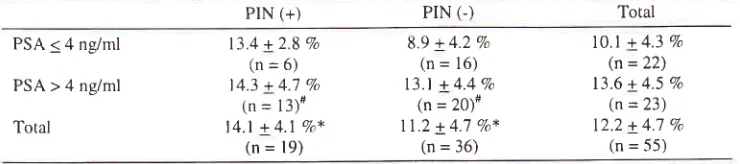
Table 5. Percentage of S-phase fiaction in low and high PSA group of BPH patients with or without PIN
PIN (+) PIN (-) Total
PSA < 4 ng/ml
PSA > 4 ng/ml
Total
13.4 + 2.8 Vo
(n=6)
14.3 + 4.'1 Vo
(n
=
l3)#l4.l + 4.1
Va*(n = 19)
8.9 + 4.2 Vo
(n = 16) 13.l + 4.4 Va
(n = 20)*
ll.2 + 4.7 Va*
(n = 36)
10.1 + 4.3 % (n = 22)
13.6 + 4.5 7o (n = 23) 12.2 + 4.7 %
(n = 55)
#rtot
[image:4.612.54.481.440.578.2] [image:4.612.50.420.624.706.2]26
Rahardjo et alTable 6. Percentage of S-phase fraction in low and high PSA group of BPH patients without PIN
BPH without PIN
PSA<4nglml(n=16)
PSA>4nglml(n=20)
8.9+4.2%*
(range:2.1-
16.'7Vo)13.l + 4.4 Vo *
(range: 10.0 - 22.7) *sigrtificanr (p = 0.008)
DISCUSSION
In Indonesia,
until
recently prostate cancer
had
not beenmuch
of
aproblem, though in the last l0
yearsthere has been an increase
in
cancerincidence.riThis
has led
to
increasing interest
in
use
of
PSA
testin-e,though
its sensitivity
andspecificity
arestill
debatable.Nevertheless,
until
today
serumPSA level is
still an
important
tool
for diagnostic
screening
of
prostatecancer.
An
increase
of
more than
4.0 n-e/ml
isindicative to perform additional diagnostic
procedureto find
prostate
cancer.l2'lr'l'+
But,
there is still
aproblem
in
selecting patients to be the
candidatesof
prostate
needle biopsy,
i.e.
if
they
clinically
shqwsigns and symptoms
of
BPH
andnormal
consistencyof
DRE. The
fact that
all
screening
modalities
including
serumPSA
level
arenot specific
will
causetoo many
unnecessarybiopsies.
This condition will
increase
not only the
logistic
burdens, but
alsomorbidity and psychological
aspects of the
patient.Beside
the
low-specificity, serum PSA level
could
also increase
in some circumstances
which are
not relatedto malignancy,
such as DRE-procedure,TRUS,
prostate massage,
prostatitis
and someinstrumentation
procedures
like cystoscopy, indwelling
catheterization andTURP.
Carcinoma
of
the prostate
is a
very important
malignant
diseasein
the United
States.rs'16In
1996,prostate cancer
was
40Vo
of all
new
malignant
diseases
in
men and gave l4Vo
mortality
due to
malignant
diseasein
men.l7In Indonesia on the
otherhand,
basedon histopathological data between
1988-1990, prostate
cancer was included
in the ten
highestmalignancy
in men,r8
i.e. the
seventh
in
1988,
theninth
in
1989 and back to seventh
again
in
1990.Although in
Jakarta prostate cancer was ranked as thesecond
highest cancer
of
genitourinary system,
thenumber
of
casesis
relatively
very
low
compared
tothe
incidence
in
the
United
States and
European countnes.Med J Indones
In
our country, there was a tendency that most BPH
patients came
in
already
severeconditions; they
hadto
be catheterized
in the emergency
room
prior to
further examinations.
This condition
is
unavoidable
since
most patients came
fiom low-economic
andlow-education group of people,
so they had
neverbeen aware
of
the
initial
symptoms
of
BPH.
Catheterrzation
in
BPH patients has been
known
toraise
the PSA
concentration and might
denotedifTerent strategy
of
interpreting
PSA.'''
Our
datashowed that
catheterization increased the PSA
level2.4 times above
the normal value (Table
2). It
wasalso obvious that
increasePSA level could be
due tohigh
prostatevolume;
this is
a constantfinding of our
reports.eProstatitis
is
the commonest
known cause
of
talsepositive PSA
in
the
West.20'21'22According
to
somereports,
clinically
detectable prostatecancer
accountsfor only
34Voof
serumPSA
elevations,2rwhile
nearly
all of the men with high PSA concentrations and
thernajority
with
normerllevels
had
at
least one
biopsy specimen codedpositive
tbr chronic
inflamation.22Inf-lammation
was defined
as inf'lammatory
cells in
the prostatic stroma,
and could
be acute
and/orchronic int-lammation. Int-lammation of
the prostate isalso
a
common histological
finding
in
prostatebiopsies.
This
subclinical inflammation
can cause
PSA elevation.tt Our study
has shownthat all
patientswithout urinary
retention
had
histological
inf-lammation,
mostly chronic infèctions. Although
there was
no control,
it could be
seenthat the PSA
tend to be higher than
normal
accepted values.Inf-lammation
most probably
increases
serum PSA
concentrations
by causing leakage
of
PSA
from
theacini
andductal lamina,
since prostateduct integrity
isdisturbed. Irani
etc/ founded that
acuteint'lammation
increased
serum
PSA
more
than
subchnicalinf'lammation.
They concluded
that unless
associatedwith
glandular epithelial disruption, density
of
prostatic interstitial int'lammatory
cell infiltrate is
notsignificantly correlated
with serum
PSA
concen-tration.
All
histological
section
comprised
a
mono-nuclear
cell
i nfiltrate.2oIn
this
current report,
more
descriptive
histo-pathological
report was
not
available
befbre 1998.
Evaluation
on
recent
data has
shown that
PIN
andother pre-malignant
findings
could
increasethe
PSA [image:5.612.47.276.114.181.2]small size
of
samples andlacking
casesof BPH with
high-grade
PIN
(Table 4).The attempt
to
correlateproliferative activity
and PSAlevel
is
relatively
new
in
BPH
study.
Increase
of
prostatevolume
could be
dueto
either
an increase incell
proliferation
with
unchanged
cell
death
orunchanged
cell proliferation
with
lower
cell
death.zaSome studies have shown
that induction
of
BPH
from
normal
prostateis obviously
associatedwith
adistinct
increased
proliferation
of epithelium
and stroma,
andfurther
increase
in
BPH
volume, however,
is
not correlatedwith
afurther
increasein
proliferation.tt
It',
should
be
noted that
PSA
is
produced
by normal
prostatic
cells
as
well
as hyperplastic and malignant
cells.26Our study
showed
that tumors
with
elevated
serumPSA level
have
higher proliferative
activity
asindicated
by
the
percentageof
S-phasecells. There
isno
universal
acceptancefor
S-phasecutoff point,
butin
most
cycling
human
cell
population, the
nuclei
in
the
S-
and G2lM
phases
may
reach
l5%o.
As
comparison,
proliferation
or
S-phasefraction
ratesof
breastcancer has been characterized as:
<87o=
low
proliferation,
8-12Vo=
intermediate
proltferation,
and>
12Vo=
high proliferation.tt But this
is
also
not
auniform
agreement.
Using
this
classification as
anexample,
our
patients
wrth
low
PSA level could
becategorized
as
intermediate proliferation
and
thepatients
with high
PSA level as high
proliferation.
High
proliferation was found
in
patients
with
high
PSA
aswell
asBPH
with PIN
(Table 5). High-grade
PIN
was
frequently
associatedwith high
cancerrisk,
therefbre
it
is
not suçrising
if
this
caseshad
high
prolrf'eratron
index. However, the
associationof
highS-phase
fraction
andhigh PSA level
in
the absenceof
PIN
suggeststhat there
was
an
increasedmitogenic
process
in
these patients.
It
is
not
known
what mechanism thatcould
increase themitogenic activity.
CONCLUSION
As
conclusion,
our
study
showedthat the
high
serumPSA level
of
our
patients
was primarily
caused
byurethral
catheterization
and
increased
prostatevolume.
Although
the
sample size wassmall
and lackof
high-grade
PIN, subclinical inflammation
andPIN
tend
to
raisePSA level
abovenormal
accepted value.Cases
with
high PSA
also
associated
with
highprolifèrative activity,
which is
suggestive tomitogenic
process
indicating
early
neoplastic
process
orinflammation. Further
study more details at thecellular
andis needed
to
elaboratemolecular level.
REFERENCES
l.
Vôller MCW, Schalken JA. Molecular genetics of benign prostatic hyperplasia, In : Kirby R et al (eds). Textbook ofBenign
Prostatic Hyperplasia.Oxford:
Isis
Medical Media, 1996.2.
UmbasR.
Pathophysiology and pathogenesisof
benign prostatic hyperplasia.Maj
Kedok Indones 1996;46(1):3 8-9
3.
Lee
CT,
OesterlingJE.
Prostate-specific antigen andcancer assessment. Kirby R, McConnell J, Fitzpatrick J,
Roehrborn C, Boyle P, eds. Textbook of benign prostatic hyperplasia. Abbott Laboratories Inc. 1996: 155-71.
4.
Lundwall A, Lilja H. Molecular cloning of human prostatespeci fi c anti gen cDNA. FEB S Len 1987 t21 4:3 17 -22.
5. Li
T,
BelingC.
Isolation and characterizationof
twospecific antigens
of
human seminal plasma.Fertil
Steril l9'73;24:134-M.6.
Lilja H. A kallikrein-like serine protease in prostatic fluidcleaces the predominant seminal vesicle protein
.
J Clin Invest 1985 :76: I 899-1 903.7.
Sipan G, Umbas R. The effect of urethral catheterization on the levelof
prostate-specific antigen. Indones J Urol199 5 ;5 :28-33 (Indonesi an).
8.
HammererPG, McNeal
JE,
StameyTA.
Correlation between serum prostate-specific antigen levels and the volumeof
the individual glandular zonesof
the human prosate. J Urol 1995;153:i 1 l-4.9.
RahardjoD.
Birowo P, Pakasi LS. Correlation between prostate volume, prostate specific antigen level, prostate specific antigen density and agein
the benign prostate hyperplasia patients. Med J Indonesta 1999;8:260-310.
HedleyDW,
FriedlanderML,
Taylor
IW,
Rugg CA, MusgroveEA.
Methodfor
analysisof
cellular DNAcontent of paraffin-embedded pathological materials using flow cytometry. J Histochem Cytochem 1983;31 :1333-5. I
l.
Himawan S, KrisnuhoniE.
Urologic cancerin
Jakarta:Pathology-based
data:
1990-1994. IndonesJ
Oncol1995;6:5'7 -64.
12.
Catalona WJ, Richie JP, deKernion JE, et al. Comparisonof prostate specific antigen density
in
the early detectionof
prostate cancer: receiver operating characteristic curves. J Urol 1994:152:2031-613.
Chen Z, Chen H, Stamey TA. Prostate specific antigen inbenign
prostatic
hyperplasia:
purification
andcharacterization. J Urol 1997 ;1 57 :2165-'7 0
14.
Bangma CH, Rietbergen JBW, Kranse R, et al. The Freeto
total PSAratio
improves the specificityof
PSA in screening for prostate cancerin
the genera'l population. JUrol 1997;157:2191-96
15.
BensonMC,
OlssonCA.
Prostate-specific antigen andprostate-specific
antigen density:
roles
in
patientevalu ati on and management. Cancer | 99 4;7 4: 1 667 -7 3
16.
NishiyaM.
Miller GJ,
LooknerDH,
Crawford ED.Prostate-specific
antigen density
in
patients
withhistologically
proven prostate
carcinoma.
Cancer23
28
t'7
l8
24
25
26
t9
27 20
2I
22
Rahardjo et al
Parker
SL,
Tong
T,
Bolden
S,
Wingo
PA.
Cancerstatistics. CA Cancer J CIin 1996;46(l):5-27.
Mangunkusumo
R.
Frekuensi tumor ganasdi
Indonesia berdasarkan pemeriksaan histopatologi. In : Susworo HR,'ljarta
HA,
Kresno SB, Poetiray EDC, Kurniawan AN,Djoerban
Z,
GondhowiardjoS,
Aziz
MF.
(Eds.).Pencegahan dan deteksi
dini
penyakit kanker. Jakarta:Indonesian Society of Oncology, 1996:84-91
Umbas
R,
Mochtar CA, RahardjoD.
Does CatheterizedPatients Denote Different Strategy
for
Prostate Biopsy?The
4th
International Conferenceof
Asian
ClinicalOncology Society
(ACOS),
Bali,
August5-7,
1999.Abstract No. 046.
Irani J, Levillain P, Goujon J-M, Bon D, Dore B, Aubert
J.
Inflammation
in
benign prostatic
hyperplasia:correlation
with
prostate specihc antigen value.J
Urol 1997:15'7:1301-3.Keetch
DW,
CatalonaWJ,
SmithDS.
Serial prostaric biopsiesin
menwith
persistently elevated serum PSAvalues. J Urol 1994;151:1571-4.
Nadler
RB,
HumphreyPA,
SmirhDS,
Catalona WJ,Ratliff
TL.
Eflectof
inflammation and benign prostaticMed J Indones
hyperplasia on elevated serum prostate specific antigen
levels. J Urol 1995;154.407-13.
Schatteman PHF, Hoekx L, Wyndaele JJ, Jeuris
W,
Van Marck E. Inflammation in prostate biopsies of men ithoutprostatic malignancy
or
clinical
prostatitis.Eur
Urol 2000;37:404-12.Claus S, Berges R, Senge
T,
Schulze H. Cell kinetic in epithelium and stromaof
benign prostatic hyperplasia. JUrol 1997;158:217-21 .
Claus
S,
WrengerM,
SengeT,
SchulzH.
Immuno-histochemical determinationof
age related proliferation ratesin
normal and benign hyperplastic human prostate.Urol Res I 993;21 :305-8.
Partin AW, Carter HB, Chan DW, Epstein, JI, Oesterling
JE, Rock RC et al. Prostate specific antigen in the staging
of
localized prostate cancer: influence
of
tumordifferentiation, tumor volume and benign hyperplasia. J
Urol 1990;143 747-52.