24
Himawan Med J IndonesPathological features of glomerulonephritis
in Jakarta
Sutisna
Himawan
Abstrak
Semua knsus biopsi ginjal yang diterima selama
l0
tahun dari 1990-1999 dikumpulkan dan diteliti. Seluruhnya terdapat 1344 kasus, terdiri atas 390 kasus pediatrik, 918 k"asus dewasa dan 36 kasus yang usianya tidak diketahui. Tujrcn penelitianini
inlah untuk mendapatkan gambaran tentanç pola dnn spektrum penyakit glomerulus di Indonesia, terutamadi
Jaknrta dan sekitarnya, dengan perhatian khusus pada kasus sindrom nefrotik, nefritis lupus dan nefropati IgA, serta membandingkannya dengan berbagai laporan terdahuludari
Indonesia dan beberapa negaralain.
Terdapat 250 kasus sindrom nefrotik pada anak dnn 479 kasus dewasa. Diagnosis histopatologik yang paling seing pada kedua golongan ialah penyakit kelainan minimal, yaitu masing-masing sebanyak 58.2Vo dan 44.7Va.Pia
lebih seing terkena daripada wanita dengan perbandingan untuk masing-mnsing golongan iatah 2.0:I
dan L4: I. Nefritis lupus meliputi 124 knsus, tiga diantaranya tidak representatif. Perbandinganpia
terhndap wanita ialah l:7.9. Frekaensi puncak tcrdapat pada dekade keempat sebanyak 47 kqsus (38.5Eù dan garnbaran histopatologik terbanyak iaLah WHO kelas IV, yaitu 71 kasus (58.7Vo). Diagnosis nefropati IgA ditegakkan pada 97 kasus. Sebaran usia dari 3 hingga 58 tahun dengan puncak insidens pada dekade keempat dengan 32 kasus (337o).Pia
lebih sertng dari wanita dengan perbandingan 1.7:I.
Gambaran histopatologik terbanyak ialah lesi sklerosis difus 34 knsus ( j5Vo) dan lesi proliferatif mesangial 33 kasus (34Vo). (Med J Indones 2002; 11: 24-9)Abstract
AII cases of renal biopsies received during a 1)-year
periodfrom
1990-1999 were collected and analyzed. There were a totat of 1344 cases, comprising 390 pediatric cases, 9 I 8 adult cases and 36 cases of unlotown age. Immunofluorescence microscopy was performed on 1089 cases (8L0Vo). The purposeof
this study is to have an overviewof
the pattem and spectrum of gLomerular diseases in Indonesia, especially in Jakarta and surroundings, with special emphasis on the cases with nephrotic syndrome, lupus nephritis and IgA nephropathy, and to compare the findings with previous reports from Indonesia and afew other countries. There were 250 cases of childhood nephrotic syndrome and 479 adult cases. The most frequent histopathological appearance in both groups was minimal change disease, i.e. 58.2Vo and 44.7Vo respectively. Males were more often affected than females with a ratio of 2.0:1for
children and L4:1for
adults. Lupus nephritis comprised 124 cases, among which three cases were not representative. The male to female ratio was I:7.9. Most cases werein
the fourth decade, i.e. 47 cases (38.580), and the most frequent histopathological appearance was WHO class IV with 7I
cases (58.7Eo). There were 97 cases of IgA nephropathy with an age range betvveen 3 to 58 years. The peak incidence was in the fourth decade with 32 cases ( 3 3 7o). The male to female ratio was L7:I.
The most frequent histopathological appearances weredffise
sclerosing lesion 34 cases (35Vo) and mesangial proliftrative lesion 33 cases (34%). (Med J Irulones 2002;l1:
24-9) Keywords: renal biopsy, pathologicalfeatures, glomerulonephritis, nephrotic syndrome, Iupus nephritis, IgA nephropathyThe
first
report
of
pe
utaneous
renal biopsy
in
Jakarta was
published
b
Sutedjo and V/ahidijatr
in
1963.
They
described thehistopathological findings in
three cases
of
children
with
nephrotic
slmdrome.However, due
to
lack
of
facilities
and various
other reasons therenal biopsies were discontinued
andonly
in 1970
it
was
started again.2Since then
it became
a routine procedurein
the Sub-departmentsof
NephrologyDepartment of Anatomic Pathology, Faculty of Medicine University of Indonesia/Dr. Cipto M angunkusumo Hospital, Jakarta, Indonesia
Presented at the L3th Asian Colloquium in Nephrology, Bali, November 23-25, 2000
of
the
Department
of
Child Health and rhe
Department
of
Internal
Medicine
of
the Faculty of
Medicine University
of
Indonesia.IDr.Cipto
Mangun-kusumo National Central
General
Hospital.
At
thattime the only private hospital which
also performed
this
procedure was
the Cikini
Hospital,
who
had developed avery solid nephrology
team.Nowadays
it
is also performed sporadically
in
a few other private
hospitals. Besides percutaneous renalbiopsy,
in
a partof
the
casesan open renal
biopsy was performed
toavoid
thepossibility of
an unrepresentative specimen.Vol
II,
No 1, January-March2002findings.
In
1976 facilities
for
immunofluorescence
microscopy was
established.Unfortunately
until
now
thereis
still
nofacility for
electronmicroscopy.
The
purpose
of
this
study
is
to
have an
overview of
the
spectrum andpattern
of
thevarious renal diseases
in
Indonesia, especially
in
Jakarta and
surroundings,during the last
decade
of
the twentieth
century,
andalso
to
compare
it
with
previous
findings
from
Indonesia, aswell
as afew reports
from other countries.
METHODS
All
casesof
renal biopsiesreceived
at theDepartment
of
Anatomic Pathology
of
the Faculty
of
Medicine
University
of
Indonesia./Dr.
Cipto
Mangunkusumo
National Central
General
Hospital during
a
lO-yearperiod
from
1990until
1999were collected
from
thefiles
and
analyzedaccording
to
theclinical
diagnosis, age, gender and pathological diagnosis.The
nomenclature and classification used were in
accordance
with
the
WHO
criteria.3'a
For
the light
microscopy examination the
paraffin
blocks were cut
in
two
micron
sections
and stained routinely with
hematoxylin and eosin
(H&E),
periodic acid-Schiff'
(PAS) and
Masson-Goldner'strichrome
(MT).
Stainingwith
periodic acid-silver
methenamine
(PASM)
wasalso applied
in
caseswhich
showedthickening
of
thecapillary walls.
While for
immunofluorescencemicros-copy the
specimenwas
tested againstIgG, IgA,
IgM,
C3
and fibrinogen.
In
this
study
evaluation
will
belimited
to
caseswith
the nephrotic
syndrome,
lupus nephritis andIgA
nephropathy.RESULTS
The total
number
of
renal biopsies received
during
al0-year period
from
1990-1999
was
1344
cases,comprising 390 pediatric
cases,918 adult
cases and36
casesof
unknown
age, among
which 214
caseswere
not
representative,
thus
only
1130 could
beevaluated.
Caseswere
regarded
not
reprgsentative if
the
microscopical
slide
showed
less than
sixglomeruli,
with
the exception
of
certain
caseslike
membranous
nephropathy
andamyloidosis
in which
adefinite
diagnosis could
be established evenwith
less tbansix glomeruli.
Immunofluorescence
microscopy was performed
on 1089 cases(i.e.8l.}Vo of
all
cases), amongwhich242
Pathologyofglomerulonephritis
25cases
were
not
representative,
thus
only
847
casescould be
evaluated.Nephrotic syndrome
More than
half
of
the renal
biopsies,
i.e. 729
cases (54.2Vo)were
sent
for
the
pathological
evaluation
of
patients
with
thenephrotic
syndrome,comprising
250pediatric cases, among
which
777
(7O.8Vo)
were representative, and 479 adult cases, amongwhich
387 (80.87o)were
representative,thus the
total
number
of
cases
which could
be
evaluated
was
564.
Thedistribution of
the pathological
diagnosesis
shown
in
Table
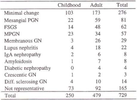
1.Table
l.
Distribution
of
pathological diagnoses among thecases with nephrotic syndrome
Childhood
Adult
Total Minimal changeMesangial PGN
FSGS
MPGN
Membranous GN Lupus nephritis IgA nephropathy Amyloidosis Diabetic nephropathy
Crescentic GN Diff. sclerosing GN Not representative
Total
The most frequent pathological diagaoses were
minimal
change,i.e. 276
cases (48.9Vo),with
respectively
103pediatic
cases
and
173 adult
cases,
followed
by
mesangial
proliferative glomerulonephritis
81
cases (l4.4%o),focal
segmentalglomerulosclerosis
62
cases(ll%o),
membranoproliferative glomerulonephritis
57 cases (10.17o) and membranous glomerulonephritisonly
29 cases (5.lVo).The
five
most frequent pathological
diagnoses
in
childhood nephrotic
syndrome are shownin
Table 2.
Table
2.
The five most frequent pathological diagnoses in cases of childhood nephrotic syndrome103
173
27622
59
8l
t4
48
6223
34
5732629
41822
268
178
044
123
4to14
73
92
165479
250
Minimal change
MPGN
103 23
58.2 t3 12.4
'1.9
Mesangial
PGN
22FSGS
t4
[image:2.612.299.528.295.462.2] [image:2.612.302.528.648.722.2]26 Himawan
The
most frequent pathological
diagnoses
were
asfollows: minimal change
103 cases (58.2Vo),membrano-proliferative glomerulonephritis
23
cases
(73Vo),mesangial
proliferative glomerulonephritis
22
cases(I2.4Vo), focal segmental
glomerulosclerosis
14 cases (7 .9Vo) and IgAnephropathy 5
cases (2.8Vo).Minimal
change disease comprised 58.2Voof
all
casesand
thenonminimal change 41.87o; with
a ratioof
1.4:1. The male tofemale ratio was 2:1.
The
five most
frequent pathological
diagnoses in adultnephrotic syndrome
are shownin Table
3.Table
3.
The hve most
frequent pathotogical diagnoses in adult nephrotic syndromeNumber
Med J Indones
Most cases were
in
the fourth decade, i.e. 45
cases(38.57o),
followed
by
the
fifth decade
35
cases(29.6Vo) and
the
second
and
third
decade each 26
cases(2l.3Vo).
The distribution
of
the
pathologica
ses using theWHO classification* is shown
in
Table
5.
Distribution
of
pathological diagnosesin
lupus nephritis according WHO classifi cationClass Number
l2
ll
l2
71 11 4 Total* 3 cases were not representative
The highest number of
caseswere classified
as ClassfV,
i.e.
7l
cases (58.7Vo),followed
by
Class
I
and Class III 12 cases (9.9Vo) each, ClassII
and Class
V
also
with a
samenumber,
i.e. l1
cases(9.lVo), and
ClassVI4
cases (3.3Vo).IgA Nephropathy
There were 97
casesof IgA nephropathy with various
histopathological
appearances asis shown
in Table
6.Table
6.
Distributionof
histopathological appearanceof IgA
nephropathy casesNumber I
II
ilI
IV VI 9.9 9.1 9.9 58.7 9.1-t. J
Minimal change Mesangial PGN FSGS
MPGN
Membranous GN
r73 55 45
3l
25 44.7 14.2 I 1.6 8.0 6.5 100 121The most frequent pathological
diagnosis
in
adultswas also minimal
change,
i.e.
173
cases (44.7Vo),followed
by mesangial proliferati ve glomerulonephri tis
55 cases (l4.2Vo),
focal
segmental glomerulosclerosis45
cases (II.6Vo),membranoproliferative
glomerulo-nephritis 31
cases (8.0Vo) and membranous glomerulo-nephritis 25 cases (6.5Vo).The ratio
of
cases
with
minimal
to nonminimal
change diseasewas 1:1.2 (173:214).
The male to female ratio was 1.4:1.
Lupus nephritis
Lupus nephritis comprised
124 cases,with
a male to femaleratio
of
1:7.9 (14:1l0).
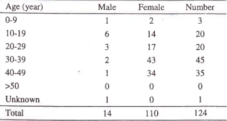
[image:3.612.320.544.211.323.2]The age
distribution
can be seenin Table
4.Table 4. Age-group distribution oflupus nephritis cases
Age (year) Male
Female
NumberDiff. sclerosing GN Mesangial PGN Focal sclerosing Minor change Chronic rejection Unclassified 34 JJ t9 6
I
4 35 34 19.6 6.2I
4.1 100 97 0-9 l0-19 20-29 30-39 40-49 >50 Unknown 1 6 3 2 1 0I
2l4
17 43 34 0 0 3 20 20 45 35 0I
The
most frequent
picture was diffuse
sclerosinglesion
which
was found
in
34 cases (35Vo),
followed
by
mesangial proliferative lesion
33
cases
(34Vo),focal sclerosing
lesion
19 cases(l9.6Vo),
minor lesion
6 cases (6.2Vo),
unclassified
4
cases(4.IVo)
and
one case(lVo)
was foundin
achronic transplant rejection.
The
le
to
female
ratio was
1.7:l
(61:36),
and the
age range was between 3 to 58 years.The
agegroup distribution is shown
in Table
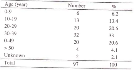
7. [image:3.612.61.286.276.359.2] [image:3.612.56.287.604.727.2]Table 7. Age group distribution
ofIgA
nephropathy casesAge (year) Number
The highest frequency
wasfound
in
thefourth
decadewith 32
cases(337o),
followed by the third
and
thefifth
decade
with an even number
of
cases,i.e.
20cases (20.6Vo), second decade 13 cases
(l3.4Vo),
first
decade
6
cases (6.ZEa),sixth
decade4
cases (4.lEo), and 2 cases(2.lEa) of unknown
age.Fxcept
IgA
deposition, other
depositions
were
alsofound
in
various combinations.
The
three
mostcommon
findings
were
IgA
andC3 which
wasfound
in
24
cases (24.8Vo),followed
by
a combination of
IgA, IgM
andC3 in
22
cases (22.6Vo), andIgA
alonein l2
cases (12.480).DISCUSSION
Nephrotic syndrome
The
most frequent histopathological
appearancein
childhood
aswell
asin
adult nephrotic
,ynd.o^"
*u,
minimal
change disease.Wilawirya5
in
his
studyof primary
nephrotic syndromein children in
Jakarta hascollected
i47
.ur",
of
newnephrotic
syndrome patients
during
a
lO_yearperiod
from
1970-1979.Of
the
541cases, 364were
biôpsiedand
of
these55
caseswere examined using
immuno_fluorescence
and electron mrcroscopy,
ùhich
*u,
performed
in
Australia.
His
study revealed
minimal
change
in 16l
cases (44.2Vo), mesangialproliferative
glomerulonephritis
139 cases (3g.2Vo),focàl
segmentalglomerulosclerosis
24
cases (6.6Vo),
membrano-proliferative
glomerulonephritis
l9
cases
(5.2Vo),membranous glomerulonephritis
g
cases(2.2Vo),
andsclerosing
glomerulonephritis
I
cases (3.6Vo).In
anearlier report
by
Kasim
et
12in
6 casesof
childhood
nephrotic
syndrome, thefigures
wereminimal
changel2
cases(46.lVo), mesangial
proliferative
glomerulo_nephritis
7 cases (26.9Va),focal
segmental glomerulo_sclerosis
and sclerosing glomerulonephritls
with
353.6Vo.In the present study the
figures
were 5g.2Vo and4l.8Vo. Thus
in this
study
the prevalenceof MCNS
is reportsfrom
Indonesia,than reported
by
thebe
performed
to
look
to
1988
they have
managed
70 black children
anclamong these
29
werehepatitis
B
virus carriers.
Of
the29,
26
had
membranous glomerulonephritis,
Imembranoproliferative glomerulonephritis,
I
focal
segmental
glomerulosclerosis
and
I
was
not
biopsied due to advanced renalfailure.
Woo et al.ro
in
1999 reported that the cornmonestfiom of
angewas
than
females.
In
childhood nephrotic syndrome
Wilawirva
et
al7
from
Jakarta
and Damanik
et
"iâ-
;;;;
Yogyakarta,
reported the maleto female
ratio
as1.3:l
and
2.4:l
respectively.
In
this
study
is
was
2.0:l for
children
and1.4:l for
adults.[.upus nephritis
Vo
0-9 10-19
20-29
30-39
0-49
>50
Unknown
6
13
20 32 20 4
2
6.2
t3.4
20.6 33 20.6
4.r
2.1
Total 9'7 100
Comgared
to
a previous
report
in lggl
by
Himawan
[image:4.612.31.258.105.225.2]28 Himawan
of
the classes.
In
the
present study
the predominant
classis
Class[V
with
58.7Vaof
the
cases,far above
the other
classes whichwere
aboutevenly distributed
between
9.1
and
9.9Vo. Whllein
the previous study
Class[V
comprised
only
39Voof
the cases,
followed
by Class III33Vo,
ClassII
l7Vo,
and ClassI
and ClassY
6Va
each.
Mok
et
al.r2
in
their
study
of
lupusnephritis
in Southern Chinese patients
found that 557o
of their
caseswere
ClassIV,
which is about
the same
as in
the present study,
followed
by Class
III
(2570),Class V
(l4Vo),
ClassII
(5Vo), and Class I(l7o).
IgA Nephropathy
In
this study
IgA
nephropathy
wasfound
in
1l.5Voof
allbiopsies.
P
aet
al.l3
and
apercentage
of
oreported that
rn Singapore
IgA
nephropathyis still
thecommonest
primary
glomerulonephritis ranging from
42Vo-457oduring the past two
decades.The
male
to
female
ratio was
1.7:1,
the
same
aspreviously
reporte
''
(1different
from
the
andwho
found a
ratio
et al
that
in their series
the
clinical
presentationof patients
with
IgA
nephropathy
were
nephrotic
syndrome35.7 1 Vo,
rapidly progressive
glomerulonephritis
3.5'7 Voand
glomerulonephritis
without nephrotic
syndrome60.75Vo.
They
found
IgA
deposits alone
in
28.5'7Vo, much more as compared to the present study (12Vo).In
summary,this
study
revealedthat
minimal
change disease is the most corrrmon typeof glomerulonephritis
in childhood as well
asin
adult
nephrotic syndrome,
i.e.
58.2Vo
and
44.7Vo
respectively. Compared
toprevious
studies from Indonesia there was a changein
the
proportion
of minimal change versus
non-minimal
change cases
in
childhood nephrotic
syndrome.In
thisstudy
the result was
58.2Voversus
4l.8Vo,
while
in
previous
studies
it
was ranging from
44.2Vo-46.4Vo versus 53.67o-55.8Vo.In
lupus
nephritis,
like
all
other
studies,
the
female preponderancewas
very striking, with
a M:F ratio of1:7.9.
Most cases
were ofWHO-Class
tV
(58.77o), and the peak incidence wasin the
fourth
decade (4570).IgA
nephropathy
was more prevalent among
maleswith a M:F ratio
of
1.7:1.The peak incidence
wasin
the
fourth
decade
(33Vo),
and
the
most
commonhistopathological
appearancewere diffuse
sclerosingglomerulonephriti s
(35Va) and mesangialproliferative
glomerulonephritis
(3 47o).Med J Indones
Acknowledgement
The author is indebted to
Dr.
Sutjahjo Endardjo, MSc
who
performed
the
immunofluorescence microscopy
examinations
and Drs. Rino Pattiata and Diah Rini
Handjari
for
their invaluable help and assistance
in
this
study.REFERENCES
1,
Sutedjo, WahidijatI. Biopsi ginjal
pada anak-anakpen-derita "nephrotic syndrome". Maj Ked Indon 1963; l2:1.
2.
Kasim JA, Himawan S, Wilawirya IGN. Renal biopsy in children with nephrotic syndrome. A morphological study(Preliminary report). Paediatr lndon 1972', 12:55-67.
3.
Churg J, Sobin LA. Renal disease. Classification and atlasof glomerular diseases Tokyo: Igaku-Shoin; 1982.
4.
Churg
J,
BernsteinJ,
Glassock RJ.Renal
disease.Classification and atlas
of
glomerular diseases. 2"d ed.New York: Igaku-Shoin; 1995.
5.
Wilawirya IGN.
Penelitian beberapa aspekklinis
danpatologi anatomis sindrom nefrotik primer pada anak di
Jakarta (disertasi). Jakarta: Univ Indonesia; 1992.
6.
Intemational Studyof Kidney
Diseasein
Children. The nephrotic syndrome in children. Prediction of histopathologyfrom clinical and laboratory characteristics at the time of diagnosis. Kidney Int 1978; 3:l 59-65.
1.
WilawiryaIGN,
Alatas H, TambunanT,
Himawan S.Gambaran histopatologis
ginjal pada sindrom
nefiotik primer di Jakarta. Presented at the Kongres Nasional IlmuKesehatan Anak IX; 1993 Jun l3-17; Semarang, Indonesia.
8.
Damanik MP, Endardjo S, Himawan S. Gambaranhisto-patologik sindroma nefrotik pada anak. Presented at the Kongres Nasional
Ilmu
KesehatanAnak
X;
1996, Bukittinggi.9.
van Buuren AJ, Bates WD, Muller N. Nephrotic syndromein
Namibian children.S
Afr Med
J
1999 89:1088-91.
Abstract.10. Woo KT, Chiang
GS, Pall A, Tan PH, Lau YK, Chin YM. The changing patternof glomerulonephritis
in Singapore
over the past two decades. Clin Nephrol 1999;52:96-102. Abstract.1
l.
Himawan
S,
Siregar
B,
Endardjo
S.
Gambaranmikroskopik cahaya dan imunofluoresensi nefritis lupus.
Beberapa pengalaman
di
Jakarta.Makalah Lengkap
Kongres NasionalVII
Ikatan Ahli Patologi Indonesia;l98l
Jun 15-19; Medan, Indonesia.12.
Mok CC, Wong RW, Lau CS. Lupus nephritis in Southem Chinese patients: clinicopathologic findings and long-term outcome. Am J Kidney Dis 1999; 34:315-23.13.
HariandjaA,
HutagalungP, Endardjo S, Himawan
S, Sidabutar R.P. lgA nephropathy in Jakarta('Ijikini
Hospital 1980-1985). Presentedat
the
6ù Asian Colloquium
in Nephrology; 1985; Kuala Lumpur, Malaysia.14.
HimawanS and
Endardjo S. Gambaran histopatologik nefropatiIgA
di
Jakarta. Kumpulan Makalah Lengkap Kongres Nasional X Ikatan Ahli Patologi Indonesia; 1990Vol I
I,
Nol,
January-
March 200215.
EffendiI,
Sidabutar RP, Flimawan S, Endardjo S. IgA nephropathyin
Jakarta, Indonesia. Presentedat
the 9ùAsian
Colloquiumin
Nephrology. 1992May
17-21;' Seoul, South Korea.P at holo gy of glo merulonephriti s
Sidabutar
RP,
LumentaNA,
Subardjono, Sumarjono,Siregar
P,
Alatas
H, et al.
Glomenrlonephritis in Indonesia. Kursus Sehari Imunologi Ginjal. 1987 Aug 20; Jakarta, Indonesia.