Biochemical study of CymJ, the putative regulator of cyclodextrin metabolism
in Klebsiella oxytoca M5a1
Tri Yudani Mardining Raras1, August Boeck2
Abstrak
Untuk melangsungkan metabolisme siklodekstrin, suatu oligoglukosida siklik, Klebsiella oxytoca (K. oxytoca) memerlukan produk dari sepuluh gen yang tergabung dalam dua operon yang berlawanan arah. Telah dilakukan penelitian mengenai fungsi CymJ, yaitu produk gen yang berada pada posisi paling jauh dari promoter pada salah satu operon cym. Overekspresi cymJ pada K.oxytoca menyebabkan penekanan ekspresi operon cym. Akan tetapi represi ini dapat dikembalikan pada kondisi normal dengan penambahan -cyclodextrin ke dalam medium. Diduga ada hubungan antara CymJ dan CymD karena ketiadaan CymD pada mutan delesi cymD mencegah efek represi oleh overekspresi CymJ. Penemuan yang tidak disangka-sangka adalah hambatan pembelahan sel yang berimbas pada terhambatnya pertumbuhan akibat overekspresi cymJ. Perubahan morphologik ini disertai dengan peningkatan sensitivitas K. oxytoca terhadap ampisilin. (Med J Indones 2007; 16:69-77)
Abstract
The products of ten genes clustered in two divergently oriented operons are required for the metabolism of cyclic oligoglucosides, the cyclodextrins, by Klebsiella oxytoca. The function of CymJ, the product of the promoter distal gene in one of the operons was studied. Over expression of cymJ in K. oxytoca led to strong reduction of the expression of the cym operons. This repression could be alleviated by addition of high concentration of -cyclodextrin into the medium. There is a possible relationship between CymJ and CymD since the absence of CymD in a cymD deletion mutant prevented the repression effect of CymJ. An intriguing finding was that the presence of CymJ in large amount in the cell caused severe cell division inhibition leading to retardation of growth. This morphological change was paralleled by a significant increase in the susceptibility of K. oxytoca to ampicillin. (Med J Indones 2007; 16:69-77)
Keywords: cym operon, putative regulator, over expression
At least ten genes have been identified up to now to code for proteins which are involved in cyclodextrin metabolism by Klebsiella oxytoca (K. oxytoca)
M5a1. They are designated as cymA-J (cyclodextrin metabolism genes).1-3
The biochemical functions of several gene products have been characterised; cymA encodes a cyclodextrin specific outer membrane porin;1 CymE is a periplasmic binding protein and CymF, CymG and CymD are other members of an ABC type transporter specific for cyclodextrins. They are functionally homologous to MalE, MalF, MalG and MalK of E. coli, respectively.1 CymH is a cytoplasmatically located cyclodextrinase.4
The functions of several gene products of the cym
operon, namely CymB, CymC, CymI and CymJ are not known. In this study we concentrated on the analysis of the function of CymJ. The cymJ gene is the promoter most distal gene in one of the two cym
operons. Computer analysis of the CymJ sequence suggested that it is 31 kD in size and that it does not contain a signal peptide.5 CymJ lacks significant sequence similarity to any other protein in the data base. It displays only very low sequence similarity to the N-terminal region of YhrO from Bacillus subtilis,6 and to AF1009 of Archaeoglobus fulgidus.7 Circumstantial evidence had indicated that CymJ could be a regulator of the cym operon. This assumption was based on experiments in which the overexpression of cymJ in wild-type cells lead to the repression of the expression of the cym operon.1 In this study we report more detailed information concerning the function of CymJ in the cym metabolism system.
1
Laboratory of Biochemistry-Molecular Biology, Faculty of Medical Science, Brawijaya University, Malang, Indonesia
2
METHODS
To characterize CymJ several biochemical and physio-logical approaches were conducted i.e construction of
cymJ overexpression system, antibiotic susceptibility study of cymJ overexpression mutant, determination of cellular localization of CymJ, biochemical, morpho-logical, and physiological studies upon overexpression of cymJ both in homologous and heterologous system as well as in cym deletion mutant.
Media and growth conditions
The rich medium used for routine growth of cells was Luria Broth (LB).7 When necessary, antibiotics were added to a final concentration of 100 g/ml (ampicillin) and 50 g/ml (kanamycin).8
In the case of K. oxytoca
M5a1, the concentration of ampicillin had to be increased to 800 g/ml because of the expression of a chromosomally encoded -lactamase.9 ''Werkman'' minimal medium was prepared as described previously10 and contained 1% (w/v) carbon source (glycerol, if not otherwise indicated).
Strains, plasmids and oligonucleotides
The bacterial strains used through this work are listed in Table 1, and Table 2 shows relevant plasmids used. During the experiment employing K. oxytoca deletion mutants, the presence of the deletion in the chromosome was confirmed via PCR tests with primers that are listed in table 2.
Table 1. Bacterial strains
Strain Genotype or relevant property Reference
K. oxytoca
M5a1 Wild type [11]
CYMA M5a1, cymA [3]
CYMCD M5a1, cymCD [5]
CYME M5a1, cymE [12]
REGB M5a1, lac, scrA:: ('uidA-(cymE'-cymA')-'lacZ) [5]
E. coli K12
K38 HfrC phoA4 pit-10 tonA22 ompF627 relA1 + [13]
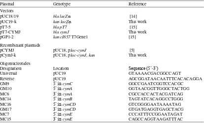
Table 2. Plasmids and oligonucleotides used in this work
Plasmid Genotype Reference
Vectors
pUC18/19 bla lacZ [14]
pUC19-k kan lacZ This work
pT7-5 bla pT7 [15]
pT7-CYMJ bla cymJ This work
pGP1-2 kancI857 T7Gene1 [15] Recombinant plasmids
pCYMJ pUC18, plac-cymJ [5] pCymJ-k pUC18, plac-cymJ, kan This work Oligonucleotides
Designation Location Sequence (5’-3’)
Universal pUC19 GTAAAACGACGGCCAGT
Reverse pUC19 AGCGGATAACAATTTCACACAGGA
GM9 5’ in cymC GGCCGAATCGGTCCACGC
GM10 5’ in cymA GGTAACGGTTGGGCTACTGG
MC6 3’ in cymA CGCCACCACTACGATCAG
MC14 5’ in cymB TAGTATCACAGGCCTGGG
MC16 5’ in cymCD GTCGGGGAATAAAATAG
GM17 3’ in cymCD GTGATGAGGTGAGCTACG
MC7 5’ in cymE CCCATTTCCGGAATAGAT
General DNA manipulation techniques
Unless otherwise mentioned, the standard DNA procedures, such as restriction endonuclease digestions, cloning and transformation procedures, were performed as described by Sambrook and Ausubel.8,16 Due to the excess of slime polysaccharides produced, K. oxytoca
had to be transformed via electroporation. Plasmids were either prepared following the procedure given by Holmes and Quigley17 or with the aid of the Qiagen Kit (Qiagen GmbH, Hilden, Germany) as described by the manufacturers.
For the polymerase chain reaction (PCR), Goldstar DNA polymerase was used employing the conditions suggested by the manufacturers (Roche Biochemicals, Penzberg, Germany). Polymerase chain reaction
experiments were performed with a Biometra personal cycler (Göttingen, Germany). Recovery of DNA-fragments from agarose was carried out by the ‘low
melting agarose’ technique18
or using the Qiagen Kit.
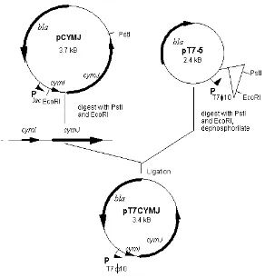
Construction of cymJ over-expression plasmid
To construct the plasmid for the over-expression of the cymJ gene, the pT7-5 vector was digested with PstI and EcoRI. The cymJ fragment was generated from plasmid pCYMJ by also digesting with PstI and EcoRI. Ligation of the two fragments resulted in pT7CYMJ (Figure 1). This plasmid was then transferred into E. coli strain K38. To ensure that the plasmid was maintained in the cells during the experiment, PCR test using reverse and universal primers (Table 2) was performed before and after the experiment.
Figure 1. Construction of pT7CYMJ.
pCYMJ and pT7-5 were digested with EcoRI and PstI. The fragment of cymI-cymJ was ligated into linearised pT7-5.
Cellular localisation of CymJ protein
The over-expression of cymJ for cellular localisation studies was performed employing the system described by Tabor and Richardson.15 For that purpose, E. coli
strain K38/pGP1-2 was transformed either with plasmid pT7CYMJ or pT7-5. Twenty ml cultures containing the transformants were grown aerobically at 30°C in LB medium containing 100 g/ml ampicillin and 50 g/ml kanamycin to an OD600 of 0.7. The cells were harvested by centrifugation at 6000 x g, washed twice with minimal medium and re-suspended in 1 ml minimal medium supplemented with 0.4% glucose, thiamine (2 g/ml) and 18 amino acids (100 g/ml) except cysteine and methionine. The suspension was used to inoculate 24 ml of the same medium which was then further incubated for one hour at the same temperature. To induce the synthesis of T7-polymerase, the culture was shifted to 42 °C for 20 min. The inhibition of the synthesis of the host RNA-polymerase was achieved by addition of rifampicin (300 g/ml) and the culture was incubated for additional 20 min. After transfer of the culture to 30 °C and incubation for 10 min, 10 Ci/ ml of [35S]-methionine (NEN Life Science Products, Bad Homburg, Germany) were added and incubation was continued for 15 min. The labelling was terminated by addition of unlabelled methionine to 1 mg/ml. After 5 min cells were collected by centrifugation and stored at -80 °C.
Cells were thawed, suspended in 50 mM potassium The separation of cytosolic proteins and membrane proteins was performed by centrifugation at 100.000 x g. The membrane associated proteins were released using sarkosyl19 or Triton X-100 (Sigma, Deisenhofen, Germany). The labelled protein was separated on 12.5 % SDS-polyacrylamide gels and the gel was autoradio-graphed. The localisation of CymJ was analysed using a phosphoimager.
Biochemical study of the cym gene expression
In this experiment cymJ was cloned into pCYMJ under the control of lac promotor and was overexpressed in
K. oxytoca REGB. This strain carries a chromosomally integrated cym intergenic region flanked by the
reporter genes lacZ and uidA. The transcription can be followed by measuring β-galactosidase and glucuronidase activities.5
Determination of beta-galactosidase activity
The -galactosidase activity of lacZ fusion strains was determined in permeabilised cells.5 Cultures were grown in LB medium until they reached an OD600 of
0.7, then 1.25 ml were taken and used to inoculate 8.75 ml prewarmed minimal medium supplemented with -cyclodextrin (final concentration 0.5%). The cultures were further incubated for one hour and the -galactosidase activity was measured.
Morphological studies on CymJ overexpression
To observe any morphological changes caused by the overexpression of cymJ, cultures were grown to an OD600 of 0.2 at 37°C with vigorous shaking in minimal medium containing 0.4 % glucose. The over-expression of cymJ was then induced by addition of 0.5 mM IPTG; after 3 h -cyclodextrin was added at the final concentration of 0.5 % (w/v). At intervals of one hour the growth of the culture was monitored spectrophotometrically and the cell morphology was studied by microscopy. Cells were visualized and photographed using a Zeiss-Axioplan microscope, using phase contrast conditions. Estimation of the difference in an average cell length between strains REGB/pCYMJ and REGB/pUC19 was achieved by measuring cell length in photographs printed at the same magnification.
For the study of the overexpression of cymJ in cym
deletion mutant, plasmid pCYMJ was transferred into
K. oxytoca CYMA, K. oxytoca CYME and K. oxytoca
CYMCD. These transformants were then grown in the same medium as above and under the same condition and treated exactly the same way as the wild type.
RESULTS
Antibiotic susceptibility of various K.oxytoca is shown in Table 3.
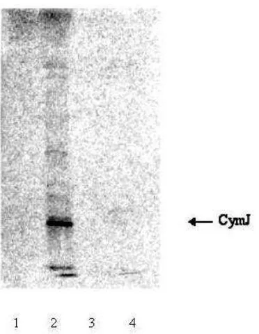
Cellular localisation of CymJ
periplasm, however, in a few preparations it was found out that some CymJ protein also co-sedimented with membrane fractions (data not shown).
Table 3. Antibiotic susceptibility of K. oxytoca REGB/pCYMJ transformants compared to K. oxytoca M5a1 and K. oxytoca
REGB/pUC19
Figure 2. Autoradiograph of an SDS gel in which soluble(s) and pellet (P) fractions of 35S-methionine-labelled cells were separated. Lane 1: S100 of K38/pGP1-2/pT7-5, Lane 2: S100 of
K38/pGP1-2/pT7CYMJ, Lane 3: P100 of K38/pGP1-2/pT7-5, Lane 4: P100 of K38/pGP1-2/pT7CYMJ
CymJ is a putative repressor of cym gene expression
It was speculated previously5 that CymJ could be a candidate for cyclodextrin-specific repressor, so it was plausible to assume that cyclodextrins may relieve the repression. However, it is shown by the result in
Figure 3, that one hour after induction of the cym
operon using 500 M -cyclodextrin the activity of
the β-galactosidase was even lower than that measured
from strain carrying the pUC19 vector indicating that the -cyclodextrin could not alleviate the repression mediated by CymJ.
Figure 3. Addition of 500 M -cyclodextrin could not alleviate the repression effect of CymJ in LB medium.
In order to avoid any possible interference which may come from compounds of the rich medium, minimal media was used in the next experiment. Intriguingly, the repression of the cym genes could be alleviated as
is shown by the increase of the β-galactosidase
activity of cells carrying overexpressed cymJ after addition of - cyclodextrin. After 30 min. it reached almost 95% of the value measured for cells carrying pUC19 (Fig. 4).
Figure 4. Release of the repression effect exerted by CymJ in minimal medium by the addition of 500 M -cyclodextrin.
min
Inhibition of cell septation upon overexpression of
cymJ
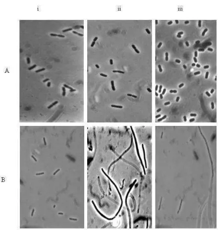
K. oxytoca REGB/pCYMJshowed wrinkled colonies on LB plates containing 100g/ml ampicillin. Observation of the cells under the microscope demonstrated that they formed long filaments. In liquid minimal medium containing 0.4% glucose, the cells showed a similar phenotype as those grown on plates. The over-expression of cymJ resulted in cells that grew slower and varied significantly in their length when compared with K.oxytoca REGB cells carrying the vector (compare Fig. 5A.i and 5B.i); they became very long, around 10 to 20 times longer than wild type cells particularly after the lac promoter had been activated with IPTG (Fig. 5B.ii).
Figure 5. Morphology of K. oxytoca REGB carrying overexpressed cymJ. Cells in their mid-exponential growth phase were imaged with a Zeiss-Axiophoto phase–contrast microscope, photographed, and printed at the same magnification. A(i) K. oxytoca REGB/pUC19 grown in minimal medium supplemented with 0,4% glucose and 100 g/ml ampicillin, (ii) 2h after induction with IPTG, (iii) 2 h after addition of 0,5% -cyclodextrin.
B(i) Strain K. oxytoca REGB/pCYMJ, (ii) 2h after induction with IPTG, (iii) 2 h after addition of 0,5% -cyclodextrin.
The filamentous cells which tended to clump and aggregate did not show any constriction. Moreover, most of the elongated cells were anucleated and only a minority contained a single nucleoid (10%). The most interesting finding was that the filamentous cells gradually reverted to the normal form, when -cyclodextrin was
added. One hour after the addition of -cyclodextrin, the filamentous cells began to form a constriction and started to divide again (Fig. 5B.iii). Finally three hours after the induction of the cym operon all the cells were completely reverted to normal morphology. We ensured that the cells under observation were not contaminants, since samples that were streaked on LB plate containing glucose grew homogenously without contaminants on plates. In addition, PCR test of the culture that was performed three hours after addition of -cyclodextrin showed that the cells still carried the corresponding plasmid.
Overexpression of cymJ in K. oxytoca cym-deletion mutant
Microscopic observation of the results of overexpression of cymJ in different K. oxytocacym deletion mutants, i.e. K..oxytoca CYMA, K. oxytoca CYME and K. oxytoca
CYMCD showed different phenomenon. Klebsiella oxytoca CYMA/pCYMJ cells showed the same pheno-type as the wild pheno-type. After addition of -cyclodextrin, the filamented cells reverted into normal cells. This means that the deletion of cymA does not affect the activity of the cymJ product (data not shown).
Klebsiella oxytoca CYME/pCYMJ exhibited a similar phenotype as wild-type cells until they reached the exponential phase; however, in contrast to K. oxytoca
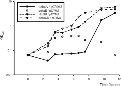
CYMA/pCYMJ, the addition of -cyclodextrin failed to stimulate the conversion of elongated cells into normally sized cells. Some of them even underwent lysis (Fig. 6). In contrast, K. oxytoca CYMCD/ pCYMJ did not exhibit a significant phenotypic change, the transformant grew much better than K. oxytoca/pCYMJ (Fig. 6). It must be emphasised that the growth curve of the K. oxytoca REGB, K. oxytoca
CYMA as well as K. oxytoca CYME overexpressing
cymJ was obtained by measuring the optical density of the cells under the condition in which the cells were in filamentous form. Therefore, the growth curve presented in Figure 6 does not reflect the growth of cells with a normal morphology.
Overexpression of cymJ from a plasmid carrying a kanamycin resistance gene (pCymJ-k)
In the homologous system
amount of glucose was increased up to 1.2% or when different concentrations of -cyclodextrin were added. However in LB medium supplemented with 0.4% glucose K. oxytoca REGB/pCymJ-k formed the elongated cells, which were also observed with the K. oxytoca
REGB/pCYMJ cells in ampicillin containing medium. However, the filamented cells were shorter and contained evenly spaced nucleoids.
Figure 6. Growth of K. oxytoca cym mutants overexpressing cymJ. Cells were grown on minimal medium supplemented with 0.4% glucose and 100 g/ml ampicillin (50g/ml for K. oxytoca CYME). First arrow represents the time when IPTG was added, the second arrow indicates the time when -cyclodextrin was added. deltaA/pCYMJ=K. oxytoca CYMA/pCYMJ deltaCD/pCYMJ= K. oxytoca CYMCD/pCYMJ, deltaE/pCYMJ=K. oxytoca CYME/pCYMJ.
In the heterologous system
The overexpression of cymJ in a heterologous system was performed using E. coli strain MC4100 which was transformed with pCymJ-k. During the lag phase, the MC4100/pCymJ-k cells showed the same filamentous form as K. oxytoca REGB/pCYMJ grown in the presence of ampicillin and became longer after the promoter was induced with IPTG. However, the addition of -cyclodextrin did not lead to the reversion, in contrary, the cells underwent lysis. The complementation with pCym1 carrying all genes involved in the cyclodextrin metabolism system also could not revert the defect.
DISCUSSION
The regulation of the cym operon
The mechanism of the regulation of the cym operon in
K. oxytoca appears to be a subject to both positive and
negative control. However, the regulator remains unidentified. There have been speculations that CymJ could be a candidate for a repressor, since the overexpression of cymJ lead to the repression of the
cym operon.5
It has been demonstrated that the expression of the cym
operon is induced by cyclodextrins, -cyclodextrin being the best inducer.1,5 The repression exerted by
cymJ can be released by the addition of 500 M of -cyclodextrin. The difference between the results obtained in rich and minimal media, however, indicates that other regulatory regimes exist which override the cyclodextrin-specific control circuit.
The fact that the components of the cyclodextrin transport system show high sequence similarity to those from the maltose uptake system leads to the speculation that the regulation of the cym operon could resemble that of the maltose regulon. The mal
genes are regulated by the activator MalT. The expression of the mal genes is induced by the binding of the inducer, maltotriose, to MalT and requires ATP.
It is clear that CymJ is involved in the regulation of the cym operon. Overexpression of cymJ in trans leads to inhibition of the expression of the cym operon genes which could be released through an addition of high concentrations of -cyclodextrin. One possible explanation may be that there is a kind of competition between CymJ andcyclodextrin upon binding to the putative regulator. Overexpression of cymJ in the cell would then effectively influence the competition. This possibility has been evident in the case of the maltose regulon. MalY represses the maltose operon by binding to the transcription activator MalT, which is activated by maltotriose; accordingly MalY competes directly with the inducer.20 Furthermore, it is known that the inducer plays an important role in the activation of the maltose operon.20
It was an unexpected result that CymD is involved in the activation of CymJ in the regulation of cym
operon. Since the absence of a functional cymD
product prevents the CymJ effect. Therefore, CymJ may require the product of CymD. CymD displays high sequence similarity to MalK. MalK is a bifunctional protein, acts as part of a transporter in the presence of maltose and in the absent of maltose binds to MalT, which leads to its inactivation.21 The function of CymD as an ATPase has not yet been proven experimentally, however, the high similarity of its amino acid sequence to MalK and the fact that it
can complement the function of MalK strongly supports this suggestion.1 There is also a lack of information on the function of CymD as a repressor. It is only known that in a heterologous system in E. coli
deletion of cymD rendered the cells unable to grow in the presence of -cyclodextrin.5 Moreover, the over-expression of cymD in the K. oxytoca REGB dramatically repressed the expression of cym operon (data not shown), which is consistent with the previous results.5
Under physiological conditions employing normal stoichiometries the components are well balanced; when cymJ is overexpressed, however, they are no longer in equilibrium and the system is fully repressed.
We also assume that at excessive overproduction of CymJ may elicit a SOS response with multiple physiological consequences like the binding of SulA to FtsZ. Furthermore, a functional defect in FtsZ could lead to the failure of DNA to segregate.22 However, these occur temporarily due to a competition between the product of the cymJ gene and an effect of -cyclodextrin that could alleviate the inhibitory effect of CymJ on cell septation. However the role of CymE with respect to the presence of abnormal amount of the CymJ remains unanswered, although the inability of the cells to revert to the normal morphology suggests that CymE or its function is required for the conversion step to proceed.
An interesting effect of the overexpression of cymJ
was the markedly increased susceptibility to -lactam antibiotics. The overexpression of cymJ in a strain with kanamycin resistance background could exclude the argument that the inhibition on the cell septation was caused by ampicillin; rather, it is a consequence of overproduction of cymJ. Klebsiella oxytoca strains generally display moderate intrinsic ampicillin resistance due to the synthesis of the chromosomally encoded OXY-1 and OXY-2 class A β-lactamases9 and consequently the organism grows flourishly in the presence of 1 mg/ml ampicillin.
The reduction of the resistance against antibiotics could be due to several factors. One of them may reside in the inhibition of export of -lactamase precursor to the periplasm.
In conclusion, Overexpression of cymJ in K. oxytoca
led to strong reduction in the expression of the cym operons. This repression could be alleviated by addition of high concentration of -cyclodextrin into the medium. There is a possible relationship between
CymJ and CymD since the absence of CymD in a cymD deletion mutant prevented the repression effect of CymJ. An intriguing finding was that the presence of CymJ in large amount in the cell caused severe cell division inhibition leading to retardation of growth. This morphological change was paralleled by a significant increase in the susceptibility of K. oxytoca
to ampicillin. starch utilization pathway present in Klebsiella oxytoca. J Mol Biol. 1996; 256: 279-91.
2. Pajatsch M, Boeck A, Boos W. Enzymatic preparation of radiolabelled linear maltodextrins and cyclodextrins of high specific activity from 14C maltose using amylomaltase, cyclodextrin glucosyltransferase and cyclodextrinase. Carbohydr Res. 1998; 307: 375-9
3. 3. Pajatsch M, Andersen C, Methes A, Boeck A, Benz R, Engelhardt H. Properties of a cyclodextrin specific, unusual porin from Klebsiella oxytoca. J Biol Chem. 1999; 274 (35):25159-66
4. Feederle R, Pajatsch M, Kremmer E, Boeck A. Metabolism of cyclodextrins by Klebsiella oxytoca M5a1: purification and characterisation of a cytoplasmically located cyclo-dextrinase. Arch Microbiol.1996;165: 206-12
5. Pajatsch M. Cyclodextrin-Metabolism in K oxytoca M5a1.
[Dissertation]. Munich: Ludwid-Maximilians University; 1999.
6. Sorokin A, Bolotin A, Purnelle B, Hilbert H, Lauber J, Dusterhoft A, et al. Sequence of the Bacillus subtilis
genome region in the vicinity of the lev operon reveals two new extracytoplasmic function RNA polymerase sigma factor SigV and SigZ. Microbiology 1997;143: 2939-43.
7. Miller JH. A short course in bacterial genetics. Cold Spring Harbor: Cold Spring Harbor Laboratory; 1992. 8. Sambrook J, Fritsch EF, Maniatis T. Molecular cloning: a
laboratory manual. Cold Spring Harbor: Cold Spring Harbor Laboratory; 1989.
9. Fournier B, Roy PH, Lagrange PH, Philippon A. Beta-lactamase gene promotors of 71 clinical strains of
Klebsiella oxytoca. Antimicrob Agents Chemother. 1996; 40: 454-9
10. Frankael DG, Neidhard FC. Use of chloramphenicol to study control of RNA synthesis in bacteria. Biochem Biophys Acta. 1961; 53: 96-100
12. Pajatsch M, Gehart M, Peist R, Horlacher R, Boos W, Boeck A. The periplasmic cyclodextrin binding protein CymE from Klebsiella oxytoca and its role in maltodextrin and cyclodextrin transport. J Bacteriol. 1998;180: 2630-5. 13. Lyons LB, Zinder ND. The genetic map of the
filamentous bacteriophage f1. Virulogy. 1972; 49: 45-60. 14. Yanisch-Perron C, Viere J, Messing J. Improved M13 phage
cloning vectors and host strains: nucleotide sequences of the M13mp18 and pUC19 vectors. Gene.1985; 33: 103-19. 15. Tabor S, Richardson CC. A bacteriophage T7RNA
polymerase/promoter system for controlled exclusive expression of specific genes. Proc Natl Acad Sci USA. 1985; 82: 1074-8.
16. Ausubel FM, Brent R, Kingston RE, Moore DD, Seidmann JG, Smith JA, et al. Current protocols in molecular biology. New York: J Wiley and Sons; 1997. 17. Holmes DS, Quigley M. A rapid boiling method for the
preparation of bacterial plasmids. Anal Biochem. 1981;114: 193-7.
18. Wieslander L. A simple method to recover intact high molecular weight RNA and DNA after electrophoretic separation in low gelling temperature agarose gels. Anal Biochem. 1979; 98: 308-9.
19. Filip C, Fletcher G, Wulff JL, Earhart CF. Solubilization of the cytoplasmic membrane of Escherichia coli by the ionic detergent sodium-lauryl sarcosinate. J Bacteriol. 1973; 115:717-22.
20. Boos W, Shuman H. Maltose/maltodextrin system in
Escherichia coli: transport, metabolism, and regulation. Microbiol and Mol Biol Rev. 1998; 62: 204-29.
21. Lippincott J, Traxler B. MalFGK complex assembly and transport and regulatory characteristic of MalK insertion mutants. J Bacteriol. 1997; 179; 1337-43.