Formation and Pathogenicity Variation of Oospores of
Phytophthora capsici
Infecting Black Pepper
DYAH MANOHARA
Indonesian Research Institute for Medicinal and Aromatic Crops, Pusat Penelitian dan Pengembangan Perkebunan, Jalan Tentara Pelajar No 3, Bogor 16111, Indonesia
Phone: +62-251-321879, Fax: +62-251-327010, E-mail: [email protected]
Phytophthora capsici Leonian is the causal agent of foot rot disease of black pepper (Piper nigrum L.). Foot rot disease is the most destructive disease which can cause significant economic losses of black pepper. Two mating types of P. capsici were found in black pepper plantations in Lampung. This research was aimed at examining the effects of temperature, light, and polycarbonate membrane on oospore formation. Also the effect of light on germination and determining both mating types of 30 randomly selected progenies. The results showed that oospores were produced through either hyphal contact or without any contact. Oospores were produced abundantly on V8 agar in the dark at 16-24 oC. Oospores germinated after 3-weeks incubation in the dark followed by 1-week incubation under TL fluorescent light. All progeny were heterothallic, and consisted of 16 isolates of A1 mating type and 14 isolates of A2 mating type. There was no correlation between mating type categories and their pathogenicities on black pepper leaves. Twenty-six progeny isolates may infect unwounded pepper leaves, whereas another four isolates may only infect wounded pepper leaves. Pathogenicities of all progenies were lower than those of their parents.
Key words: Phytophthora capsici, oospore, black pepper, Lampung
_____________________________________________
Phytophthora capsici is a pathogenic causal agent of foot rot disease on black pepper (Piper nigrum L.). The attack of P. capsici on the root and collar region causes sudden plant death. Foot rot disease on black pepper was found for the first time in Lampung in 1885 (Muller 1936). At present this disease has spread to all black pepper plantations in Indonesia and is the main constraint in black pepper production.
P. capsici is a heterothallic fungus. Sexual reproduction can occur when there are two compatible mating types (A1 and A2) for oospore formation. Two mating types of P. capsici
from black pepper plants were reported to have been found in Lampung (Manohara and Sato 1992). Ko (1980) used polycarbonate membrane to prove that oospore formation of P. cinnamomi, P. parasitica, and P. palmivora could take place without contact hyphal of the two compatible mating types.
The formation of oospores of Phytophthora is affected by several environmental factors such as temperature and light. Harnish (1965) and Brasier (1969) stated that light inhibited the formation of oospores but stimulated sporangia formation. Optimum temperature for the formation of oospores of Phytophthora differed, depending on the species.
Oospore germination is affected by light, oospore maturity (age), temperature, enzyme treatment and chemical treatment (Ribeiro 1983). In general, the quantity of germinating oospores will increase in line with the increase of an oospore’s age. According to Hord and Ristaino (1991), the germination rate of oospores of P. capsici originating from chili pepper plants improved by 3% at the age of 14 days, and became 20% at the age of 60 days. The light and temperature requirement during the oospore germination process varied, depended on the species of Phytophthora. Oospores are very important in epidemiology and etiology of Phytophthora because they are able to survive
in extreme environmental conditions. The oospore is also the stage where genetic recombination occurs which can create a new individual more pathogenic than its parents (Hausbeck and Lamour 2004). The objectives of this research are to study: (i) the effects of temperature and light on oospore formation, (ii) oospore formation with polycarbonate membrane, (iii) oospore germination, and (iv) mating types and pathogenicities of progeny isolates.
MATERIALS AND METHODS
Fungal Material. P. capsici isolates were obtained from diseased black pepper plants in North Lampung, i.e. isolate N2 (A1 mating type), and in South Lampung, i.e. isolate N4 (A2 mating type). The two isolates are assumed to have the same polyploidy i.e. diploid. These isolates were grown and maintained in V8 agar (200 ml V8, 1.0 g CaCO3, 20 g agar, and 800 ml aquadest).
The Effect of Temperature on Oospore Production. Culture disks of each of the isolates, N2 and N4 (diameter 0.5 cm), were placed in pairs facing each other on V8 agar with 5-cm distance from each other, and then incubated at 16, 20, 24, 28, and 32 oC. Every treatment was replicated three
times. Colony growth was observed every day until both isolates made contact. After the culture reached the age of 14 days, the number of oospores formed was counted by taking two samples of media disks (diameter 0.5 cm) from each Petri dish. The sample was placed on object glass, stained using lactophenol trypan blue with simultaneous heating, and then covered with a cover glass. The number of oospores was counted employing light microscope at 200 x magnification. Every disk was examined at three randomly chosen places.
The Effect of Light on Oospore Production. Culture disks of isolates N2 and N4 (diameter 0.5 cm) were placed facing
MICROBIOLOGY INDONESIA, August 2007, p 61-64 Volume 1, Number 2
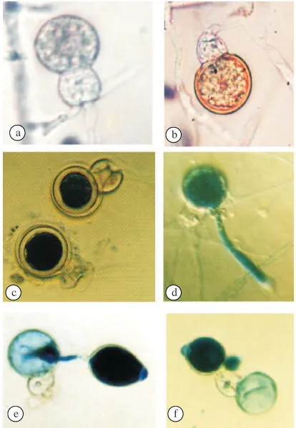
Fig 1 The oospores of P. capsici and their germination. a. young oospore, b immature oospore, c. mature oospore stained with lactophenol tryhpan blue, d. hypha formation of germinating oospores, e and f. sporangium was formed by germinating oospores. Table 1 Incubation treatment for the germination of oospores of
P. capsici
Incubation in the dark (20 oC) Incubation under the light (26-28 oC)
2 weeks 2 weeks 2 weeks 2 weeks 3 weeks 3 weeks 3 weeks 3 weeks
1 week 2 weeks 3 weeks 4 weeks 1 week 2 weeks 3 weeks 4 weeks
Table 2 The effect of temperature on the formation of oospores of P. capsici
Incubation temperature (oC) Colony contact (day) Number of oosporesa 1 6
2 0 2 4 2 8 3 2
4 3 3 3 4
44.16 85.67 46.67 14.33 1.00
athe average of 18 microscope view area with 200x magnification.
each other on V8 agar at a 5-cm distance, and then incubated under several light treatments using a TL fluorescent lamp with 600 lux intensity and placed at a 40-cm distance from the Petri dish lid. The durations of light exposure are 0, 6, 12, 18, and 24 h respectively. Temperature inside the incubation room ranged between 26-28 oC. Each treatment was replicated
three times. The determination of oospore number was carried out as given above.
Oospore Production with Polycarbonate Membrane. Isolates N2 and N4 were grown on V8 agar and incubated for 3 days in the dark. Culture disks of isolate N2 (diameter 1.0 cm) were obtained from the edge of a growing colony then placed in the middle of a sterile Petri dish, covered with a polycarbonate membrane(CPR, 0.2 µm, 90 mm diameter;
Nucleopore Corporation, Pleasanton, California, USA). On top of this was placed another culture disk of isolate N4. For comparison, an opposite treatment was conducted using the same isolates (Ko 1980). Incubation was carried out in the dark at 20 oC. After 7 days, the number of oospores formed
during incubation was observed.
Oospore Germination. Oospores were released from agar media by crushing in a blender at 5000 rpm for 5 min. The oospore suspension was then centrifuged at 2000 rpm for 5 min. The sediment was filtered employing a gradual sieving filter with pore diameter of 75, 38, and 20 µm respectively.
The remaining oospores left in 20 µm filter were then
separated and washed with 0.5% (wt/vol) KMNO4for 20 min. The oospore suspension was then immersed in warm water (36-38 oC) for 2.5 h (Wahyuno et al. 1995). The oospore
suspension was then diluted to obtain about 20 oospores per inoculating loop tip, and then streaked on basalt media (20 mg glucose, 100 mg lecithin, and 10 ml basalt solution). The incubation treatment consisted of 8 lighting combinations (Table 1). The percentage of oospore germination was estimated visually.
Mating Types and Pathogenicities of Progeny Isolates on Black Pepper Leaves. Thirty progeny isolates were obtained from randomly selected germinating oospores (single spore isolation), isolated on V8 agar. The mating types of progeny isolates were determined using pairing method by pairing the isolates with known-tester isolates (A1 or A2 mating type) on V8 agar. The presence of oospores was observed. Pathogenicities of progeny isolates were tested by inoculating them on black pepper leaves. These progeny isolates were grown on V8 agar and incubated for 7 days under continuous light. An inoculum disk (diameter 0.5 cm) was placed on the lower side of unwounded black pepper leaf surface and then one drop of sterile water was added. These leaves were placed inside a plastic container whose base had been covered by moist tissue paper. Measurement of leaves spots formed was conducted after 4-day incubation at room temperature.
RESULTS
Optimum Temperature for Oospore Formation. Oospore formed within the temperature range of 16-32 oC (Table 2).
When incubated at 16-24 oC, sexual reproduction structures
began to appear one day after both colonies made contact. However at 28 and 32 oC, the structures were formed 4 days
after both colonies made contact. The antheridia were amphigynous and colorless (hyaline). Oogonia had spherical or subspherical form and were colorless (hyaline). Oospores were colorless (hyaline) at the young stage and became brownish when mature (Fig 1a,b). Oospores were generally globose in shape with thick walls (Fig 1c). The optimal temperature for oospore formation was 20 oC (Table 2).
The Role of Light in Oospore Production. Oospore production was very sensitive to light. The contact of both colonies occurred 4 days after incubation, and on the fifth day, several reproduction structures had been formed
a b
c d
e f
Table 3 The effect of the duration of light exposure on the formation of oospores of P. capsici
Light exposure (h) Number of oosporesa
athe average of 18 microscope view area with 200x magnification
Table 4 Formation of oospores of P. capsici with polycarbonate membrane
aPolycarbonate membrane was inserted between the two isolates, bthe average of 18 microscope view area with 200x magnification.
Table 5 Oospore germination of P. capsici
Incubation treatment (in the dark). The treatment of a 6-hour exposure to light inhibited oospore formation, while exposure to light for more than 6 hours caused no oospore formation (Table 3).
The Effect of Polycarbonate Membrane. This is impenetrable by the hyphae of P. capsici. This can be proven by inserting the membrane between culture disks of P. capsici
and fresh V8 media. It turned out that oospores continued to be formed on isolate disk N2 and N4 even though polycarbonate membrane had been inserted between them (Table 4). This result revealed that oospores formation took place without direct contact between the hyphae of two compatible mating types. Oospore formation seemed to be induced by the presence of compounds that could penetrate the membrane. Compared with isolate N4, the isolate N2 always produced the most oospores regardless of the position of pairing (Table 4).
Oospore Germination. Oospore germinated when they were first incubated for 3 weeks in the dark, followed by exposure to continuous light for 1 week or more (Table 5). Oospores germinated to form germ tubes that penetrated oospore walls or passed through antheridia. The number of germ tubes produced could be more than one. The germ tubes grew longer and became hypha, or created sporangium at the tip. (Fig 1d, e, f).
The treatment of incubation in the continuous dark caused no oospore germination (data are not shown). A longer light treatment increased the ability of oospore to germinate. The number of germinating oospores increased in line with the duration of light exposure for up to the 3 weeks (Table 5). Four-weeks of exposure to light resulted in
a lower number of germinating oospores compared to 3-week light exposure (Table 5). Further assessment found that this treatment produced many empty oospores, probably resulting from lysis.
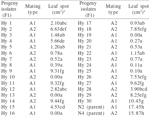
Mating Types and Pathogenicities of Progeny Isolates. All progeny isolates were heterothallic; they formed oospores with their compatible tester. Sixteen progeny isolates belonging to the A1 mating type formed oospores with isolate N4, while the remaining 14 isolates belong to the A2 mating type, formed oospores with isolate N2 (Table 6).
The symptom of infection on black pepper leaves could be seen as small spots which were brown-blackish color and which appeared 24 h after inoculation with parental isolates (N2 or N4). On the 4th day, the leaves spot areas are 17.45 cm2
and 15.87 cm2 respectively. Eight progenies (Hy 9, Hy 11, Hy
14, Hy 18, Hy 26, Hy 27, Hy 29, and Hy 30) caused symptoms similar to their parents on the 2nd day after inoculation. On
the 4th day, the leaves spot area ranged between 7.53-10.45
cm2. Four progenies (Hy 10, Hy 13, Hy 16, and Hy 19) did not
show any symptoms. However, if the leaves were wounded before inoculation, these four isolates were then able to infect and produce small spots after 4-days incubation. From the 30 progenies tested, it was evident that none of the isolates had the same or more pathogenicity compared to their parental isolates. Most of them (18 progenies) produced symptom less than 3.00 cm2 (Table 6).
DISCUSSION
The formation of oospores of P. capsici in vitro is clearly affected by temperature and light. Light was a constraining factor for oospore formation. In the dark, oospores could be produced within the temperature range of 16-32 oC. The
optimum temperature for oospore formation was 20 oC. This
temperature was lower than the optimum temperature for vegetative growth, i.e. 24-28 oC (Manohara 1988). This
findings are similar to Brasier’s (1969) results which revealed that the optimum temperature for vegetative growth of
Table 6 The variety of 30 progeny isolates of P. capsici and their pathogenicities on black pepper leaves
Progeny Progeny
Number followed by the same letters are not significantly different at 5% DMRT, aunwound inoculated
P. palmivora was 27.5-30 oC, while sexual reproduction
occurred in the range of 16-20 oC.
Oospore formation could occur without hyphal contact between the two mating types. It was probable that the two isolates produced compounds that induced each other to form gametangium and then oospores. This compound was then declared to be a sexual hormone. The A1 mating type produced α1 sexual hormone whereas the A2 mating type
produced α2 sexual hormone (Ko 1980). Furthermore Shen
et al. (1983) reported that this sexual hormone was not specific. For example, P. infestans with an A1 mating type will produce oospores if paired with an A2 mating type such as P. parasitica, P. capsici, and P. palmivora besides with
P. infestans itself. Jee et al. (2002) found that phytol was highly stimulatory to oospore formation of P. cactorum and
P. parasitica. Elliot (1983) suspected that the role of the sexual hormone was in the inhibition of steroid synthesis which controls sexual reproduction. Production of sexual hormone was affected by temperature and light. Yu et al. (1981) reported that the optimum temperature for P. colocasiae
to produce the hormone was 25 oC, at which temperature it
would be able to stimulate P. parasitica to form oospores in large quantity.
My research reported in this paper showed that oospores of P. capsici from black pepper plants would be mature after 3-weeks incubation in the dark, but that germination would require light. One-week exposure to light resulted in 29.02% germination. Increasing the duration of the exposure to up to 3 weeks would increase the percentage to 52.10%. Germinating oospores formed germ tubes that would develop into hyphae and mycelia or sporangia which would in turn form zoospores. According to Banihashemi (2004), oospore germination is determined by the level of maturity of oospores as the internal factor, and an external factor such as exposure to light, whose duration depended on the species of Phytophthora.
Observation by Banihashemi (2004) proved that P. cactorum needed dark conditions during the formation and maturation of oospores. The germination would then occur if incubated under continuous fluorescent illumination, but would not occur without the exposure to light. The germination of oospores of P. cactorum was only 3% if incubated in the dark, while-light exposure would increase it to 38%. Oospores of P. infestans germinated up to 70% under cool white fluorescent light (Chang and Ko 1991). The opposite happened in the germination process of oospores of P. capsici obtained from chili pepper plants. Germinating oospores would be abundant if light treatment was applied during both formation and germination process (Hord and Ristaino 1991).
My work reported here, shows that the pathogenicities of progenies seemed to have no correlation with mating types. Pathogenicities of all progenies were lower than those of their parents. The same result was described by Mayton et al. (2000) who did the mating with P. infestans. From 53 progenies, 52 isolates had lower pathogenicities compared to their parents. Abu-El Samen et al. (2003) found a genotypic variability among the asexual progenies of P. infestans.
Although oospores as the sexual reproduction have been considered as the important resting spore, the stage where genetic recombination occurs and the primary source of inoculum in the field, but little information is reported about the influence of some factors which stimulate the formation, germination and the pathogenicity of their progenies. Thus it is an important precedent for research in P. capsici especially on black pepper.
REFERENCES
Abu-El Samen PM, Secor, GA, Gudmestad. 2003. Genetic variation among asexual progeny of Phytophthora infestans detected with RAPD and AFLP markers. Plant Pathol 52:314-325.
Banihashemi Z. 2004. Light-dependent oospore germination of
Phytophthora cactorum in the presence of susceptible host plant.
J Phytopathol 152:683-686.
Brasier CM. 1969. Formation of oospores in vivo by Phytophthora palmivora. Trans Br Mycol Soc 52:273-279.
Chang TT, Ko WH. 1991. Factors affecting the germination of oospores of Phytophthora infestans. J Phytopathol 133:29-35. Elliott CG. 1983. Physiology of sexual reproduction in Phytophthora.
In: Erwin DC, Bartnicki-Garcia S, Tsao PH (eds). Phytophthora: its Biology, Taxonomy, Ecology, and Pathology. St. Paul: American Phytopathological Society. p 71-80.
Harnish WN. 1965. Effect of light on production of oospores and sporangia in species of Phytophthora. Mycologia 57:85-90. Hausbeck MK, Lamour KH. 2004. Phytophthora capsici on vegetable
crops: research progress and management challenges. Plant Dis
88:1282-1303.
Hord MJ, Ristaino JB. 1991. Effects of physical and chemical factors on the germination of the germination of oospores of
Phytophthora capsici in vitro. Phytopathology 81:1541-1546. Jee HJ, Tang CS, Ko WH. 2002. Characterization of phytochemicals stimulatory to sexual reproduction in Phytophthora cactorum
and P. parasitica. Bot Bull Acad Sin 43:203-210.
Ko WH. 1980. Hormonal regulation of sexual reproduction in
Phytophthora. J Gen Microbiol 116:459-463.
Manohara D. 1988. Ekobiologi Phytophthora palmivora (Butler). Penyebab penyakit busuk pangkal batang lada (Piper nigrum L.) [Dissertation]. Bogor: Institut Pertanian Bogor.
Manohara D, Sato N. 1992. Morphological and physiological observation on Phytophthora isolates from black pepper. Indust Crops Res J 4:14-19.
Mayton H, Smart CD, Moravec BC, Mizubuti ESG, Muldoon AE, Fry WE. 2000. Oospore survival and pathogenicity of single oospore recombinant progeny from a cross involving US-17 and US-8 genotype of Phytophthora infestans. Plant Dis 84:1190-1196. Muller HRA. 1936. Het Phytophthora-voetrot van pepper (Piper
nigrum L.) in Nederlanch-Indie. Mededelingen van het Institut voor Plantziekten, No. 88, p 79.
Ribeiro OK. 1983. Physiology of asexual sporulation and spore germination in Phytophthora. In: Erwin DC, Bartnicki-Garcia S, Tsao PH (eds). Phytophthora: Its Biology, Taxonomy, Ecology and Pathology. St. Paul: American Phytopathological Society. p 55-70. Shen CY, Bower LA, Erwin DC, Tsao PH. 1983. Formation of sex organs in the A1 mating type of Phytophthora infestans induced chemically by A2 isolates of other species of Phytophthora. Can J Bot 61:1462-1466.
Wahyuno D, Manohara D, Mogi S. 1995. Menginduksi perkecambahan oospora Phytophthora capsici. In: Strengthening Research on Disease of Industrial Crops in Indonesia [Annual Report]. Bogor: Research Institute for Spice and Medicinal Crops. p 37-42. Yu JY, Chang HS, Ko WH. 1981. Factor affecting the induction of
sexual reproduction in Phytophthora parasitica by Phytophthora colocasiae. J Gen Microbiol 123:249-252.