i
ABSTRAK
ii
ABSTRACT
iii
ACKNOWLEDGEMENT
iv
DEDICATION
First and for most I would like dedicate to my late father, Daud Bin Hassan, my siblings and especially my mother, Norisah@Merisah Binti Salleh. They have given
v 2.2.3 Biodegradable / Bioresorbable 8 2.3 Metal 8
2.4 Polymer 9 2.5 Ceramic 10 2.6 Bioceramics 10
2.7 Hydroxyapatite 11
2.7.1 Atomic Structure of Hydroxyapatite 11 2.7.2 Properties of Hydroxyapatite 12 2.7.3 Applications of Hydroxyapatite 13 2.7.3.1 Bioceramic Coatings 13
vi
2.7.3.3 Bone and Tooth Implant 14
2.8 Porous Hydroxyapatite 14
2.8.1 Preparation method of porous hydroxyapatite 15 2.8.1.1 Ceramic Foaming Technique 15
2.8.1.2 Gel Casting of Foam 15
2.8.1.3 Starch Consolidation 16
2.8.1.4 Slip Casting 16
2.8.1.5 Electrophoretic Deposition Technique 17
2.9 Polymeric Sponge Method 17
3.2.2 Preparation of porous hydroxyapatite 20
3.3 Sintering 25
3.4 Characterization of Porous Hydroxyapatite 26 3.4.1 Scanning Electron Microscope (SEM) 26 3.4.2 Fourier Transform InfraRed (FTIR) 27 3.4.3 Themogravimetric analysis (TGA) 28
3.4.4 X-ray Diffraction (XRD) 29
3.4.5 Porosity Measurement 31
3.5 Characterization 31
3.5.1 FTIR 31
3.5.2 TG-DTA 32
3.5.3 XRD 32
3.5.4 SEM 32
vii
4. RESULT AND DISCUSSION 33
4.1 Introduction 33
4.2 Characterization of Hydroxyapatite Powder 33 4.2.1 XRD of Hydroxyapatite Powder 33 4.2.2 Particle Size Analyzer of Hydroxyapatite Powder 34 4.2.3 TGA of Hydroxyapatite Powder 35 4.3 Characterization of Cellulosic Sponge 36 4.3.1 TG/ DTA of Cellulosic Sponge 36 4.3.2 SEM of Cellulosic Sponge 37
4.4 Porous Hydroxyapatite 37
4.4.1 Effect of Heating Rate on Porosity and Apparent Density 37 4.4.2 Effect of Stirring Time on Porosity and Apparent Density 39 4.4.3 Effect of Powder Loading on Apparent Density and Porosity 41 4.4.4 FTIR Spectrum of Porous Hydroxyapatite 43 4.4.5 XRD Pattern of Porous Hydroxyapatite 46 4.4.6 SEM of Porous Hydroxyapatite 49
5. CONCLUSION AND RECOMMENDATION 53
5.1 Conclusion 53
5.2 Recommendation for Further Study 54
REFERENCES 55
APPENDICES
viii
LIST OF TABLES
2.1 Biomaterial Classifications 5 2.2 Orthopedic Biomaterials and Their Primary Uses 6 2.3 Classes of Biomaterials According to Tissue Response 7 2.4 Clinical Uses of Bioceramics 11 2.5 Physical Properties of Synthetic Calcium Phosphate 13
4.1 Parameter of Prepared Porous HA and Properties of Samples A and B 38 4.2 Parameter of Prepared Porous HA and Properties of Samples C and D 40 4.3 Parameter of Prepared Porous HA and Properties of Samples E, F, G 42
ix
LIST OF FI
GURES
2.1 Investment-Cast Titanium Alloy Knee and Hip Implant Prostheses 9 2.2 Hydroxyapatite Structure Projected Down The c-axis on The Basal Plane 12
2.3 Slip Casting Processing 16
2.4 Powder Transformation in Sintering Process 18
3.1 Cellulose Sponge 20
3.2 Flowchart for Preparation of Porous Hydroxyapatite 22
3.3 As received cellulosic sponge 23
3.4 Cellulosic Sponge Cut into Circular Shape of 1 cm (10mm) Diameter 23
3.5 Humidified Cellulosic Sponge 23
3.6 Humidified Cellulosic Sponge Ready for Drying 23
3.7 Dried Cellulosic Sponge 23
3.8 Slurry Impregnated Cellulosic Sponges 24
3.9 Dried Impregnated Cellulosic Sponge, Top View and Front View 24 3.10 Sintered Porous Hydroxyapatite, Top View and Front View 24
3.11 Sintering Cycle of Porous Hydroxyapatite (5°C/min) 25
3.12 Sintering Cycle of Porous Hydroxyapatite (20°C/min) 25
3.13 Scanning Electron Microscope 27
3.14 Basic Component of an FTIR Spectrometer 27
3.15 FTIR Spectrometer 28
3.16 Thermogravimetric Analysis 29
3.17 Schematic of X-ray Diffractometer 30
3.18 X-ray Diffractometer 30
4.1 XRD Result for Hydroxyapatite Powder 33
4.2 Particle Size of Hydroxyapatite Powder 34
4.3 TGA of Hydroxyapatite Powder 35
4.4 TGA of Cellulosic Sponge 36
4.5 SEM Picture for Cellulosic Sponge 37
x
and B (Heating Rate of 20°C/min)
4.7 Average Porosity of Sample A (Heating Rate of 5°C/min) and B 38 (Heating Rate of 20°C/min)
4.8 Average Apparent Density of Sample C (Stirring Time of 4 Hours) 40 and D (Stirring Time of 20 Hours)
4.9 Average Porosity of Sample C (Stirring Time of 4 Hours) and D 40 (Stirring Time of 20 Hours)
4.10 Average Apparent Density of Sample E (Powder Loading of 55 %), 42 F (Powder Loading of 65 %), G (Powder Loading of 75 %) and H
(Powder Loading of 85 %)
4.11 Average Porosity of Sample E (Powder Loading of 55 %), F 42 (Powder Loading of 65 %), G (Powder Loading of 75 %) and H
(Powder Loading of 85 %)
4.12 FTIR Spectra of Porous Hydroxyapatite at Different Stirring Time 43 4.13 FTIR Spectra of Porous Hydroxyapatite at Different Heating Rate 44 4.14 FTIR Spectra of Porous Hydroxyapatite at Different Powder Loading 45 4.15 XRD Pattern of Porous Hydroxyapatite at Different Stirring Time 46 4.16 XRD Pattern of Porous Hydroxyapatite at Different Powder Loading 47 4.17 XRD Pattern of Porous Hydroxyapatite at Different Sintering Rate 48 4.18a SEM Image of Porous Hydroxyapatite at Different Sintering Rate. 49
Sample A - 5°C/min
4.18b SEM Image of Porous Hydroxyapatite at Different Sintering Rate. 49 Sample B - 20°C/min
4.19a SEM Image of Porous Hydroxyapatite at Different Sintering Rate 49 with Magnification of 1000x. Sample A - 5°C/min
4.19b SEM Image of Porous Hydroxyapatite at Different Sintering Rate 49 with Magnification Of 1000x. Sample B - 20°C/min
4.20a SEM Image of Porous Hydroxyapatite at Different Stirring Time. 50 Sample C - 4 Hours
4.20b SEM Image of Porous Hydroxyapatite at Different Stirring Time. 50 Sample D - 20 Hours
xi
4.21b SEM Image of Porous Hydroxyapatite at Different Stirring Time 50 with Magnification of 1000x. Sample D - 20 Hours
4.22a SEM Image of Porous Hydroxyapatite at Powder Loading. 51 Sample E-55%
4.22b SEM Image of Porous Hydroxyapatite at Powder Loading. 51 Sample F - 65%
4.22c SEM Image of Porous Hydroxyapatite at Powder Loading. 51 Sample G- 75%
4.22d SEM Image of Porous Hydroxyapatite at Powder Loading. 51 Sample H - 85%
4.23a SEM Image of Porous Hydroxyapatite at Powder Loading with 51 Magnification of 1000x. Sample E - 55%
4.23b SEM Image of Porous Hydroxyapatite at Powder Loading with 51 Magnification of 1000x. Sample F - 65%
4.23c SEM Image of Porous Hydroxyapatite at Powder Loading with 52 Magnification of 1000x. Sample G - 75%
xii
LIST OF ABB
REVI
AT IO
NS
HA - Hydroxyapatite CHA - Carbo-hydroxyapatite TCP - Tricalcium Phosphate
SEM - Scanning Electron Microscope FTIR - Fourier Transform InfraRed TGA - Themogravimetric analysis XRD - X-ray Diffraction
Al2O3 - Alumina Oxide Ti - Titanium ZrO2 - Zirconia
PMMA - Poly(methyl methacrylate)
UHMWPE - Ultrahigh-molecular weight polyethylene PLA - Polylactic acid
PLGA - Poly(lactic-co-glycolic acid) PCL - Polycaprolactone
1
C
HAPTER 1
INTRODUCTION
Hydroxyapatite (HA) is one of the most biocompatible ceramics because of its significant chemical and physical resemblance to the mineral constituents of human bone and teeth (Swain, 2009). The excellent biocompatibility that it has, make it meets the requirement of any materials designed for bone repair and augmentation (Sopyan, et al, 2007). Its major advantage is that it is biologically active in a skeletal site, i.e. bone opposition, rather than fibrous encapsulation, is produced around the implant. It is this feature of favourable bioactivity which distinguishes hydroxyapatite from the various alloys and polymers used in skeletal implants and allows biological, ‘cementless’, fixation with enhanced long-term survival (Bonfield, 2006).
2
Hydroxyapatite can be produced into various forms like porous and dense bodies (Nicholson, 2002). Porous hydroxyapatite shows strong bonding to the bone compared to dense bodies. The pores provide a mechanical interlock leading to firm fixation of the material (Sopyan, et al, 2007). The strength of the hydroxyapatite implants will increase as the bone tissue grows well into the pores. When compared to the dense hydroxyapatite, hydroxyapatite in form of porous is more resorbable and more osteoconductive (Swain, 2009). The pores also provide a way for living bone to attach itself permanently to an implant (Ain, et al, 2008).
There are a lot of method to produce porous hydroxyapatite, including incorporation of volatile organic particles in hydroxyapatite powder, polymeric sponge method, gel casting foams, starch consolidation, microwave processing, slip casting and electrophoretic deposition technique (Sopyan and Kaur, 2009). In this project, polymeric sponge method is used to produce the porous hydroxyapatite. Polymeric sponge method is performed by impregnating porous cellulosic substrates (sponge) with hydroxyapatite slurry. Slurry is prepared by adjusting hydroxyapatite powder loading.
1.1 Problem Statement
3
anemia, osteomalacia and neurological dysfunction. Titanium that regarded as inert has been associated with pulmonary disease (Ratner, 2004).
The limitations of those materials was overcome by the introduction of synthetic hydroxyapatite, a calcium phosphate compound which approximates to the bone mineral phase that comprises about 45% by volume and 65% by weight of human cortical bone (Anonymous, 2010). Then the hydroxyapatite in form of porous are then further developed because their interconnected pores can provide a favorable environment for bone ingrowth and osseointegration (Jo, et al, 2009). Pores are important, they are conduits for blood flow (blood is generated in bone marrow) and they allow bones to be strong without being too heavy (Ain, et al, 2008).
1.2 Objective
The objectives of this project are:
(i). To characterize the physical and chemical properties of hydroxyapatite powder and cellulose sponge used for preparing porous hydroxyapatite. (ii). To produce porous hydroxyapatite via polymeric sponge method and
characterize its physical and chemical properties.
(iii). To optimize the slurry composition in order to achieve the best properties (porosity, compressive strength and crystallinity) of porous hydroxyapatite. (iv). To evaluate the effect of heating rate on porosity, compressive strenght and
crystallinity of porous bodies.
1.3 Scope of Study
4
5
CHAPTER
2
LITERATURE REVIEW
2.1 Biomaterial
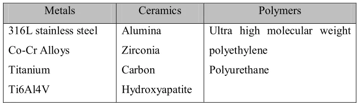
A biomaterial can be defined as any material used to make devices to replace a part or a function of the body in a safe, reliable, economic and physiologically acceptable manner. The purpose of using biomaterials is to improve human health by restoring the function of natural living tissues and organs in the body (Park and Lakes, 2007). Table 2.1 shows the biomaterials classifications and examples.
Table 2.1: Biomaterial classifications (Anonymous, 2001).
Metals Ceramics Polymers
Ultra high molecular weight polyethylene
Polyurethane
6
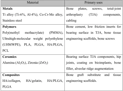
dentures. Silicones are polymer material that used in cosmetic surgery such as breast augmentation (Anonymous, 2001). Table 2.2 shows the summary of the three types of the materials (metals, ceramics and polymers) used in orthopedic application.
Table 2.2: Orthopedic biomaterials and their primary use (Basu, et al, 2009).
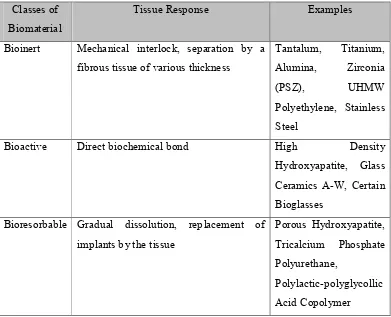
2.2 Types of Implant Tissue Response
No material implanted in living tissues is inert; all materials elicit a response from the host tissue. (Hench and June, 1993). In general, materials can be placed into three classes that represent the tissue response they are elicit, which are inert, bioresorbable and bioactive (Ain, et al, 2008) as shown in Table 2.3.
Material Primary uses
Metals
Ti alloy (Ti-6%, Al-4%), Co-Cr-Mo alloy, Stainless steel
Bone plates, screws, total-joint
arthroplasty (TJA) components,
7
Table 2.3: Classes of biomaterials according to tissue response (Ain, et al, 2008). Classes of
Biomaterial
Tissue Response Examples
Bioinert Mechanical interlock, separation by a
fibrous tissue of various thickness
Bioactive Direct biochemical bond High Density
Hydroxyapatite, Glass Ceramics A-W, Certain Bioglasses
Bioresorbable Gradual dissolution, replacement of implants by the tissue
8 2.2.2 Bioinert
Bioinert materials are biocompatible materials but cannot induce any interfacial biological bond between implants and bone. When a bioinert material is implanted, a capsule-like layer forms on the surface of the implant to keep it isolated from the living part of the body. For example, bioinert ceramics such as alumina or zirconia, develop fibrous capsules at their interface when implanted. However, the thickness of an interfacial fibrous layer depends upon motion and the extent of required fit at the interace. Therefore, bioinert materials are not useful for long-term application (Basu, et al, 2009).
2.2.3 Biodegradable / Bioresorbable
Bioresorbable materials are the type of biocompatible materials that are gradually resorbed before they finally disappear and are totally replaced by new tissues in vivo. This kind of material that is bioresorbable, degrades with time inside the body’s environment. The degradation rate should be such that the regeneration rate of new tissue will be same as the material resorption rate. Tricalcium Phosphate (TCP) and bone cement are the two examples of bioresorbable materials (Basu, et al, 2009).