MORPHOLOGICAL IDENTIFICATION AND DIVERSITY
ANALYSIS OF FOSSIL DIATOMS FROM DIATOMITE
SANGIRAN CENTRAL JAVA INDONESIA
YENNY CHUSNA KHUSTINA
DEPARTMENT OF BIOLOGY
FACULTY OF MATHEMATICS AND NATURAL SCIENCE
BOGOR AGRICULTURAL UNIVERSITY
STATEMENT ABOUT MINI-THESIS, INFORMATION
SOURCE, AND COPYRIGHT HAND OVER
By this writing I verify that mini-thesis entitled Morphological Identification and Diversity Analysis of Fossil Diatoms from Diatomite Sangiran Central Java Indonesia is my own work under the supervisions of advising committee and never been proposed in any type to any institution before. All information sources from published and unpublished works from other authors are stated clearly in the text and listed in the references at the last part of this mini-thesis.
By this writing I give the copyright of my mini-thesis to Bogor Agricultural University.
ABSTRACT
YENNY CHUSNA KHUSTINA. Morphological Identification and Diversity Analysis of Fossil Diatoms from Diatomite Sangiran Central Java Indonesia. Supervised by DEDY DURYADI SOLIHIN and NIKEN TM PRATIWI.
Diatomite Sangiran is sediment rich of fossil diatoms. The aim of this research was to analyze the diversity of fossil diatoms from Sangiran, Indonesia based on morphological characteristics. Samples were taken from lower, middle, and upper sediment layers based on their different physical features with three replications each. Diatomite extraction was completed following modification of Setty (1966) and frustule counting was accomplished by census method. There were total 50 species found in diatomite layers. Diversity analysis showed that highest species richness (21-22), diversity (1.35-1.47), and evenness index (0.44-0.48) were belong to lower layer. Highest frustule abundance (9.66x10⁷-1.43x10⁸ frustules/gram) and dominance index (0.67-0.72) were belong to middle layer. On the other hand, centrales:pennales ratios (0.73-1.11) were belong to upper layer. The sediment layers signified a good indication of gradual changes from marine to freshwater environment. Dendrogram analysis using MINITAB.v.15.1.2 software denoted similarity between lower sediment layer and the younger layers was 72.12%; while similarity between middle and upper layers 92.63%.
Keywords: diatom, diatomite, diversity, morphology, Sangiran
ABSTRAK
YENNY CHUSNA KHUSTINA. Identifikasi Morfologi dan Analisis Keanekaragaman Fosil Diatom dari Diatomite Sangiran Jawa Tengah Indonesia. Dibimbing oleh DEDY DURYADI SOLIHIN dan NIKEN TM PRATIWI.
Diatomite Sangiran merupakan sedimen yang kaya fosil diatom. Tujuan penelitian ini adalah menganalisis keanekaragaman fosil diatom dari Sangiran, Indonesia berdasarkan karakteriktik morfologis. Sampel diambil dari lapisan bawah, tengah, dan atas berdasarkan ciri fisik yang berbeda dengan masing-masing tiga ulangan. Ekstraksi diatomite dilakukan mengikuti modifikasi metode Setty (1966) dan penghitungan frustul dilakukan dengan metode sensus. Total 50 spesies ditemukan dari ketiga lapisan diatomite. Analisis keanekaragaman menunjukkan kekayaan spesies (21-22), indeks keanekaragaman (1.35-1.47), dan keseragaman (0.44-0.48) tertinggi dimiliki oleh lapisan bawah. Kelimpahan frustul (9.66x10⁷-1.43x10⁸ frustul/gram) dan indeks dominansi (0.67-0.72) tertinggi dimiliki oleh lapisan tengah. Rasio centrales:pennales (0.73-1.11) tertinggi dimiliki oleh lapisan atas. Lapisan-lapisan sedimen tersebut mengindikasikan perubahan lingkungan secara berkala dari laut menjadi tawar. Analisis dendrogram menggunakan software MINITAB.v.15.12 menunjukkan kemiripan lapisan bawah dengan dua lapisan yang lebih muda adalah 72.12%, sedangkan lapisan tengah dan atas mempunyai kemiripan sebesar 92.63%.
a Mini-thesis
Intended to Acquire Bachelor Degree of Science in
Department of Biology
MORPHOLOGICAL IDENTIFICATION AND DIVERSITY
ANALYSIS OF FOSSIL DIATOMS FROM DIATOMITE
SANGIRAN CENTRAL JAVA INDONESIA
YENNY CHUSNA KHUSTINA
DEPARTMENT OF BIOLOGY
FACULTY OF MATHEMATICS AND NATURAL SCIENCE
BOGOR AGRICULTURAL UNIVERSITY
Title : Morphological Identification and Diversity Analysis of Fossil Diatoms from Diatomite Sangiran Central Java Indonesia Name : Yenny Chusna Khustina
NIS : G34080041
Approved by
Dr Ir Dedy Duryadi Solihin, DEA Supervisor I
Dr Ir Niken TM Pratiwi, MSi Supervisor II
Acknowledged by
Dr Ir Iman Rusmana, MSi Head of Department
ACKNOWLEDGEMENTS
knowledges, and advices from my supervisors, Dr Dedy Duryadi Solihin and Dr Niken TM Pratiwi to whom I tremendously grateful. Special thanks are due to Prof Yahdi Zaim from Geology Department of Bandung Technology Institute, Dr David Williams from Natural History Museum London, my examiner Nina Ratna Djuita MSi from Biology Department of Bogor Agricultural University, Dr Tri Retnaningsih from Biology Department of Diponegoro University, Dr Triadiati, Dra Taruni Sri Prawasti MSi, and Mr. Bob Yuris Chandra for supports, advices, and trusts so I can complete my research, especially Dr Nurlisa A Butet who patiently edited my writing. Sincerely thanks to all staffs of Sangiran Museum (Pak Budi, Mbak Pipit, Mbak Lia), Microtechnique staffs (Mbak Tini and Mbak Ani), Biomicro 1 staffs (Dr Maya, Bu Siti, Mbak Ina, Mbak Dede, Kak Reza, Mbak Aay, Mbak Alim, Santika) for their great help and patience. I would also like to thank BUMN Scholarship for financially supported my study at IPB.I wish to express love and gratitude to my beloved parents for all prays and advices, my great brothers (Mas Alfi and Dhik Qohhar) who always believe in me, and my family (Mbah Kakung, Budhe Sanah, Budhe Saodah, Mas Ichus) for all supports and encouragements. Special thanks to my housemates Pustaka Ummah (Dian, Ega, Wiwi, Mbak Putri, Nina, Nurika, Teh Delita, Mbak Ria, Mbak Poo, Mbak Didi), my best friends IPA3 (Aula, Paras, Bambang, Dany, Yuna, Erna, etc), HKRB fellows (Novita, Robin, Yulifa, Intan, etc), my friend Beatrix Kotzyi, my juniors Bio 46 (Hera, Heca, Eca, Surya, Ipeh, Mario, Dimas, etc), and my lovely friends in Bio 45 (A’Agus, Afnan, Desi, Hafiz, Whendi, Aldi,Uun, Latifah, Gina, Azizah, Ai, etc) and especially my best friends ever, Dyah and Citra who show such a strong life and always stand up encouraging me to believe and pursue my dreams.
Last but by no means least, for every person I met, every moment I had, every experience I’ve been through, this mini-thesis is for all of them. Hope this mini-thesis will be useful.
TABLE OF CONTENT
ACKNOWLEDGEMENTS viii
LIST OF FIGURES x
LIST OF APPENDICES x
INTRODUCTION 1
Background 1
Objective 1
MATERIALS AND METHOD 2
Sampling Period and Location 2
Materials and Equipments 2
Methods 2
RESULTS 4
Lower Diatomite Layer 4
Middle Diatomite Layer 4
Upper Diatomite Layer 6
DISCUSSION 8
CONCLUSION 10
SUGGESTION 10
REFERENCES 10
APPENDICES 12
LIST OF FIGURES
1 Diatom species and frustule amount found at lower diatomite layer of
Sangiran Formation 5
2 Species which has the most abundant frustules from lower diatomite
layer 5
3 Diatom species and frustule amount found at middle diatomite layer of
Sangiran Formation 6
4 Species which has the most abundant frustules from middle diatomite
layer 6
5 Diatom species and frustule amount found at upper diatomite layer of
Sangiran Formation 7
6 Species which has the most abundant frustules from upper diatomite
layer 7
7 Dendrogram of cluster variables of three diatomite layers (upper,
middle, and lower) 8
LIST OF APPENDICES
1 Alphabetical list of diatom taxa from lower diatomite layer (Plate 1) 12 2 Alphabetical list of diatom taxa from middle diatomite layer (Plate 2) 14 3 Alphabetical list of diatom taxa from upper diatomite layer (Plate 3) 16
1
INTRODUCTION
Background
Diatoms are unicellular golden brown algae inhabiting light-exposed zones of entire aqueous and semi-aqueous environments. Diatoms consist of two orders, Pennales (bilateral symmetry) and Centrales (radial symmetry). External box-like skeleton (frustule) of diatom comprise of two overlapping silica valves. Classification of diatoms is based on frustule shape and patterns-types of surface ornamentation such as pores, areolae, spines, ridges, etc (Barron 1987). Dead diatom frustules form sediments at the base of their habitats (lake, river, sea, ocean, etc). These sediments still existed million years later and become diatomite through fossilization.
Color indicates purity of diatomite. White represents high purity of diatomite. Diatomite color is commonly off-white to gray, it is rarely black. Diatomite can be further described as chalk-like, soft, friable, and very finely porous or very low in density (floating on water until saturated) (Moyle and Dolley 2003). According to Soeprobowati et al. (2010), diatoms can be fossilized as diatomite because their silica contents are more than 90%. Diatomite in Indonesia can be found at majority of fossil-rich sites, such as Sangiran.
Fossil-rich site of Sangiran, known as Sangiran Dome, is located in Central Java Province, 12-20 km north of Solo City. A series of mud volcano eruptions made the Dome’s center breached out. The Dome is subdivided into four formations: Kalibeng, Pucangan, Kabuh, and Notopuro (Itihara et al. 1985). Bettis et al. (2009) stated that nowadays those formations are known as Puren, Sangiran, Bapang, and Pohjajar. Research by Itihara et al. (1985) showed that diatoms can be found at Sangiran (Pucangan) Formation. Bettis et al. (2009) and Bettis et al. (2004) studied the age of Sangiran Formation using 40Ar/39Ar analysis resulting that the age is between 1.66 and 1.90 ± 0.02 million years ago (Ma).
Knowledge about diatoms has many applications and uses, such as the presence of petroleum (Smol and Stoermer 2010). Diatoms preserved in marine sediments are commonly used to reconstruct paleoenvironments and paleoceanographic events for Holocene and Quaternary (Jordan and Stickley 2010). Van Den Hoek et al. (1995) stated that fossil diatom assemblages can be used to determine whether the sediments are of marine or non-marine origin. To achieve this, of course, it must be assumed that species had the same ecological preferences in the past that can be observed today.
Many research in Sangiran focused on fossil molluscs, foraminiferas, vertebratas, and hominids. However, research focused on morphological identification and diversity analysis of fossil diatom are rare and the publications are difficult to be found.
Objective
2
MATERIALS AND METHOD
Sampling Period and Location
This research was conducted on January-July 2013. Diatomite samples were taken from Pablengan-Krikilan Village, Sangiran Dome, Sragen Regency, Central Java, Indonesia. Preparation of samples and slides was conducted in Microtechnique Laboratory of Biology Department, Faculty of Mathematics and Natural Science. Sample identification and counting was conducted in Biomikro 1 Laboratory of Department of Aquatic Resources Management, Faculty of Fisheries and Marine Science, Bogor Agricultural University, Indonesia.
Materials and Equipments
Materials used in this research were three diatomite layers from Sangiran, hydrogen peroxide (H2O2) 7.5% and 15%, distilled water, hydrochloric acid
(HCl) 12.5% and 25%, nitric acid (HNO3) 12.5% and 25%, and entellan.
Equipments used were scales, digital camera, oven, acid room, 250 mL and 1.5 L beaker glass, water bath, stopwatch, 1 mL syringe, hot plate, sample bottle, counter, Primo Star iLED microscope, and AxioCam ERc 5s microscope camera.
Methods
Sample Preparation
Samples were taken from lower, middle, and upper diatomite layers from Sangiran Formation. The layers were differentiated based on their physical features. Lower layer is a grey-dark diatomite with high hardness because it were mixed with other sediment of Sangiran Formation. Middle layer is a white and less hard diatomite, showing that this layer has high purity of diatom. Upper layer is a white-brownish and less hard diatomite mixed with alluvial sediment.
Samples taken from the three layers were prepared following modification of Setty (1966). Those samples were dried overnight at 125ºC in the oven and then cooled to room temperature. After drying and cooling, sample from lower layer were seperated because of its high hardness. Samples from upper and middle layers were taken out 200 mg each and added with 33 mL 7.5% hydrogen peroxide (H2O2) using beaker glass 250 mL. This procedure was completed in an
acid room. Samples were boiled for 10 minutes in a hot waterbath. After samples were cooled, 200 mL distilled water were added to the samples and allowed to stand for 24 hours. Supernatant were carefully discarded prior to addition of 33 mL 12.5% hydrochloric acid (HCl) inside an acid room. The suspension were boiled again for 7 minutes and 30 seconds. After boiling and cooling, 200 mL distilled water were added for 24 hours. Supernatant were carefully discarded and 200 mL distilled water were added for 24 hours. Following that, 33 mL 12.5% nitric acid (HNO3) and 200 mL distilled water were added for 24 hours.
3 Acid liquid for lower layer sample were hydrogen peroxide (H2O2) 15% for
20 minutes, hydrochloric acid (HCl) 25% for 15 minutes, and nitric acid (HNO3)
25%. The remaining steps were the same as upper and middle layer above. Slides Preparation
Slides were prepared following modification of Setty (1966). The residue of sample preparation method were mixed with distilled water 40 mL. Using syringe, 0.05 mL suspension were deposited on cover glass and dried on hot plate. This volume represented 0.025 mg of the sediments. Then, a really small drop of entellan was added on each corner of cover glass and patched carefully on labeled object glass. Three cover glasses from each sample were prepared as replications. Identification and Counting
Diatom identification was completed using Prescott (1954), Yamaji (1966), Desikachary (1986, 1988, 1989). Counting was carried out using census method. Frustules in fair condition (seen at least ≥ 50% of the original shape) and poor condition were counted differently to reduce error counting (fair frustule was counted as 1 and poor frustule as 1/2). Acquired data were converted into one result. Because each replication represented 0.025 mg of the sediment, number of frustule per gram were acquired by formula:
Number of frustule per gram = counting result x 40000
Frustule observation was completed using Primo Star iLED microscope and AxioCam ERc 5s microscope camera by 400x magnification. Images of found diatoms were captured using Axio Vision Rel. 5.8 software.
Data Analysis
Data from identification and counting result was analyzed according to number of species, abundance, diversity and evenness index using Shannon-Wiener diversity index, and dominance index using Simpson dominance index (Dash 2001). Ratios of centrales:pennales (c:p) were analyzed following Cooper (1995) by comparing number of centrales and pennales diatom species.
Shannon-Wiener diversity index formula (H’): H’ = - ∑ piln pi
pi = proportion of each species (ni/N) ni = number of each species
N = total number of all species
According to Wilhm and Dorris (1968), values of diversity index using log function are divided into 3 categories, i.e low (H’<1), moderate (1≤ H’≤ 3), and high (H’>3). These values are equal with values using ln function, i.e low (H’< 2.3026), moderate (2.3026≤ H’≤ 6.9078), and high (H’>6.9078).
Evennes index formula (J): J = H ’
LnS
4 maximum value 1 is in the case of complete dominance (Dash 2001).
Dendrogram based on similarity among each sediment layer was constructed using MINITAB.v.15.1.2. Used variable of similarities were species and its average frustule density.
RESULTS
Lower Diatomite Layer
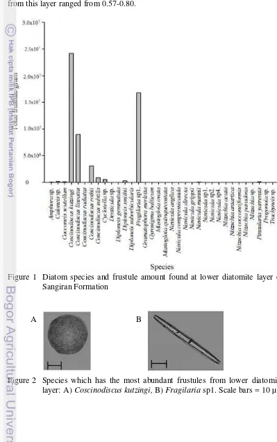
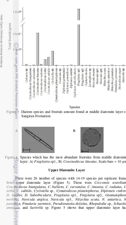
Lower diatomite layer contained the largest number of species among the layers. Total 34 number of species, ranging from 21-22 species per replicate, were found from lower diatomite layer (Figure 1). The species were Amphora sp., Caloneis sp., Cocconeis scutellum, Coscinodiscus kutzingi, C. lineatus, C. radiatus, C. rothii, C. subtilis, Cyclotella sp., Denticula sp., Diploneis gemmatula, D. Smithii, D. Suborbicularis, Fragilaria sp1., Gramatophora merletta, Gyrosigma balticum, Mastogloia ovata, Mastogloia quinquecostata, Navicula anglica, N. compressicauda, N. directa, N. grippii, N. mannii, Navicula sp1., Navicula sp2., Navicula sp4., Nitzschia acuta, Nitzschia antartica, Nitzschia cocconeiformis, Nitzschia paradoxa, Nitzschia sp., Pinularia yarrensis, Progonoia sp., and Trachyneis sp. Figure 1 shows that lower diatomite layer has Coscinodiscus kutzingi as the most abundant species with average frustule amounted to 2.42x10⁷ frustules/gram, and followed by Fragilaria sp1. as the second (Figure 2) with average frustule amounted to 1.68x10⁷ frustules/gram. The layer had diatom abundance, counted by total frustule per replicate, ranging from 5.30x10⁷-5.88x10⁷ frustule/gram. The diversity index ranged from 1.35-1.47; while the evenness index ranged from 0.44-0.48. Dominance index of the layer ranged from 0.29-0.32. Ratios of c:p ranged from 0.38-0.40.
Middle Diatomite Layer
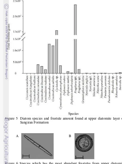
5 frustules/gram. The second abundant species, Coscinodiscus lineatus (Figure 4), has much fewer average frustule amount compared with Fragilaria sp1. i.e 1.03x10⁷ frustules/gram. The layer had diatom abundance, counted by total frustule per replicate, ranging from 9.66x10⁷-1.43x10⁸ frustule/gram. The diversity index ranged from 0.68-0.77; while the evenness index ranged from 0.22-0.27. Dominance index of the layer ranged from 0.67-0.72. Ratios of c:p from this layer ranged from 0.57-0.80.
Figure 1 Diatom species and frustule amount found at lower diatomite layer of Sangiran Formation
Figure 2 Species which has the most abundant frustules from lower diatomite layer: A) Coscinodiscus kutzingi, B) Fragilaria sp1. Scale bars = 10 µ m.
6
Figure 3 Diatom species and frustule amount found at middle diatomite layer of Sangiran Formation
Figure 4 Species which has the most abundant frustules from middle diatomite layer: A) Fragilaria sp1., B) Coscinodiscus lineatus. Scale bars = 10 µm.
Upper Diatomite Layer
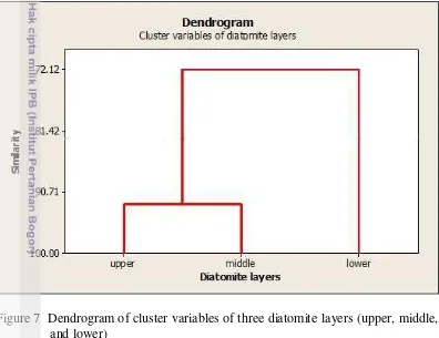
There were 26 number of species with 14-19 species per replicate found from upper diatomite layer (Figure 5). Those were Cocconeis scutellum, Coscinodiscus biangulatus, C. bulliens, C. curvatulus, C. lineatus, C. radiatus, C. rothii, C. subtilis, Cyclotella sp., Cymatodiscus planetophorus, Diploneis crabro, D. Smithii, D. Suborbicularis, Fragilaria sp1., Fragilaria sp2., Gramatophora merletta, Navicula anglica, Navicula sp3., Nitzschia acuta, N. antartica, N. paradoxa, Pinularia yarrensis, Pseudoeunotia doliolus, Rhopalodia sp.,Schuettia annulata, and Surirella sp. Figure 5 shows that upper diatomite layer has
7 Fragilaria sp1. as the most abundant species with average frustule amounted to 2.37x10⁷ frustules/gram followed by Cyclotella sp. as the second (Figure 6) with average frustule amounted to 1.55x10⁷ frustules/gram. The layer had diatom abundance, counted by total frustule per replicate, ranging from 4.49x10⁷ -1.19x10⁸ frustule/gram. The diversity index ranged from 1.03-1.11; while the evenness index ranged from 0.35-0.42. Dominance index of this layer ranged from 0.41-0.45 and ratios of c:p ranged from 0.73-1.11.
Figure 5 Diatom species and frustule amount found at upper diatomite layer of Sangiran Formation
Figure 6 Species which has the most abundant frustules from upper diatomite layer: A) Fragilaria sp1., B) Cyclotella sp. Scale bars = 10 µm.
8
DISCUSSION
Dendrogram of cluster variables of three diatomite layers (upper, middle, and lower) was constructed to show similarity among the layers. Figure 7 shows that upper and middle diatomite layers were closely similar in 92.63%. On the other hand, lower diatomite layer was different from two other layers with similarity 72.12%. It was implied that the environment of lower diatomite layer was different from the younger layers.
Figure 7 Dendrogram of cluster variables of three diatomite layers (upper, middle, and lower)
Sedimentation process always follows superposition law. Upper layer is naturally formed last, consequently it is the youngest sediment compared to other layers (Soeprobowati 2010). Given to this condition, lower diatomite layer is the oldest. The layers could show different characters because of the differences in forming age.
9 contained Fragilaria sp1. as the most abundant species and Cyclotella sp. as the second. Hasle et al. (1996) also stated that most Cyclotella species belong to freshwater. Therefore, upper diatomite layer origin could be presumed as freshwater.
The existence of diatom can be described by several approaches, such as diversity and richness. Diversity indices usually consist of diversity, evenness, and dominance index (Dash 2001 and Magurran 1988). This study revealed that order of layers from lowest to highest in diversity index was middle (0.68-0.77), upper (1.03-1.11), and lower layer (1.35-1.47); while in evenness index was middle (0.22-0.27), upper (0.35-0.42), and lower layer (0.44-0.48). However, all layers were considered to have low diversity (H’< 2.3026) and minimum evenness (J close to 0). On the other hand, that in dominance index was lower (0.29-0.32), upper (0.41-0.45), and middle layer (0.67-0.72). Lower and upper layers were considered to have low dominance (D close to 0); while middle layer has high dominance (D close to 1). Frustule abundance of lower, upper, and middle layers were 5.30x10⁷-5.88x10⁷, 4.49x10⁷-1.19x10⁸, and 9.66x10⁷-1.43x10⁸, respectively, with middle layer has the highest frustule abundance. In species richness, the lowest was found in the upper layer (26 species), followed by middle layer (27 species), then lower layer (34 species). There were total 50 species found from the three diatomite layers.
Ratios of centrales:pennales can indicate planktonic or benthic habitats. Lower ratio indicates a tendency to benthic habitat; in contrast, higher ratio tends to be more planktonic habitat (Cooper 1995). Lower diatomite layer has the lowest c:p ratios (0.38-0.40), middle layer has ratios 0.57-0.80, and upper layer has the highest ratios (0.73-1.11). Therefore, order of layers based on habitats was lower layer (more benthic), middle layer (more benthic-planktonic), and upper diatomite layer (more planktonic).
Each part of diatomite layers from Sangiran Central Java showed different characters. This suggest an indication of environmental change. First, lower layer is originated from more benthic marine mixed with freshwater diatoms which has highest indices of diversity, evenness, and species richness. However, lower layer has lowest dominance index and frustule abundance. Then, middle layer was preserved from more benthic-planktonic freshwater slightly mixed with marine diatoms which has highest dominance index and frustule abundance. Meanwhile, the middle layer has a fair species richness but lowest in diversity and evenness index. Finally, upper layer is of more planktonic freshwater diatoms which has lowest species richness but fair in diversity, evenness, dominance, and frustule abundance. However, the dendrogram showed high similarity between upper and middle layer comparing to lower layer.
10
CONCLUSION
A total of 50 species were found from diatomite layers of Sangiran. Diversity analysis showed that highest species richness, diversity, and evenness index were found in lower layer. Highest frustule abundance and dominance index were in the middle layer. However, upper layer had the highest centrales:pennales ratios. The layers showed a good indication from marine to freshwater environment change.
SUGGESTION
More study is necessary to analyze interpretation of diversity indices for fossil diatoms. This is because most of diversity indices interpretations are of modern and live plankton. More research is also needed to analyze how erosions of uplifted hinterland affecting the diatomite sedimentation.
REFERENCES
Barron JA. 1987. Miocene to Holocene Planktic Diatoms. Cambridge (GB): Cambridge University Press.
Bettis EA III, Zaim Y, Larick RR, Ciochon RL, Suminto, Rizal Y, Reagan M, Heizler M. 2004. Landscape development preceding Homo erectus immigration into Central Java, Indonesia: the Sangiran Formation Lower Lahar. Paleogeogr, Paleoclimatol, Paleoecol. 206:115-131. doi:10.1016/j.paleo.2004.01.016.
Bettis EA III, Milus AK, Carpenter SJ, Larick R, Zaim Y, Rizal Y, Ciochon RL, Tassier-Surine SA, Murray D, Suminto et al. 2009. Way out of Africa: Early Pleistocene paleoenvironments inhabited by Homo erectus in Sangiran, Java. J Human Evol.56:11-24. doi:10.1016/jhevol.2008.09.003.
Cooper SR. 1995. Cheapsake Bay watershed historical land use: impact on water quality and diatom communities. Ecol Ap. 5 (3):703-723.
Dash MC. 2001. Fundamentals of Ecology. 2nd ed. New Delhi (IN): Tata McGraw-Hill.
Desikachary TV. 1986. Atlas of Diatoms: Marine Fossil Diatoms from India and Indian Ocean Region. Madras (IN): Madras Science Foundation.
Desikachary TV. 1988. Atlas of Diatoms: Marine Diatoms of The Indian Ocean Region. Fascicle V. Madras (IN): Madras Science Foundation.
Desikachary TV. 1989. Atlas of Diatoms: Marine Diatoms of The Indian Ocean Region. Fascicle VI. Madras (IN): Madras Science Foundation.
11 Itihara M, Sudijono, Kadar D, Shibasaki T, Kumai H, Yoshikawa S, Aziz F, Soeradi T, Wikarno, Kadar AP et al. 1985. Geologi and Stratigraphy of the Sangiran Area. [Bandung]: Geological Research and Development Centre. Jordan RW, Stickley CE. 2010. Diatoms as Indicators of Paleoceanographic
events, in Smol JP and Stoermer EF, editors, The Diatoms: Aplications for the Envoronmental and Earth Sciences. 2nd ed. Cambridge (GB): Cambridge University Press.
Magurran AE. 1988. Ecological Diversity and Its Measurement. Cambridge (GB): Cambridge University Press.
Moyle PR, Dolley TP. 2003. Contributions to industrial-mineral research. With or without salt – a comparison of marine and continental-lacustrine diatomite deposits. Bull USGS. 2209(4):1-7.
Prescott GW. 1954. The Freshwater Algae. Iowa (US): WMC Brown Company Publishers.
Setty MGAP. 1966. Preparation and method of study of fossil diatoms. J Micropaleo 12:511-514.
Smol JP, Stoermer EF. 2010. The diatoms: Aplications for the environmental and earth sciences. 2nd ed. Cambridge (GB): Cambridge University Press.
Soeprobowati TR, Tandjung SD, Sutikno, Hadisusanto S, Gell P. 2010. Stratigrafi diatom danau Rawapening: Kajian paleolimnologi sebagai landasan pengelolaan danau. Di dalam: Prosiding seminar limnologi V; Bogor, 28 Juli 2010. Bogor: Puslit Limnologi Lembaga Ilmu Pengetahuan Indonesia (LIPI). hlm 102-115.
Van Den Hoek C, Mann DG, and Jahns HM. 1995. Algae an Introduction to Phycology. Cambridge (GB): Cambridge University Press.
Wilhm JL, Dorris TC. 1968. Biological parameters for water criteria. Biosci 18(6):477-481. doi:10.2307/1294272.
12
13 Plate 1
2 3
1 4 5
6 7 8 9 10
11 12 13 14 15
16 17 18 19 20 21
22 23 24 25 26
27 28 29 30 31 32
14
Appendix 2 Alphabetical list of diatom taxa from middle diatomite layer (Plate 2) 1. Caloneis sp.
2. Coscinodiscus biangulatus 3. Coscinodiscus curvatulus 4. Coscinodiscus lineatus 5. Coscinodiscus radiatus 6. Coscinodiscus rothii 7. Coscinodiscus subtilis 8. Cyclotella sp.
9. Cymatodiscus planetophorus 10. Denticula sp.
11. Diploneis gemmatula 12. Diploneis gruendlerii 13. Diploneis smithii
14. Diploneis suborbicularis 15. Fragilaria sp1.
15 Plate 2
1 2 3
7 6
5 4
9
8 10
11 12 13 14 15
16
23 22
21 20
19
17 18
16
Appendix 3 Alphabetical list of diatom taxa from upper diatomite layer (Plate 3) 1. Cocconeis scutellum
2. Coscinodiscus biangulatus 3. Coscinodiscus bulliens 4. Coscinodiscus curvatulus 5. Coscinodiscus lineatus 6. Coscinodiscus radiatus 7. Coscinodiscus rothii 8. Coscinodiscus subtilis 9. Cyclotella sp.
10. Cymatodiscus planetophorus 11. Diploneis crabro
12. Diploneis smithii
13. Diploneis suborbicularis 14. Fragilaria sp1.
15. Fragilaria sp2.
16. Gramatophora merletta 17. Navicula anglica
18. Navicula sp3. 19. Nitzschia acuta 20. Nitzschia antartica 21. Nitzschia paradoxa 22. Pinularia yarrensis 23. Pseudoeunotia doliolus 24. Rhopalodia sp.
17 Plate 3
1 2 3 4 5
6 7 8 9
15 14
13 12
11
10
16 17 18 19
23 24
20 21
26 25
18
Appendix 4 Diatomite Layers of Sangiran
19
CURRICULUM VITAE
Author was born in Rembang, January 29th 1991 as the second child of Zaenal Arifin and Mas’adah.
In 2008, author was graduated from SMAN 2 Rembang and accepted in Department of Biology, Faculty of Mathematics and Natural Sciences, Bogor Agricultural University (IPB) through Undangan Seleksi Masuk IPB (USMI).
During the college, author was active in many organizations, such as Dormitory Council A1 as Event Staff 2008-2009, Himpunan Mahasiswa Biologi (Himabio) as Bioworld staff 2009-2010, Masyarakat Rumput Community as an active member 2008-2009, and Himpunan Mahasiswa Rembang di Bogor (HKRB) as an active member 2008-recent.
Author also took part as committee in many college events such as Pesta Sains Nasional, Biology on Experiment, Biologi Interaktif, and many others. During the study, author recieved Peningkatan Prestasi Akademik (PPA) scholarship 2008-2010 and Badan Usaha Milik Negara (BUMN) scholarship 2010-2012. Beside that, author also took part on several competitions, such as became one of Olimpiade Sains Nasional (OSN) Pertamina finalist for West Java in 2011, IPB student creativity program (PKM), and others.