1. Introduction
Studies of maximal vertical jumping have revealed important insights regarding how the neuromuscular system controls and coordinates movement. For example, the effect of muscle strengthening (Bobbert and van Soest, 1994; Nagano and Gerritsen, 2001), starting posture (Selbie and Caldwell, 1996), tendon compliance (Bobbert, 2001; Anderson and Pandy, 1993), a countermovement (Bobbert et al. 1996; Finni, et al., 2000; Fukashiro, et al., 1995; Nagano, et al., 1998), muscle stimulation dynamics (Bobbert and van Zandwijk, 1999a,b; Zandwijk et al. 2000),
muscle coordination patterns (Pandy et al. 1990) and segment interactions (Bobbert and van Soest, 2001) have all been investigated using the maximal vertical jump. Relatively few studies however have investigated the standing horizontal jump, or made comparisons between the vertical jump and the horizontal jump. Such comparisons are of interest because of the potential to better understand the factors that inluence control of jump direction.
Ridderikhoff et al. (1999) compared the vertical jump with horizontal jumps without a countermovement at different projection angles and demonstrated via the use of a forward dynamics
Direction Control in Standing Horizontal
and Vertical Jumps
Senshi Fukashiro
*, Thor F. Besier
**, Rod Barrett
***, Jodie Cochrane
**,
Akinori Nagano
****and David G. Lloyd
***Graduate School of Interdisciplinary Information Studies, University of Tokyo
3-8-1 Komaba, Meguro-ku, Tokyo 153-8902 Japan fukashiro@idaten.c.u-tokyo.ac.jp
**School of Human Movement and Exercise Science, The University of Western Australia
Australia
***School of Physiotherapy and Exercise Science, Grifith University
Australia
****Computational Biomechanics Unit, RIKEN
Saitama, Japan
[Received July 12, 2005 ; Accepted August 30, 2005]
The purpose of this study was to perform a detailed k inematic, k inetic, and electromyographic comparison of maximal effort horizontal and vertical jumping. It was of particular interest to identify factors responsible for the control of jump direction. Eight male subjects performed maximal horizontal jumps (HJ) and vertical jumps (VJ) from a standing posture with a counter movement. Three-dimensional motion of the trunk, pelvis, and bilateral thigh, shank, and foot segments were recorded together with bilateral ground reaction forces and electromyographic (EMG) activity from seven right leg muscles. Relative to the VJ, the trunk is displaced further forward at the beginning of the HJ, through greater ankle joint dorsilexion and knee extension. The activity of the biarticular rectus femoris and hamstrings were adapted to jump direction and helped to tune the hip and knee joint torques to the requirements of the task. The primary difference in joint torques between the two jumps was for the knee joint, with the extension moment reduced in the HJ, consistent with differences in activation levels of the biarticular rectus femoris and hamstrings. Activity of the mono-articular knee extensors was adapted to jump direction in terms of timing rather than peak amplitude. Overall results of this study suggest that jump direction is controlled by a combination of trunk orientation at the beginning of the push-off and the relative activation levels of the biarticular rectus femoris and hamstring muscles during the push-off.
Keywords: jumping, EMG, coordination
simulation model that realistic horizontal jumps may be achieved using the muscle stimulation patterns from a vertical jump by simply rotating the body mass forward in relation to the base of support prior to extending the legs. The authors referred to this as a rotation-extension strategy. Results suggested that the required adaptations to the net joint moments that occur when jumping forward compared to upward can be produced by intrinsic muscle properties alone (ie. stiffness and damping). This simpliies the neural control of tasks such as jumping because the same muscle stimulation is effective, although not optimal, for a range of jump directions.
In a comparison of countermovement jumps performed in different directions, Jones and Caldwell (2003) explored hypotheses concerning the role of mono-articular and bi-articular muscles. It was hypothesized that bi-articular muscle activity would be modulated to control the direction of the ground reaction force for jumps in different directions, whereas mono-articular muscle activity would not be affected by jump direction. These hypotheses are based on the idea that bi-articular muscles are activated to tune the distribution of moments amongst joints in order to meet task requirements (Jacobs and Ingen Schenau, 1992), whereas mono-articular muscles are activated when they can contribute positive work at the joint they span (Jacobs, Bobbert & van Ingen Schenau, 1993). While Jones and Caldwell (2003) showed activity of the biarticular rectus femoris, hamstrings and gastrocnemius changed with jump direction, changes in activity patterns of some mono-articular muscles were also observed in contrast with their hypothesized role. Jones and Caldwell (2003) also reported that the centre of mass had unique linear and angular momenta at the beginning of the push-off for each jump direction, which suggested that direction control begins during the countermovement phase.
The purpose of the present study was to perform a detailed kinematic, kinetic, and electromyographic comparison between HJ and VJ throughout the countermovement, push-off phase and initial light phase. More speciically, it was of interest to determine to what extent a rotation-extension strategy described by Ridderikhoff et al. (1999) for horizontal jumps performed without a countermovement, is adopted in horizontal jumps performed with a countermovement. It was hypothesized that jump direction would be inluenced by a combination of
body coniguration at the beginning of the push-off, as well as the relative activation of bi-articular muscles during the push-off.
2. Methods
Eight male intermediate level Australian Football players (mean (SD) body height: 178.3 (7.5) cm, body mass: 70.4 (6.8) kg, age: 24.9 (2.4) yrs) performed three successive standing horizontal and vertical jumps. Subjects submitted written informed consent forms before testing to comply with the ethics committee of The University of Western Australia. Subjects were instructed to jump as ‘far’ as possible in HJ’s, and as ‘high’ as possible in VJ’s, from an erect standing position. Subjects were instructed to keep their hands on their waist during all the jumps and were allowed to perform a countermovement.
Clusters of three retro-relective markers were irmly attached to the pelvis, thigh, shank, and foot segments of each subject using double-sided tape. Markers were also attached to the sternum, left and right acromion processes, and thoracic spinous process (opposite the sternum marker) to represent the trunk segment (Figure 1). Three-dimensional segment motion was recorded using a six-camera, 50-Hz VICON motion analysis system (Oxford Metrics Ltd., Oxford, UK) and ground reaction force data were collected synchronously from two AMTI force plates at 2000 Hz (Advanced Mechanical Technology Inc., Watertown, MA). A kinematic and kinetic model was developed using BodyBuilder software (Oxford Metrics Ltd., Oxford, UK) to determine trunk angle relative to the horizontal, as well as hip, knee, and ankle joint angles and moments. The lower limb model has been described in detail elsewhere (Besier et al. 2003) and produces repeatable kinematic and kinetic data by utilizing functional methods to deine hip and knee joint centres. Sagittal plane hip, knee, and ankle joint angles, moments and power data were averaged across three trials for each subject.
semimembranosus (SEMIMEM), m. biceps femoris (BIFEM), m. gastrocnemius medialis (GASMED), and m. gastrocnemius lateralis (GASLAT). The skin was shaved and exfoliated using coarse plastic gauze, then cleaned with alcohol prior to electrode placement. Raw EMG signals from each muscle were high pass iltered at 30 Hz using a zero-lag Butterworth ilter to remove movement artifact. The EMG data were then full wave rectiied before being smoothed with an 8 Hz zero-lag low pass Butterworth ilter. The resulting EMG proiles were then normalized to a maximal isometric voluntary contraction (MVC) for each muscle group, which was collected prior to the jumping trials.
The dependent measures evaluated were categorized as center of mass (COM) motion variables, ground reaction force variables, joint rotation variables and muscle activation variables. The COM motion variables included; vertical and horizontal components of the COM velocity at take-off, the projection angle of the COM, and
maximal height jump reached by the COM during the jump. Ground reaction force variables were the peak vertical and horizontal GRF in the push-off as well as the minimum ‘unweighting’ force (arrow a in Figure 2) during the countermovement. Joint rotation variables consisted of the peak hip, knee, and ankle joint angles during push-off and take-off as well as the peak sagittal plane hip, knee, and ankle joint moments during the push-off. The peak trunk lexion angle during push-off was also assessed. Muscle activation variables consisted of the peak normalized EMG signals of RF, VASMED, VASLAT, SEMIMEM, BIFEM, GASMED and GASLAT during push-off.
Two-tailed paired t-tests were used to determine differences in each dependent measure between the horizontal and vertical jumps. Signiicance was accepted at p<0.05.
3. Results
Mean values for COM motion, ground reaction force, joint rotation and muscle activation variables for the HJ and VJ are reported in Table 1. As expected, the horizontal take-off velocity was signiicantly higher in the HJ compared to the VJ, whereas the vertical take-off velocity, maximal jump height and projection angle of the COM were signiicantly higher for the VJ.
Notable differences were observed in ground reaction force (GRF) proiles between HJ and VJ (Figure 2). The peak vertical GRF was greater in the VJ compared to the HJ (Figure 2A), and the horizontal GRF was much greater in the HJ than in the VJ (Figure 2B).
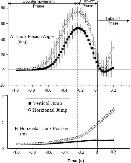
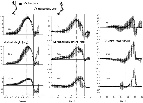
Larger peak angular displacement of the trunk segment relative to the horizontal was observed in the HJ than in the VJ (Figure 3). Although only a small angular displacement of the trunk segment was observed in the VJ after take off, a substantial angular displacement of the trunk segment was observed after take off in the HJ (Figure 3A). Note also that the trunk translates in the horizontal direction during the push-off in the HJ (Figure 3B). Joint kinematics of the hip, knee, and ankle were remarkably similar between both jumps during the push-off (Figure 4A), but with ~10 degrees less knee lexion in the HJ compared to the VJ. Hip, knee, and ankle joint angles were similar at take-off between the two jumps. Following take off, the hip, knee, and ankle
Pelvis
Femur
Tibia Foot
Trunk
zzzz
Y
X Z
Y
X Z
Y
X Z
Y
X Z Y
X Z
joints began to lex in the HJ in anticipation of the forward landing, whereas these joint angles remained constant during the VJ.
Although the hip, knee, and ankle joint angles were similar between HJ and VJ before take off, the net joint moments displayed more noticeable differences (Figure 4B). Hip and knee joints exhibited greater peak extension moments during the VJ than the HJ and were also greater for most of the push-off phase. The timing of peak moment generation at the knee and ankle joints was also different between jumps. In the VJ, the knee moment peak was reached prior to the ankle moment and this sequence was reversed in the HJ. Although the peak ankle joint extension was similar between jumps, the peak ankle moments occurred much earlier in the HJ than the VJ, and reduced to near zero values ~0.1 sec prior to take-off.
Joint powers for the VJ and SJ are displayed in
Figure 4C. Joint power at the knee joint was similar between jumps. However, hip and ankle joint powers Table 1 Mean values (SD) (n = 8) for centre of mass motion, ground reaction force, joint rotation and muscle activation variables for the horizontal and vertical jump (* = p<0.05).
Figure 2 Proiles of ground reaction force, (a) vertical component and (b) horizontal component. The instant of take off is indicated as 0.0 sec. 1.0 sec before take off and 0.4 sec after take off is shown.
-1.0 -0.8 -0.6 -0.4 -0.2 0 0.2 -200
0 200 400 600 800 1000
A: Vertical component, Fy
B: Horizontal component, Fx
Time (s)
-1.0 -0.8 -0.6 -0.4 -0.2 0.0 0.2 -100
0 100 200 300
a
Vertical Jump Horizontal Jump Ground Reaction Force (N)
Variable Horizontal Vertical
Centre of Mass motion variables
Vertical take-off velocity (m.s-1) 2.63 (0.13) 2.83 (0.09) *
Horizontal take-off velocity (m.s-1) 2.40 (0.12) 0.06 (0.18) *
Projection angle (deg) 47.6 (2.64) 88.8 (1.88) * Jump height (m) 0.35 (0.03) 0.41 (0.03) *
Ground reaction force (GRF) variables (N)
Peak Vertical GRF 761 (133) 832 (111) * Peak Horizontal GRF 259 (48) 60 (21) * Unweighting GRF 463 (79) 436 (85)
Joint variables
Peak trunk flexion angle (deg) 76.1 (5.5) 54.8 (7.1) * Peak hip flexion angle (deg) 95.9 (7.7) 91.9 (4.2) Peak knee flexion angle (deg) 90.8 (7.8) 100.5 (7.3) * Peak ankle flexion angle (deg) 40.6 (3.0) 36.2 (3.7) * Hip angle at take-off (deg) 7.6 (7.0) 22.7 (5.5) * Knee angle at take-off (deg) 22.4 (8.0) 13.9 (10.1) Ankle angle at take-off (deg) -15.7 (5.9) -20.1 (10.3) Peak hip moment (Nm) 102.2 (33.0) 112.8 (40.5) * Peak knee moment (Nm) 105.2 (24.6) 118.7 (34.7) * Peak ankle moment (Nm) 95.3 (21.2) 96.1 (24.4)
Muscle activation variables (% MVC)
RF 65.9 (11.4) 126.7 (45.7) *
VASMED 166.8 (65.6) 169.8 (52.6) VASLAT 156.7 (47.3) 160.7 (53.7)
SEMIMEM 55.3 (22.2) 17.7 (8.2) *
BIFEM 109.5 (56.7) 73.6 (59.0) *
GASMED 225.4 (76.3) 195.4 (66.6)
GASLAT 124.3 (7.8) 121.2 (28.7)
Figure 3 Proiles of the angle (a) of the trunk segment (upright = 0 deg) and the position (b) of the trunk segment. Forward inclination of the trunk segment is indicated as positive.
0 1 2
-1.0 -0.8 -0.6 -0.4 -0.2 0 0.2
-20 0 20 40 60 80
A: Trunk Flexion Angle (deg)
Time (s)
B: Horizontal Trunk Position (m)
-1.0 -0.8 -0.6 -0.4 -0.2 0 0.2
Countermovement
Phase Push-offPhase
Take-off Phase
differed greatly between the two jump conditions, with large variation between subjects. Peak hip extension power generated during the HJ was ~30% less than the VJ, as a result of reduced hip angular velocity and reduced net moment at the hip. The power generated at the ankle in the VJ was more than four times that of the HJ, due to a large reduction in the joint moment towards the end of push off in the HJ condition.
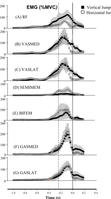
Differences in EMG proiles were also observed between the HJ and the VJ (Figure 5). Compared to the VJ, the peak RF activity during HJ was lower (Figure 5A) and the hamstring activation was greater (Figure 5D/E). Just before take off, VASMED and VASLAT activity reduced in the HJ, whereas substantial muscle activation was maintained even after the instant of take off in the VJ (Figure 5B/C). GASMED and GASLAT were activated similarly
during push off in both jumps, however, greater activation was observed after take off in the VJ compared to the HJ (Figure 5F/G).
4. Discussion
This study sought to identify factors responsible for the control of jump direction by comparing the kinematic, kinetic, and muscle activation data for maximal vertical and horizontal jumps. A focus of the study was to determine the speciic nature of differences in the body coniguration at the start of and during the push-off. It was also of interest to determine whether muscle activation patterns during the push-off in the horizontal jump were adapted consistent with hypothesis concerning the differential roles of mono- and bi-articular muscles.
Hip
B: Net Joint Moment (Nm) C: Joint Power (W/kg)
A: Joint Angle (deg)
4. 1. Differences in body coniguration
The coniguration of the body segments at the start of the push-off has previously been shown to explain how an effective horizontal jump can be obtained using the neural input from a vertical jump (Ridderikhoff, et al., 1999). This strategy (termed rotation-extension) simply involves rotating the centre of mass forward of the feet prior to an explosive leg extension. In the present study, the main difference in the coniguration of the segments between HJ and VJ was for the trunk angle at the beginning of the push-off, which was about 25° more
lexed in the HJ. The trunk segment was positioned more anteriorly in the HJ, so that subsequent extension of the lower extremity contributed to further anterior displacement of the COM due to a larger horizontal component of the GRF. The reason for the more anterior trunk displacement during the push-off in the horizontal jump was related to the ankle being more dorsi-lexed and the knee being more extended during push-off. These results suggest that, unlike for pure rotation-extension, ankle and knee joint angle proiles are different for maximal vertical and horizontal jumps. However these differences help to locate the body mass appropriately so that it can be projected in the desired direction during the extension phase.
4.2. Differences in muscle activation patterns and joint moments
Similar to Jones and Caldwell (2003), the results of the present study indicate that the activation levels of bi-articular muscles differ according to jump direction. Hamstring activity increased, and rectus femoris activity decreased from the VJ to the HJ consistent with the hypothesis that the activity of biarticular muscles is modulated to control the direction of the ground reaction force (Jacobs and Ingen Schenau, 1992). These results suggest that bi-articular muscle activity during the push-off plays a part in adapting the joints moments when jumping in different directions. This is not to say that intrinsic muscle properties do not play an important role as demonstrated by Ridderikhoff, et al., (1999), but rather that the neural control strategy differs according to jump direction, presumably to optimize the jump performance.
The main adaptation to joint moments from VJ to HJ was at the knee, where the extension moment was lower for the HJ, presumably due to lower rectus femoris and greater hamstrings activity. Hip moments were greater in the HJ during the countermovement phase as predicted due to lower rectus femoris and higher hamstring activity which is related to the higher gravitational torque associated with greater trunk lexion in the HJ. However, hip moments were found to be greater for the VJ through push-off, perhaps because of inertial differences between the two jumps. No differences in the peak activity of the mono-articular knee extensors were evident between the two jumps consistent with the Figure 5 Electromyography of lower extremity muscles:
(A) m. rectus femoris, (B) m. vastus medialis, (C) m. vastus lateralis, (D) m. semimembranosus, (E) m. biceps femoris, (F) m. gastrocnemius medialis, and (G) m. gastrocnemius lateralis.
0 100 200
(E) BIFEM 0
100
200 EMG (%MVC)
(A) RF
0 100 200
(B) VASMED
0 100 200
(C) VASLAT
0 100
(D) SEMIMEM
0 100 200 300
(F) GASMED
-1.0 -0.8 -0.6 -0.4 -0.2 0.0 0.2 0.4
0 100 200
(G) GASLAT
Time (s)
hypothesis that these muscles are primarily work generators (Jacobs, Bobbert & van Ingen Schenau, 1993). However, similar to Jones and Caldwell (2003) qualitative comparisons suggest that the activity of these muscles is more prolonged in the VJ. We interpret this result to indicate that monoarticular muscle activity is adapted to jump direction more in terms of timing rather than peak amplitude.
5. Conclusion
Results of this study suggest that jump direction is inluenced by trunk segment position at the beginning of the push-off as well as the relative activation of biarticular muscles during the push-off, which help to adapt the joint moments to the requirements of the task.
Acknowledgment
This research was supported by Ministry of Education, Culture, Sports, Science and Technology in Japan (No: 12680015), and the Australian Football Research and Development Board. The authors gratefully acknowledge the assistance of Donna-Lee Ferguson for processing the data.
References
Anderson, F. C. and Pandy, M. G. (1993). Storage and utilization of elastic strain
energy during jumping. Journal of Biomechanics. 26, 1413-1427.
Besier, T. F., Sturnieks, D. L., Alderson, J. A. and Lloyd, D. G. (2003). Repeatability of gait data using a functional hip joint centre and an optimal knee helical axis. Journal of Biomechanics. 2003. 36, 1159-1168.
Bobbert, M. F., Gerritsen, K. G. M., Litjens, M. C. and van Soest, A. J. (1996). Why is counter movement jump height greater than squat jump height? Medicine and Science in Sports and Exercise. 28, 1402-1412.
Bobbert, M. F. and van Soest, A. J. (1994). Effects of muscle strengthening on vertical jump height: a simulation study.
Medicine and Science in Sports and Exercise. 26, 1012-1020. Bobbert, M.F. and van Soest, A.J. (2001). Why do people jump
the way they do? Exercise and Sport Sciences Reviews, 29, 95-102.
Bobbert, M.F. and van Zandwijk, J.P. (1999a). Dynamics of force and muscle stimulation in human vertical jumping.
Medicine and Science in Sports and Exercise, 31, 303-310. Bobbert, M.F. and van Zandwijk, J.P. (1999b). Sensitivity
of vertical jumping performance to changes in muscle stimulation onset times: a simulation study. Biological Cybernetics, 81, 101-108.
Bobbert, M.F. (2001). Dependence of human squat jump performance on the series elastic compliance of the triceps surae: a simulation study. Journal of Experimental Biology, 204, 533-542.
Doorenbosch, C.A. and van Ingen Schenau, G.J. (1995). The role of mono- and biarticular muscles during contact control leg
tasks in man. Human Movement Science, 14, 279-300. Finni, T., Komi, P. V. and Lepola, V. (2000). In vivo human
triceps surae and quadriceps femoris muscle function in a squat jump and counter movement jump. European Journal of Applied Physiology. 83, 416-426.
Gregoire, L., Veeger, H. E., Huijing, P. A. and van Ingen Schenau, G. J. (1984). Role of mono- and biarticular muscles in explosive movements. International Journal of Sports Medicine. 5, 301-305.
Jacobs, R., Bobbert, M. F. and van Ingen Schenau, G. J. (1996). Mechanical output from individual muscles during explosive leg extensions: the role of biarticular muscles. Journal of Biomechanics. 29, 513-523.
Jones, S.L. & Caldwell, G.E. (2003). Mono- and biarticular muscle activity during jumping in different directions.
Journal of Applied Biomechanics, 19, 205-222.
Nagano, A. and Fukashiro, S. (2000). Biomechanical comparison of the role of biarticular rectus femoris in standing broad jump and vertical jump. Japanese Journal of Biomechanics in Sports and Exercises. 4, 8-15.
Nagano, A. and Gerritsen, K. G. M. (2001). Effects of neuromuscular training on vertical jumping performance – a computer simulation study. Journal of Applied Biomechanics. 17, 27-42.
Nagano, A., Ishige, Y. and Fukashiro, S. (1998). Comparison of new approaches to estimate mechanical output of individual joints in vertical jumps. Journal of Biomechanics. 31, 951-955.
Pandy, M. G.and Zajac, F. E. (1991). Optimal muscular c o ord i n at ion s t r at eg ie s for ju mpi ng. Jo u r n a l of Biomechanics. 24, 1-10.
Prilutsky, B. I. and Zatsiorsky, M. (1994). Tendon action of two-joint muscles: transfer of mechanical energy between joints during jumping, landing, and running. Journal of Biomechanics. 27, 25-34.
Prilutsky, B.I. and Gregor, R.J. (2000). Analysis of muscle coordination strategies in cycling. IEEE Transactions on Rehabilitation Engineering, 8, 362-370.
Prilutsky, B.I. and Gregor, R.J. (1997). Strategy of muscle coordination of two- and one-joint leg muscles in controlling and external force. Motor Control, 1, 92-116.
Prilutsky, B.I. (2000). Coordination of two- and one-joint muscles: functional consequences and implications for motor control. Motor Control, 4, 1-44.
Ridderikhoff, A., Batelaan, J. H. and Bobbert, M. F. (1999). Jumping for distance: control of the external force in squat jumps. Medicine and Science in Sports and Exercise. 31, 1196-1204.
Selbie, W. S. and Caldwell, G. E. (1996). A simulation study of vertical jumping from different starting postures. Journal of Biomechanics. 29, 1137-1146.
van Ingen Schenau, G. J., Bobbert, M. F. and Rozendal, R. H. (1987). The unique action of bi-articular muscles in complex movements. Journal of Anatomy. 115, 1-5.
van Ingen Schenau, G.J. (1990). On the action of biarticular muscles, a review. Netherlands Journal of Zoology, 40, 521-540.
van Ingen Schenau, G.J., Bobbert, M.F. and van Soest, A.J. (1990). The unique action of biarticular muscles in leg extensions. In J.M. Winters and S.L.-Y. Woo (Eds). Multiple Muscle Systems: Biomechanics and Movement Organisation
van Ingen Schenau, G.J., Pratt, C.A. and McPherson, J.M. (1994). Differential use and control of mono- and biarticular muscles. Human Movement Science, 13, 495-517.
van Soest, A. J., Schwab, A. L., Bobbert, M. F. and van Ingen Schenau, G. J. (1993). The inluence of the biarticularity of the gastrocnemius muscle on vertical-jumping achievement.
Journal of Biomechanics. 26, 1-8.
van Zandwijk, J.P., Bobbert, M.F. and Harlaar, J. (2000). Predictions of mechanical output of the human m. triceps surae on the basis of electromyographic signals: the role of stimulation dynamics. Journal of Biomechanical Engineering, 122, 380-386.
Name:
Senshi Fukashiro
Afiliation:
Graduate School of Interdisciplinary Infor-mation Studies, University of Tokyo
Address:
3-8-1 Komaba, Meguro, Tokyo 153-8902, Japan Brief Biographical History:
1993 PhD, University of Tokyo
1993- Executive Council of Japanese Society of Biomechanics 1999-2005 Executive Council of International Society of Biomechanics
1998-2004 Executive Council of Japan Society of Physical Education, Health and Sport Sciences
2002- President of Tokyo branch of Japan Society of Physical Education, Health and Sport Sciences
2002-2004 Associate Editor of Journal of Japan Society of Physical Education, Health and Sport Sciences
2003- Associate Editor of International Journal of Sport and Health Science
Main Works:
• S. Fukashiro (1990): Science of Jumping. Taishukan-shoten, Tokyo.
• S. Fukashiro, S. Sakurai, Y. Hirano and M. Ae (2000): Sport Biomechanics. Asakura-shoten, Tokyo.
• S. Fukashiro and A. Shibayama (2000): Handbook of Mathematics and Physics in Sports. Asakura-shoten, Tokyo. • S. Fukashiro, M. Noda and A. Shibayama (2001): In vivo
determination of muscle viscoelasticity in the human leg. Acta Physiol. Scand. 172:241-248.
• S, Fukashiro, T. Abe, A. Shibayama and W.F. Brechue(2002): Comparison of viscoelastic characteristics in triceps surae between Black and White athletes. Acta Physiol Scand. 175(3):183-187.
Membership in Learned Societies: • Japanese Society of Biomechanics • International Society of Biomechanics


