The efficiency of lactic acid bacteria against pathogenic
fungi and mycotoxins
Adam Perczak
1, Piotr Goliński
1, Marcin Bryła
2, and Agnieszka Waśkiewicz
1Department of Chemistry, Poznan University of Life Sciences, Poznań
1, Department of Food Analysis, Prof. Waclaw
Dabrowski Institute of Agricultural and Food Biotechnology, Warsaw
2, Poland
[Received in October 2017; Similarity Check in October 2017; Accepted in March 2018]
Mycotoxins are produced by some fungal species of the genera
Aspergillus
,
Penicillium
, and
Fusarium
and are common
contaminants of a wide range of food commodities. Numerous strategies are used to minimise fungal growth and mycotoxin
contamination throughout the food chain. This review addresses the use of lactic acid bacteria, which can inhibit fungal
growth and participate in mycotoxin degradation and/or removal from contaminated food. Being beneficial for human
and animal health, lactic acid bacteria have established themselves as an excellent solution to the problem of mycotoxin
contamination, yet in practice their application in removing mycotoxins remains a challenge to be addressed by future
research.
KEY WORDS: Aspergillus
; biological methods;
Fusarium
; inhibition; LAB;
Penicillium
Mycotoxin contamination of feed and food is a
significant issue worldwide. Mycotoxins are a large group
of secondary metabolites produced by the
Aspergillus
,
Penicillium
, and
Fusarium
genera, and pose serious risks
for human and animal health (1-4). Fungal growth and
mycotoxin production may occur in the field and/or during
storage, if the temperature and humidity are favourable
(4-8).
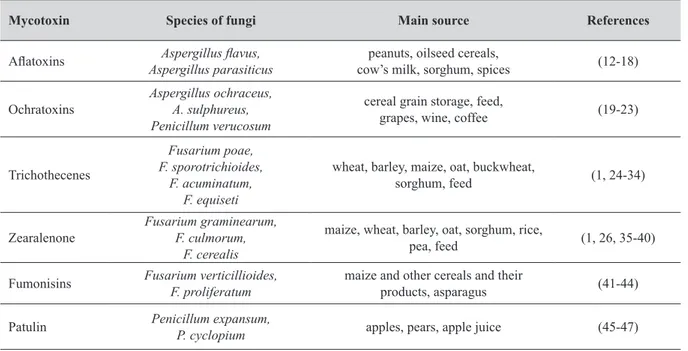
The main sources of mycotoxins are cereal grains
(including wheat, barley, oats, corn, and rice) and their
products, nuts, almonds, fruits, coffee, spices, and legumes
(5, 6, 9-11) (Table 1).
Correspondence to: Agnieszka Waśkiewicz, Department of Chemistry, Poznan University of Life Sciences, Wojska Polskiego 75, Poznań, Poland, E-mail: agat@up.poznan.pl
Table 1 The main sources of mycotoxins
Mycotoxin Species of fungi Main source References
Aflatoxins Aspergillus parasiticusAspergillus flavus, cow’s milk, sorghum, spicespeanuts, oilseed cereals, (12-18) Ochratoxins Aspergillus ochraceus,A. sulphureus,
Penicillum verucosum
cereal grain storage, feed,
grapes, wine, coffee (19-23)
Trichothecenes
Fusarium poae, F. sporotrichioides,
F. acuminatum, F. equiseti
wheat, barley, maize, oat, buckwheat,
sorghum, feed (1, 24-34)
Zearalenone Fusarium graminearum,F. culmorum, F. cerealis
maize, wheat, barley, oat, sorghum, rice,
pea, feed (1, 26, 35-40) Fumonisins Fusarium verticillioides,F. proliferatum maize and other cereals and their products, asparagus (41-44) Patulin Penicillum expansum,P. cyclopium apples, pears, apple juice (45-47)
contaminate eggs, milk, and meat and accumulate in
different organs or tissues (9, 11, 48).
Long-term exposure to mycotoxins has also been
associated with carcinogenic, mutagenic, teratogenic,
oestrogenic, haemorrhagic, immunotoxic, nephrotoxic,
hepatotoxic, neurotoxic, and immunosuppressive adverse
health effects (48-51).
Because of these risks, the EU has set down limits for
several important mycotoxins in food and feed: aflatoxins
(AFs), ochratoxin A (OTA), fumonisins (FBs), zearalenone
(ZEA), trichothecenes [principally deoxynivalenol (DON),
T-2, and HT-2 toxins], and patulin (PAT) (9).
A number of studies have investigated the options of
eliminating these compounds (52-54), and arrived at the
conclusion that the best way to solve the problem is to
prevent mycotoxin formation. To do that, the following
rules need to be observed: (i) plant materials should be
stored in a cold and dry environment, (ii) plants should be
harvested without delay, (iii) crops should be rotated in the
field to prevent adaptation of pathogenic microorganisms
to a specific monoculture, and (iv) agricultural crops should
be handled carefully to prevent mechanical damage, which
renders them vulnerable to contamination (52, 53, 55).
There where prevention fails, chemical, physical, or
biological methods of detoxification step in (56-58). Some
of these methods, such as the use of ozone (59-62), alkaline
hydrogen peroxide (63), or gamma irradiation (64, 65) have
more promising results than ammoniation (66) or heat
treatment (67-69).
However, contamination with several mycotoxins at the
same time lessens the efficiency of detoxification, as some
mycotoxins are less sensitive to the method than others.
Recently, a new approach to the removal of mycotoxins
emerged, and microorganisms such as propionic
fermentation bacterium
Saccharomyces cerevisiae
, and
lactic acid bacteria (LAB) have come into focus. This article
reviews the current uses of the latter as promising probiotics
in mycotoxin removal.
Lactic acid bacteria
Lactic acid bacteria (LAB) are Gram-positive,
nonsporulating, air and acid tolerant, organotrophic,
fermentative rods or cocci producing lactic acid. They do
not use oxygen as an electron acceptor. Not possessing
catalase, they synthesise superoxide dismutase, removing
reactive oxygen species. All the lactic acid bacteria are
anaerobic, while some of them tolerate low levels of oxygen
in the environment. Currently, only a few are considered to
be probiotic, and, together with prebiotics, these have been
used in nutrition and treatment of people and farm animals,
such as pigs. When homofermentative, LAB ferment 85 %
of glucose into lactic acid. In heterofermentation (70) the
yield is 50 % plus ethanol and CO
2.
including
Lactococcus, Enterococcus, Oenococcus,
Pediococcus, Streptococcus, Leuconostoc
, and
Lactobacillus
species (71). Yet, LAB have been used to preserve food and
beverages since the beginnings of agriculture (72). Different
strains of LAB have been passed down from generation to
generation through culinary traditions and fermented food.
Currently, LAB play a significant role in the world food
production, performing major bioconversions in fermented
dairy products, vegetables, and meat. They are also essential
for the production of silage, coffee, wine, cocoa, sourdough,
and many indigenous fermented foods (73-75). LAB
improve flavour, texture, and shelf-life of food products
(76).
LAB inhibit fungal growth
Lactic acid bacteria have the ability to control the
growth of various fungi. Inhibition of toxigenic fungi has
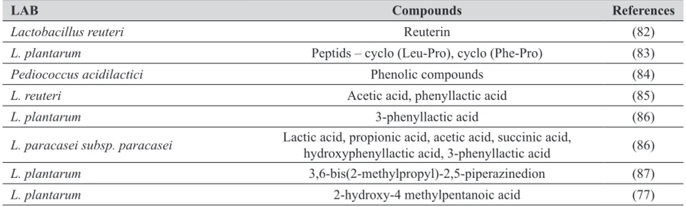
been demonstrated many times over (77-81). Generally,
this antagonistic effect is owed to low-molecular-weight
compounds produced by the LAB, such as organic acids
(acetic and lactic acid), hydrogen peroxide, proteinaceous
compounds, reuterin, hydroxyl fatty acids, and phenolic
compounds (Table 2). Organic acids can be native to food
or added to it. They are products of carbohydrate metabolism
and are safe to use for food preservation. Lactic acid lowers
pH, which inhibits the growth of various microorganisms
or even kills susceptible bacteria (89). In heterofermentation,
LAB can produce acetic acid and trace amounts of propionic
acid, both of which have a higher content of undissociated
forms at a given pH of the lactic acid. In addition to their
effect on the fungus membrane, they also inhibit the
absorption of amino acids (89). Low pH also increases the
antifungal activity of various salts of propionic acid (90).
A particularly interesting component involved in the
inhibition of fungal growth is reuterin, a compound of
glycerol fermentation produced by various LAB genera
under anaerobic conditions (91). Reuterin suppresses the
activity of ribonuclease, the enzyme involved in the
biosynthesis of DNA (98). It inhibits the growth of the
Fusarium
and
Aspergillus
species. Therefore, to enhance
these effects, simply add glycerol to LAB cultures.
Lactic acid bacteria can produce various types of fatty
acids that improve the sensory quality of fermented
products. One such fatty acid, caproic acid, has a strong
antifungal activity. It may be synergistic with propionic,
butyric, or valeric acids (92).
The best period of incubation to inhibit the growth of
toxin-forming fungi is about 48 h and the best temperature
is from 25 to 30 °C (93). These conditions favour the
production of organic acids, which in turn, inhibit the
growth of pathogenic fungi.
AFG
2), while
A. flavus
is usually found on cereals and
produces only the B
1and B
2aflatoxins (13, 18) (Table 1).
AFB
1in dairy mammal feed is strongly associated with
aflatoxin M
1(AFM
1) in milk (104). Other, less common
species include
A. nomius
,
A. toxicarius, A. tamarii, A.
pseudotamari
, and
A. bombycids
(15, 140, 141).
Conditions favouring aflatoxin production are humidity
above 13 % and temperature between 24 and 37 °C (142),
which are mostly encountered in the countries with
subtropical and tropical climates (143-145). In recent years,
aflatoxins in maize have also been reported in southern
Europe. This is probably due to climate change and adaptive
abilities of the
Aspergillus
spp. (146).
Agricultural commodities get contaminated with
aflatoxigenic fungi before and at the harvest, processing,
transport, and storage (147), especially peanuts, cereals,
and their products (148, 149), as well as animal feeds (16,
150-152).
Motameny et al. (117) investigated the removal of AFB
1from a gastrointestinal model with
L. rhamnosus, L.
plantarum
,
and
L. acidophilus
and found that
L. plantarum
was the most successful (28 %), followed by
L. acidophilus
(22 %) and
L. rhamosus
(18 %). Elsanhoty et al. (120)
compared the ability of viable and heat-treated
L.
acidophilus
,
L. rhamnosus
,
L. sanfranciscensis,
and
Bifidobacterium angulatum
to remove AFBs (AFB
1, AFB
2,
AFG
1, and AFG
2) from PBS liquid medium. Among the
four tested strains,
L. rhamnosus
was the most efficient in
the initial binding of all these aflatoxins and confirmed
superior efficiency after 4 washes, which suggests that it
forms the most stable complexes with these aflatoxins.
Hernandez-Mendoza et al. (124) studied the binding of
AFB
1by
Lactobacillus reuteri
and
L. casei
at different pH
(6, 7.2, and 8) and incubation time (0, 4, and 12 h)
.
Both
strains showed the highest AFB
1-binding capacity at pH 7.2
after 4 and 12 h of incubation (67.8 and 55.6 % for
L. casei
and 80 and 80 % for
L. reuteri
, respectively).
Corassin et al. (112) compared the AFB
1-binding ability
of
L. delbrueckii
spp.
bulgaricus, L. rhamnosus, and B.
lactis
in combination with heat-killed
S. cerevisiae
. This
combination ensured complete mycotoxin binding (100 %).
Khoury et al. (113) compared the AFM
1-binding
efficiency of
L. bulgaricus
and
S. thermophilus
in PBS.
L.
Removal of mycotoxins with the LAB
Numerous studies have demonstrated that many LAB
species can remove mycotoxins. Removal efficiency ranges
from small amounts to almost complete removal (94-99).
The most efficient species are
Lactobacillus rhamnosus, L.
acidophilus, L. plantarum, L. lactis, Streptococcus
thermophilus
,
and
Bifidobacterium bifidum.
Each species
acts differently and on different mycotoxins. The most
versatile seems to be
L. rhamnosus
, which efficiently
removes several mycotoxins at once (96, 100-103).
Reduction is even higher at pH 4 (100). Other crucial
parameters include LAB cell viability and mycotoxin
concentrations (104). There are several mechanisms of
removal, but the most efficient is binding to the bacterial
cells (105). LAB cell surfaces bind various molecules such
as toxins and metal ions (106, 107). Their cell walls contain
peptidoglycan matrices, neutral polysaccharides, teichoic
and lipoteichoic acid, and a protein S layer. However,
binding is based on the adsorption capacity of mycotoxins
to the cells and not on enzyme activity. This is where
peptidoglycan and exopolysaccharides play an important
role (108). In fact, thermally inactivated LAB exhibit higher
removal capacity, due to changes on the cell surface.
Mycotoxin binding is permanent only if the LAB are dead,
whereas the living bacteria may release some of the
mycotoxin content with time (109). Bueno et al. (110)
proposed a mathematical model to illustrate the attachment
of AFB
1to LAB and
S. cerevisiae
, taking into account two
processes: adsorption and desorption. This model shows
that AFB
1binds to a number of sites in LAB.
Another method of mycotoxin removal is adhesion
(111). Its efficiency correlates with the bacterial
concentration, but some of the toxin content is released with
time and is therefore not permanent.
Table 3 lists the LAB that can remove mycotoxins.
Aflatoxins
This group of compounds is formed mainly by the
species
Aspergillus flavus
and
A. parasiticus
, commonly
found in soil and in stored agricultural produce (137-139).
A. parasiticus
often contaminates oilseeds and produces
B
1, B
2, G
1, and G
2aflatoxins (AFB
1, AFB
2, AFG
1, and
LAB Compounds References
Lactobacillus reuteri Reuterin (82)
L. plantarum Peptids – cyclo (Leu-Pro), cyclo (Phe-Pro) (83)
Pediococcus acidilactici Phenolic compounds (84)
L. reuteri Acetic acid, phenyllactic acid (85)
L. plantarum 3-phenyllactic acid (86)
L. paracasei subsp. paracasei Lactic acid, propionic acid, acetic acid, succinic acid, hydroxyphenyllactic acid, 3-phenyllactic acid (86)
L. plantarum 3,6-bis(2-methylpropyl)-2,5-piperazinedion (87)
Mycotoxins Bacteria Matrices References
Aflatoxin
Lactobacillus bulgaricus phosphate buffer saline, skim milk;UHT skim milk; milk and yogurt
(94) (112) (113)
Lactobacillus plantarum
phosphate buffer saline, skim milk; silage extract medium; phosphate buffer saline; ruminant gastrointestinal model;
maize grain; (94) (114) (115, 116) (117) (118)
Lactobacillus gasseri phosphate buffer saline, skim milk (94)
Lactobacillus rhamnosus
phosphate buffer saline, skim milk; UHT skim milk;
silage extract medium;
phosphate buffer saline, dough, baladi bread;
in vitro digestion model; phosphate buffer saline; ruminant gastrointestinal model;
MRS broth (94, 119) (112) (114) (120) (121) (122) (117) (100) Lactobacillus casei
phosphate buffer saline; female rats;
maize grain;
in vitro digestion model;
(115, 123, 124) (125) (118) (121)
Lactobacillus fermentum phosphate buffer saline; (115, 122)
Lactobacillus acidophilus
in vitro digestion model; ruminant gastrointestinal model;
maize grain;
phosphate buffer saline, skim milk
(121) (117) (118) (119)
Lactobacillus brevis maize grain (118)
Lactobacillus delbruekki maize grain (118)
Lactobacillus reuteri phosphate buffer saline;female rats; phosphate buffer saline, skim milk
(125) (124) (119)
Lactobacillus johnsonii phosphate buffer saline, skim milkphosphate buffer saline; (124)(119) Lactobacillus sanfranciscenis phosphate buffer saline, dough, baladi bread (120)
Lactococcus lactis phosphate buffered salineLAPTg medium; (126)(116) Streptococcus thermophilus milk and yogurt (113)
Enterococcus avium Phosphate buffer saline, skim milk (94)
Enterococcus faecium phosphate buffer salineLAPTg medium; (126)(127) Pediococcus pentosaceus phosphate buffer saline, skim milk;phosphate buffer saline (122)(94)
Bifidobacterium lactis phosphate buffer saline, skim milk;UHT skim milk (112)(94) Bifidobacterium bifidum phosphate buffer saline, skim milkphosphate buffer saline; (124)(119)
Bifidobacterium longum in vitro digestion model (121)
Bifidobacterium angulatum phosphate buffer saline, dough, baladi bread (120)
Ochratoxin A
Leuconostoc mesenteroides MRS agar, PDA agar, coffee meal extract agar (95)
Lactobacillus brevis MRS agar, PDA agar, coffee meal extract agar (95)
Lactobacillus plantarum MRS agar, PDA agar, coffee meal extract agarsodium phosphate buffer; (128)(95) Lactobacillus helveticus MRS medium (102)
Mycotoxins Bacteria Matrices References
Ochratoxin A
Lactobacillus bulgaricus sodium phosphate buffer;MRS medium; dried skim milk
(128) (102) (129)
Lactobacillus casei yeast medium, MRS broth;MRS medium (130)(102) Lactobacillus lactis sodium phosphate buffer;MRS medium (128)(102) Lactobacillus plantarum MRS mediumsourdough (131)(102) Lactobacillus brevis MRS mediumsourdough; (131)(102) Lactobacillus rhamnosus MRS medium (102)
Lactobacillus sanfrancisco sourdough (131)
Lactobacillus sanfransciscensis MRS medium (102)
Lactobacillus sakei yeast medium, MRS broth (130)
Lactobacillus acidophillus sodium phosphate buffer;MRS medium (128)(102) Oenococcus oeni MLO culture medium (132)
Streptococcus salivarius subsp.
thermophilus dried skim milk (129)
Streptococcus salivarius yeast medium, MRS broth (130)
Bifidobacterium bifidum dried skim milk (129)
Bifidobacterium longum sodium phosphate buffer (128)
Bifidobacterium animalis sodium phosphate buffer (128)
Fumonisins
Lactobacillus paraplantarum corn infusion (133)
Lactobacillus lactis corn infusion (96)
Lactobacillus bulgaricus corn infusion (96)
Lactobacillus rhamnosus corn infusion (96)
Lactococcus lactis subsp. cremoris corn infusion (133)
Leuconostoc mesenteroides corn infusion (96)
Streptococcus thermophilus corn infusion (133)
Zearalenon
Lactobacillus paracasei phosphate buffer saline, mice (97)
Lactobacillus plantarum phosphate buffer saline, mice;silage extract medium (114)(97) Lactobacillus rhamnosus phosphate buffer saline (102, 134)
Streptococcus thermophilus ruminal fluid (135)
Trichotecenes
Lactobacillus plantarum MRS broth (98)
Lactobacillus pentosus ultrapure water (104)
Lactobacillus paracasei ultrapure water (104)
Lactobacillus casei MRS broth (98)
Lactobacillus brevis MRS broth (98)
Lactococcus lactis MRS broth (98)
Patulin
Lactobacillus rhamnosus phosphate buffer salineapple juice; (103)(136) Lactobacillus acidophilus MRS broth (99)
Lactobacillus delbrueckii ssp.
lactis phosphate buffer saline;MRS broth (136)(99)
Lactobacillus plantarum MRS broth (99)
Enterococcus faecium phosphate buffer salineapple juice; (103)(127)
Bifidobacterium bifidum phosphate buffer saline (136)
The same species was also used to investigate its AFM
1binding in yogurt processing over 6 h. Again,
L. bulgaricus
won with 58.5 %, over
S. thermophilus
which bound 37.7 %
of AFM
1. It was also found that the binding efficiency
increased with time.
Sezer et al. (116) investigated the efficiency of LAB (
L.
lactis
and
L. plantarum
) and their bacteriocins in removing
AFB
1from liquid culture.
L. plantarum
was more efficient
than
L. lactis
(46 % vs 27 %, respectively), but efficacy was
even higher when combined with bacteriocins. When the
two strains were combined, AFB
1removal reached 81 %.
Zinedine et al. (100) studied LAB efficiency in
removing AFB
1from the Moroccan sourdough bread. The
winner was
L. rhamnosus
with 44.89 % AFB
1removal at
pH 6.5 and 30 °C.
Ochratoxin A
There are three major OTA-producing species,
Aspergillus ochraceus
,
A. carbonarius
, and
Penicillium
verrucosum
(153, 154). Other species reported to produce
OTA include
A
.
niger, A. sclerotioniger, A. lacticoffeatus,
A. foetidus,
A. westerdijkiae, A. steynii
and
A. tubingensis
(155, 156).
OTA is common in stored cereal grain, starch-rich food
such as cereals (including wheat, barley, maize, rice, oat,
and rye), and edible legume seeds (20). It does not attack
plants during vegetation, save for the grapevines (23) (Table
1).
A number of studies investigated its removal by LAB
(95, 102, 128-130, 132). Piotrowska and Żakowska (102)
reported removal by
L. rhamnosus
as high as 87.5 %.
L.
acidophilus
removed
70.5 %,
L. lactis
59.6 %,
L. brevis
56.2 %,
L. plantarum
56.2 %,
L. sanfranciscensis
52.0 %,
L. helveticus
(31.0 %),
L. delbrueckii subsp. bulgaricus
28.3 %, and
L. casei
16.6%. In another study, Piotrowska
and Żakowska (131) investigated OTA removal from flour.
L. plantarum
was the most efficient (56 % removal),
followed by
L. sanfrancisco
(51.0 %). A combination of
L.
plantarum, L. sanfrancisco, L. brevis,
and
S. cerevisiae
,
however, yielded even higher removal of 68 % after 40 h
of incubation.
Fuchs et al. (128) examined the reactions and the
relationship between the amount of added mycotoxins (500
and 1000 ng) and LAB species in a liquid medium. Even
though they did not establish a clear relationship, the most
efficient in removing OTA was
L. acidophilus
(97 %),
followed by
Bifidobacterium longum
(58 %),
L. plantarum
(44 %),
L. lactis
(34 %),
L. casei
(31 %), and
L. bulgaricus
(29 %).
Mateo et al. (132) tested three factors to investigate the
dynamics of OTA removal:
Oenococcus oeni
(10 strains),
OTA level in medium (2 and 5 µg L
-1), and incubation time
(0, 5, 10, and 14 days). All ten strains eliminated OTA from
the medium but the highest reduction was 63 % after 14
with 2 µg L
-1of OTA and 58 % after 10 days of incubation
with the 6G strain in a medium spiked with 5 µg L
-1of OTA.
In another experiment Kapetanakou et al. (130) used
Streptococcus salivarius, Lactobacillus sakei
,
and
L. casei
to reduce varying amounts of OTA, taking into account pH.
Reduction increased slightly with the amount of added
mycotoxins. The highest removal of 20 % was observed
for the two
Lactobacillus
species at pH 5. The best result
for
S. salivarius
was about 10 % at pH 4.
Fumonisins
Fumonisins have been identified and described
relatively recently. They were first isolated from the strain
Fusarium verticillioides
(formerly
F. moniliforme
) in 1988
in South Africa (158). Other producers of fumonisins are
F. proliferatum
,
F. napiforme
,
F. oxysporum
,
F. dlamini
,
F.
nygamai
, and
Aspergillus niger
(which produce fumonisins
B
2, B
4, and B
6but not B
1) (44, 159-161).
Of the 28 fumonisin analogues, only three are natural
contaminants of food and feed: FB
1(which makes 70-80 %
of the three fumonisins), FB
2(15-25 %), and FB
3(3-8 %)
(162)
.Fumonisins typically contaminate maize crops, but
were also reported in other cereals (42) and asparagus (163)
(Table 1).
Niderkorn et al. (96) tested the ability of several
bacterial species to remove FB
1and FB
2from a medium at
pH 4. FB
1was best removed by
Leuconostoc mesenteroides
(82 %),
Pediococcus pentosaceus
(79 %),
L. plantarum
(74 %), and
L. rhamnosus
(74 %). FB
2was completely
(100 %) removed by
L. lactis
, whereas
L. mesenteroides,
S. thermophilus, P. pentosaceus, L. casei, L. helveticus, L.
bulgaricus, L. plantarum,
and
L. rhamnosus
removed over
90 %.
Niderkorn et al. (133) also combined
L. paraplantarum
,
S. thermophilus
, and various treatment methods to eliminate
FB
1and FB
2. The best binding result (37 %) was observed
with
S. thermophilus
in trichloroetic acid. Under the same
conditions
L. paraplantarum
bound 19 % of the mycotoxin.
With HCl
S. thermophilus
bound
24 %. Binding with other
treatments did not exceed 15 %. FB
2binding rate was much
higher than that of FB
1, and the highest was observed with
trichloroacetic acid (76 % for
S. thermophilus
and 65 % for
L. paraplantarum
) and HCl (65 % for
S. thermophilus
and
51 % for
L. paraplantarum
). These findings indicate that
the method of detoxification, pH, and bacterial concentration
play the key role in fumonisin removal. Methods that
degrade cell wall surface structures increase the mycotoxin
binding area. Binding can be further improved by increasing
the concentration of peptidoglycans.
Zearalenone
Zearalenone (ZEA), also known as the F-2 toxin, is the
third most common mycotoxin in plants, maize in particular
(1, 26, 35). It is one of the strongest non-steroid oestrogens
found in nature (164) produced by certain
Fusarium
species
,
equiseti
,
and
F. cerealis
(32, 165, 166).
Fusaria
are among
the most pathogenic toxin-forming fungi. Unlike other
mycotoxins, ZEA reaches its maximum levels at 16 %
humidity and temperature below 25 °C (167), usually before
harvest. High levels were also detected in animal feed
containing improperly stored maize (26, 32). Apart from
maize, zearalenone can contaminate wheat, barley, oat,
sorghum, rice, and peas (26, 38, 39, 40) (Table 1).
El-Nezami et al. (134) tested ZEA removal from
culturing media with
L. rhamnosus
. In one experiment, it
was about 60 % and in another (102) 64 % from phosphate
buffer and lipase with heat-killed bacteria. Acid-killed
bacteria removed 59 % of the mycotoxin.
Niderkorn et al. (135) tested the ability of
S. thermophilus
to bind ZEA and its metabolites (α- and β-ZOL) in ruminal
fluid. Feed (50 % maize grain and 50 % alfalfa hay) alone
bound 73 % of ZEA and its metabolites almost immediately
and 69 % after 18 h. When
S. thermophilus
was added to
the feed, binding rose to 91 % at first and dropped to 67 %
after 18 h. When feed was eliminated as an experimental
factor,
S. thermophilus
alone bound 83 % and 46 % of ZEA
and its metabolites, respectively.
In another study (168),
L. plantarum
was combined
with the Tunisian montmorillonite clay as absorbent. Clay
alone bound 87 %, of ZEA,
L. plantarum
alone bound 78 %,
while the combination bound as much as 94 % after 24 h.
Čvek et al. (111) reported that ZEA binding rose with
LAB concentrations in MRS agar (99.12 % for
L. plantarum
and 84.71 % for
L. rhamnosus
at the concentration of 8
log
10CFU mL
-1) and dropped with incubation time (60-70 %
after 72 h).
Trichothecenes
Fungi producing trichothecenes B (deoxynivalenol and
its derivatives as well as nivalenol) mostly affect wheat and
other crops (169). They include
Fusarium culmorum
and
F. graminearum
, which are also responsible for the
biosynthesis of ZEA (1, 26, 170). Conditions favouring
trichothecenes production are 21-25 °C and >0.95 % water
activity, depending on
Fusarium
species (32, 171).
The primary sources of deoxynivalenol (DON) in the
food chain are cereals, including wheat, barley, maize, and
oat (2, 25-27, 30-33). It was also found in buckwheat,
sorghum, and processed food such as flour, bread, pasta,
beer, and malt (29, 34) (Table 1).
Franco et al. (104) investigated its removal by
L.
plantarum, L. pentosus,
and
L. paracasei
. The study was
conducted in three variants; (i) with viable cells, (ii) with
pasteurised cells, and (iii) with sterilised cells. Sterilised
unviable cells showed the best results when used alone;
L
plantarum
bound 67 % of the toxin,
L. pentosus
47 %, and
L. paracasei
57 %.
Zou et al. (98) investigated the removal of DON and
T-2 from MRS agar with
L. lactis, L. brevis, L. casei
,
and
best results were observed at 48 h, while at 72 h the binding
rate did not change.
L. plantarum
was the most successful
in reducing both DON and T-2 levels (from 1 to about
0.8 µg mL
-1).
Patulin
Patulin is the best known mycotoxin, toxic to both plants
and animals and associated with fruit and fruit preserves
(45-47). It was first isolated from
Penicillium patulum
in
1940. The Joint Food and Agriculture Organization - World
Health Organization Expert Committee on Food Additives
has limited the maximum tolerable daily intake of this
mycotoxin to <0.4 mg kg
-1of body weight per day.
Patulin is a dangerous mycotoxin produced under
improper storage conditions of various products. Therefore,
numerous studies have been conducted to reduce it. Hatab
et al. (136) tested the efficiency of viable and unviable
Bifidobacterium bifidum, B. animalis, L. rhamnosus,
and
L. lactis
at 37 °C for 24 h varying the pH. The best results
were obtained at pH 4 with unviable bacteria, as follows
(in the descending order):
B. bifidum
(54.8 %),
L. rhamnosus
(52 %),
L. lactis
(35.6 %), and
B. animalis
(21.3 %). The
same authors (103) also investigated the efficiency of
L.
lactis, L. rhamnosus, L. helveticus, B. animails, B. bifidum,
and
Enterococcus faecium
in patulin reduction in apple
juice varying two factors: temperature (30 and 37 °C) and
patulin concentrations (100, 150, and 200 µg mL
-1). The
most efficient reduction (about 80 %) was observed with
L. rhamnosus
(strain
6224) at patulin concentration of
100 µg mL
-1and temperature of 30 °C.
Hawar et al. (99) reported the highest reduction rate
from 100 to about 50 µg mL
-1at pH 2 and the lowest at
pH 9 (to about 85 µg mL
-1). They also found that the
reduction rate dropped with higher CFU.
CONCLUSIONS
Many studies have demonstrated varying efficiency of
LAB in removing mycotoxins from a variety of matrices.
Removal mainly relies on mycotoxin binding to LAB cells
and inactivation by antifungal products such as acetic acid.
Rendering LAB cells unviable with high temperature
or acids seems to increase their mycotoxin-binding
efficiency. This is quite likely related to the LAB cell wall
components, mainly peptidoglycans and exopolysaccharides.
The binding mechanisms, however, are not yet fully
understood, and remain to be investigated by future
research.
The most efficient LAB strains could be applied in
various cereal products and livestock feed to increase food
safety. Washing the products with suitable LAB preparations
could also bind and remove mycotoxins. Preparations could
also be used in cases of fungal infection in animals.
Livestock may be fed these compounds at an early stage of
act as pharmaceuticals.
Despite the promising research findings, several
questions need to be answered by future tests. As raw
materials are subjected to ever more complex technological
processes to meet consumer requirements, these questions
include optimal timing, pH, methods for inactivating
bacterial cells, and LAB concentrations that would yield
best results. Future studies should also focus on identifying
the exact mechanisms of mycotoxin binding to render it
permanent. In the future, LAB will be used more widely in
processing raw food liable to contamination with
mycotoxins. At this stage, reducing mycotoxins in practice
seems like a challenge to be addressed by new technological
schemes.
REFERENCES
1. Goliński, P., Waśkiewicz, A., Gromadzka, K. Mycotoxins and mycotoxicoses under climatic conditions of Poland. Polish J Vet Sci 2009;12:581-8. PMID: 20169938
2. Antonissen G, Martel A, Pasmans F, Ducatelle R, Verbrugghe E, Vandenbroucke V, Li S, Haesebrouck F, van Immerseel F, Croubels S. The impact of Fusarium mycotoxins on human and animal host susceptibility to infectious diseases. Toxins (Basel) 2014;6:430-52. doi: 10.3390/toxins6020430
3. Bhatnagar D, Yu J, Ehrlich KC. Toxins of filamentous fungi. Chem Immunol 2002;81:167-206. doi: 10.1159/000058867
4. Marin S, Ramos AJ, Cano-Sancho G, Sanchis V. Mycotoxins: Occurrence, toxicology, and exposure assessment. Food Chem Toxicol 2013;60:218-37. doi: 10.1016/j.fct.2013.07.04
5. Adeyeye SAO. Fungal mycotoxins in foods: A review. Cogent
F o o d A g r i c 2 0 1 6 ; 2 : 1 2 1 3 1 2 7 . d o i : 10.1080/23311932.2016.1213127
6. Tola M, Kebede B. Occurrence, importance and control of mycotoxins: A review. Cogent Food Agric 2016;2:1191103.
doi: 10.1080/23311932.2016.1191103
7. Bryden WL. Mycotoxin contamination of the feed supply chain: implications for animal productivity and feed security. Animal Feed Sci Technol 2012;173:134-58. doi: 10.1016/j. anifeedsci.2011.12.014
8. Han Z, Nie D, Ediage EN, Yang X, Wang J, Chen B, Li S, On SL, De Saeger S, Wu A. Cumulative health risk assessment of co-occurring mycotoxins of deoxynivalenol and its acetyl derivatives in wheat and maize: case study, Shanghai, China. Food Chem Toxicol 2014;74:334-42. doi: 10.1016/j.fct.2014.10.018
9. Bennett JW, Klich M. Mycotoxins. Clin Microbiol Rev
2003;16:497-516. doi: 10.1128/CMR.16.3.497-516.2003 10. Calado T, Venancio A, Abrunhosa L. Irradiation for mold and
mycotoxin control: a review. Compr Rev Food Sci Food Saf
2014;13:1049-61. doi: 10.1111/1541-4337.12095
11. de Nijs M, Mengelers MJB, Boon PE, Heyndrickx E, Hoogenboom LAP, Lopez P, Mol HGJ. Strategies for estimating human exposure to mycotoxins via food. World Mycotoxin J 2016;9:831-45. doi: 10.3920/WMJ2016.2045
12. Gnonlonfin GJB, Adjovi YC, Tokpo AF, Agbekponouc ED, Ameyapohc Y, de Souzac C, Brimerd L, Sannib A. Mycobiota and identification of aflatoxin gene cluster in marketed spices
foodcont.2013.04.021
13. Iqbal SA, Khalil IA, Shah H. Aflatoxin contents of stored and artificially inoculated cereals and nuts. Food Chem 2006;98:699-703. doi: 10.1016/j.foodchem.2005.06.034
14. Lewis L, Onsongo M, Njapau H. Aflatoxin contamination of commercial maize products during an outbreak of acute aflatoxicosis in eastern and central Kenya. Environ Health Perspect 2005;113:1763-7. doi: 10.1289/ehp.7998
15. Mushtaq M, Sultana B, Anwar F, Khan MZ, Ashrafuzzaman M. Occurrence of aflatoxins in selected processed foods from Pakistan. Int J Mol Sci 2012;13:8324-37. doi: 10.3390/ ijms13078324
16. Offiah N, Adesiyun A. Occurrence of aflatoxins in peanuts, milk, and animal feed in Trinidad. J Food Prot 2007;70:771-5. doi: 10.4315/0362-028X-70.3.771
17. Ratnavathi CV, Komala VV, Kumar BS, Das IK, Patil JV. Natural occurrence of aflatoxin B1 in sorghum grown in different geographical regions of India. J Sci Food Agric 2012;92:2416-20. doi: 10.1002/jsfa.5646
18. Romagnoli B, Menna V, Gruppioni N, Bergamini C. Aflatoxins in spices, aromatic herbs, herb-teas and medicinal plants marketed in Italy. Food Control 2007;18:697-701. doi: 10.1016/j.foodcont.2006.02.020
19. Fazekas B, Tar AK, Zomborszky-Kovács M. Ochratoxin a contamination of cereal grains and coffee in Hungary in the year 2001. Acta Vet Hung 2002;50:177-88. doi: 10.1556/ AVet.50.2002.2.7
20. Llorent-Martínez EJ, Ortega-Barrales P, Fernández-de Córdova ML, Ruiz-Medina A. Quantitation of ochratoxin a in cereals and feedstuff using sequential injection analysis with luminescence detection. Food Control 2013;30:379-85. doi: 10.1016/j.foodcont.2012.07.036
21. Meucci V, Costa E, Razzuoli E. Occurrence of ochratoxin A in blood of Italian slaughtered pigs. Toxicol Lett 2005:158(1 Suppl):S116.
22. Terra MF, Prado G, Pereira GE, Ematné HJ, Batista LR. Detection of ochratoxin A in tropical wine and grape juice from Brazil. J Sci Food Agric 2013;93:890-4. doi: 10.1002/ jsfa.5817
23. Varga J, Kozakiewicz Z. Ochratoxin A in grapes and grape-derived products. Trends Food Sci Technol 2006;17:72-81. doi: 10.1016/j.tifs.2005.10.007
24. Aniołowska M, Steininger M. Determination of trichothecenes and zearalenone in different corn (Zea mays) cultivars for human consumption in Poland. J Food Composit Anal 2014;33:14-9. doi: 10.1016/j.jfca.2013.09.008
25. dos Santos JS, Souza TM, Ono EYS, Hashimoto EH, Bassoi MC, de Mirande MZ, Itano EN, Kawamura O, Hirooka EY. Natural occurrence of deoxynivalenol in wheat from Paraná State, Brazil and estimated daily intake by wheat products. F o o d C h e m 2 0 1 3 ; 1 3 8 : 9 0 - 5 . d o i: 1 0 . 1 0 1 6 / j . foodchem.2012.09.100
26. Goliński P, Waśkiewicz A, Wiśniewska H, Kiecana I, Mielniczuk E, Gromadzka K, Kostecki M, Bocianowski J, Rymaniak E. Reaction of winter wheat (Triticum aestivum
L.) cultivars to infection with Fusarium spp.: mycotoxin contamination in grain and chaff. Food Addit Contam Part A Chem Anal Control Expo Risk Assess 2010;27:1015-24.
doi: 10.1080/19440041003702208
27. Gonzalez Pereyra ML, Pereyra CM, Ramirez ML, Rosa CAR, Dalcero AM, Cavaglieri LR. Determination of mycobiota
M i c r o b i o l 2 0 0 8 ; 4 6 : 5 5 5 - 6 1 . d o i :
10.1111/j.1472-765X.2008.02347.x
28. Montes R, Segarra R, Castillo MA. Trichothecenes in breakfast cereals from the Spanish retail market. J Food Compost Anal 2012;27:38-44. doi: 10.1016/j.jfca.2012.05.004
29. Pacin A, Ciancio Bovier E, Cano G, Taglieri D, Pezzani HC. Effect of the bread making process on wheat flour contaminated by deoxynivalenol and exposure estimate. Food Control 2010;21:492-5. doi: 10.1016/j.foodcont.2009.07.012
30. Sampietro DA, Fauguel CM, Vattuone MA, Presello DA, Catalan CAN. Phenylpropanoids from maize pericarp: resistance factors to kernel infection and fumonisin accumulation by Fusarium verticillioides. Eur J Plant Pathol 2013;135:105-13. doi: 10.1007/s10658-012-0069-3
31. Tajehmiri A, Aliabadi MA, Darsanaki RK. Occurrence of deoxynivalenol in cereals and cereal based products: a short review. Scientific J Biol Sci 2014;3:1-5.
32. Wiśniewska H, Stępień Ł, Waśkiewicz A, Beszterda M, Góral T, Belter J. Toxigenic Fusarium species infecting wheat heads in Poland. Cent Eur J Biol 2014;9:163-72. doi: 10.2478/ s11535-013-0262-1
33. Yazar S, Omurtag GZ. Fumonisins, trichothecenes and zearalenone in cereals. Int J Mol Sci 2008;9:2062-90. doi: 10.3390/ijms9112062
34. Yazdanpanah H, Shafaati A, Foroutan SM, Zarghi A, Aboul-Fathi F, Khoddam A, Shaki F, Nazari F. Occurrence of deoxynivalenol in foods for human consumption from Tehran, Iran. Iran J Pharmaceut Res 2014;13(Suppl):87-92.
PMCID: PMC3977057
35. Cano-Sancho G, Marin S, Ramos AJ, Sanchis V. Occurrence of zearalenone, an oestrogenic mycotoxin, in Catalonia (Spain) and exposure assessment. Food Chem Toxicol 2012;50:835-9. doi: 10.1016/j.fct.2011.11.049
36. Döll S, Dänicke S. The Fusarium toxins deoxynivalenol (DON) and zearalenone (ZON) in animal feeding. Prev Vet Med 2011;102:132-45. doi: 10.1016/j.prevetmed.2011.04.008
37. Hewitt TC, Flack CL, Kolodziejczyk JK, Chacon AM, D’Ovidio KL. Occurrence of zearalenone in fresh corn and corn products collected from local Hispanic markets in San Diego County, CA. Food Control 2012;26:300-4. doi: 10.1016/j.foodcont.2012.01.035
38. Seeling K, Dänicke S. Relevance of the Fusarium toxins deoxynivalenol and zearalenone in ruminant nutrition. A review. J Anim Feed Sci 2005;14:3-40. doi: 10.22358/ jafs/66965/2005
39. Streit E, Schatzmayr G, Tassis P, Tzika E, Marin D, Taranu I, Tabuc C, Nicolau A, Aprodu I, Puel O, Oswald IP. Current situation of mycotoxin contamination and co-occurrence in animal feed-focus on Europe. Toxins (Basel) 2012;4:788-809. doi: 10.3390/toxins410078
40. Waśkiewicz A, Gromadzka K, Wiśniewska H, Goliński P. Accumulation of zearalenone in genotypes of spring wheat after inoculation with Fusarium culmorum. Cereal Res Commun 2008;36(Suppl 6):401-4.
41. Feng YZ, Lu XH, Tao B, Pang MH, Liu YC, Dong JG. Natural occurrence of fumonisins B1 and B2 in corn from three main
production provinces in China. J Food Prot 2011;8:1374-8. doi: 10.4315/0362-028X.JFP-11-103
42. Ghiasian SA, Rezayat SM, Kord-Bacheh P, Maghsood AH, Yazdanpanah H, Shephard GS, van der Westhuizen L, Vismer HF, Marasas WFO. Fumonisin production by Fusarium
Mycopathologia 2005;159:31-40. doi:
10.1007/s11046-004-3899-5
43. Martins FA, Ferreira FMD, Ferreira FD, Bando E, Nerilo SB, Hirooka EY, Machinski Jr M. Daily intake estimates of fumonisins in corn-based food products in the population of Parana, Brazil. Food Control 2012;26:614-8. doi: 10.1016/j. foodcont.2012.02.019
44. Waśkiewicz A, Stępień Ł, Wilman K, Kachlicki P. Diversity of pea-associated F. proliferatum and F. verticillioides
populations revealed by FUM1 sequence analysis and fumonisin biosynthesis. Toxins (Basel) 2013;5:488-503. doi: 10.3390/toxins5030488
45. Beltran E, Ibanez M, Sancho VJ, Hernández F. Determination of patulin in apple and derived products by UHPLC-MS/MS. Study of matrix effects with atmospheric pressure ionisation sources. Food Chem 2014;142:400-7. doi: 10.1016/j. foodchem.2013.07.069
46. Bragulat MR, Abarca ML, Cabañes FJ. Low occurrence of patulin- and citrinin-producing species isolated from grapes. L e t t A p p l M i c r o b i o l 2 0 0 8 ; 4 7 : 2 8 6 - 9 . d o i : 10.1111/j.1472-765X.2008.02422.x
47. Marin S, Mateo EM, Sanchis V, Valle-Algarra FM, Ramos AJ, Jiménez M. Patulin contamination in fruit derivatives, including baby food, from the Spanish market. Food Chem 2011;124:563-8. doi: 10.1016/j.foodchem.2010.06.072
48. Zain ME. Impact of mycotoxins on humans and animals. J Saudi Chem Soc 2011;15:129-44. doi: 10.1016/j. jscs.2010.06.006
49. Hussein HS, Brasel JM. Toxicity, metabolism, and impact of mycotoxins on humans and animals. Toxicology 2001;167:101-34. doi: 10.1016/S0300-483X(01)00471-1
50. Assunção R, Silva MJ, Alvito P. Challenges in risk assessment of multiple mycotoxins in food. World Mycotoxin J 2016;9:791-811. doi: 10.3920/WMJ2016.2039
51. Pierron A, Alassane-Kpembi I, Oswald IP. Impact of mycotoxin on immune response and consequences for pig health. Anim Nutr 2016;2:63-8. doi: 10.1016/j. aninu.2016.03.001
52. Magan N, Aldred D, Mylona K, Lambert RJ. Limiting mycotoxins in stored wheat. Food Addit Contam Part A Chem Anal Control Expo Risk Assess 2010;27:644-50. doi:
10.1080/19440040903514523
53. Magan N, Aldred D. Post-harvest control strategies: minimizing mycotoxins in the food chain. Int J Food M i c r o b i o l 2 0 0 7 ; 11 9 : 1 3 1 - 9 . d o i : 1 0 . 1 0 1 6 / j . ijfoodmicro.2007.07.034
54. Kabak B, Dobson ADW. Biological strategies to counteract the effects of mycotoxins. J Food Prot 2009;72:2006-16. doi: 10.4315/0362-028X-72.9.2006
55. Kabak B, Dobson AD, Var I. Strategies to prevent mycotoxin contamination of food and animal feed: a review. Crit Rev F o o d S c i N u t r 2 0 0 6 ; 4 6 : 5 9 3 - 6 1 9 . d o i :
10.1080/10408390500436185
56. Karlovsky P, Suman M, Berthiller F, De Meester J, Eisenbrand G, Perrin I, Oswald IP, Speijers G, Chiodini A, Recker T, Dussort P. Impact of food processing and detoxification treatments on mycotoxin contamination. Mycotoxin Res 2016;32:179-205. DOI:
mycotoxins. Anim Nutr 2016;2:127-33. doi: 10.1016/j. aninu.2016.07.003
58. Tsitisigiannis DI, Dimakopoulou M, Antoniou PP, Tjamos EC. Biological control strategies of mycotoxigenic fungi and associated mycotoxins in Mediterranean basin crops.
Phytopathol Mediterr 2012;51:158-74. doi: 10.14601/ Phytopathol_Mediterr-9497
59. Inan F, Pala M, Doymaz I. Use of ozone in detoxification of aflatoxin B1 in red pepper. J Stored Prod Res 2007;43:425-9. doi: 10.1016/j.jspr.2006.11.004
60. McKenzie KS, Sarr AB, Mayura K, Bailey RH, Miller DR, Rogers TD, Norred WP, Voss KA, Plattner RD, Kubena LF, Phillips TD. Oxidative degradation and detoxification of mycotoxins using a novel source of ozone. Food Chem Toxicol 1997;35:807-20. PMID: 9350226
61. McDonough MX, Campabadal CA, Mason LJ, Maier DE, Denvir A, Woloshuk C. Ozone application in a modified screw conveyor to treat grain for insect pests, fungal contaminants, and mycotoxins. J Stored Prod Res 2011;47:249-54. doi: 10.1016/j.jspr.2011.04.001
62. de Alencar ER, Faroni LRD, Soares NFF, da Silva WA, da Silva Carvalho MC. Efficacy of ozone as a fungicidal and detoxifying agent of aflatoxins in peanuts. J Sci Food Agric 2012;92:899-905. doi: 10.1002/jsfa.466
63. Fouler SG, Trivedi AB, Kitabatake N. Detoxification of citrinin74 and ochratoxin A by hydrogen peroxide. J AOAC
Int 1994;77:631-7. PMID: 8012212
64. di Stefano V, Pitonzo R, Avellone G. Effect of gamma irradiation on aflatoxins and ochratoxin a reduction in almond samples. J Food Res 2014;3:113-8. doi: 10.5539/jfr.v3n4p113
65. Ghanem I, Orfi M, Shamma M. Effect of gamma radiation on the inactivation of aflatoxin B1 in food and feed crops.
Braz J Microbiol 2008;39:787-91. doi:
10.1590/S1517-838220080004000035
66. Weng CY, Martinez AJ, Park DL. Efficacy and permanency of ammonia treatment in reducing aflatoxin levels in corn.
F o o d A d d i t C o n t a m 1 9 9 4 ; 11 : 6 4 9 - 5 8 . d o i : 10.1080/02652039409374266
67. Oliveira G, da Silva DM, Pereira RGFA, Paiva LC, Prado G, Batista LR. Effect of different roasting levels and particle sizes on ochratoxin a concentration in coffee beans. Food Control 2013;34:651-6. doi: 10.1016/j.foodcont.2013.06.014
68. Bittner A, Cramer B, Harrer H, Humpf H-U. Structure elucidation and in vitro cytotoxicity of ochratoxin alpha amide, a new degradation product of ochratoxin A. Mycotoxin Res 2015;31:83-90. doi:
10.1007/s12550-014-0218-y
69. Rempe I, Kersten S, Valenta H, Dänicke S. Hydrothermal treatment of naturally contaminated maize in the presence of sodium metabisulfite, methylamine and calcium hydroxide; effects on the concentration of zearalenone and deoxynivalenol. Mycotoxin Res 2013;29:169-75. doi: 10.1007/s12550-013-0166-y
70. Kandler O. Carbohydrate metabolism in lactic acid bacteria. Antonie van Leeuwenhoek 1983;49:209-24. PMID: 6354079
71. Stiles ME, Holzapfel WH. Lactic acid bacteria of foods and their current taxonomy. Int J Food Microbiol 1997;36:1-29. doi: 10.1016/S0168-1605(96)01233-0
72. Miller N, Wetterstrom W. The beginnings of agriculture: The ancient near east and north Africa. In: Kiple K, Ornelas K,
Cambridge (UK): Cambridge Univ Press; 2000. p. 1123-39.
73. Nuraida L. A review: Health promoting lactic acid bacteria in traditional Indonesian fermented foods. Food Sci Human Wellness 2015;4:47-55. doi: 10.1016/j.fshw.2015.06.001
74. Wood B. Microbiology of Fermented Foods. London:
Blackie; 1998.
75. Zannini E, Waters DM, Coffey A, Arendt EK. Production, properties, and industrial food application of lactic acid bacteria-derived exopolysaccharides. Appl Microbiol Biotechnol 2016;100:1121-35. doi:
10.1007/s00253-015-7172-2
76. Indira K. Jayalakshmi S, Gopalakrishnan A, Srinivasan M. Biopreservative potential of marine Lactobacillus spp. Afr J Microbiol Res 2011;5:2287-96.
77. Ndagano D, Lamoureux T, Dortu C, Vandermoten S, Thonart P. Antifungal activity of 2 lactic acid bacteria of the Weissella genus isolated from food. J Food Sci 2011;76:M305-11. doi: 10.1111/j.1750-3841.2011.02257.x
78. Gerez CL, Carbajo MS, Rollán G, Torres Leal G, Font de Valdez G. Inhibition of citrus fungal pathogens by using lactic acid bacteria. J Food Sci 2010;75:M354-9. doi: 10.1111/j.1750-3841.2010.01671.x
79. Gerez CL, Torres MJ, Font de Valdez G, Rollán G. Control of spoilage fungi by lactic acid bacteria. Biol Control 2013;64:231-7. doi: 10.1016/j.biocontrol.2012.10.009
80. Oranusi S, Braide W, Oguoma OI. Antifungal properties of lactic acid bacteria (LAB) isolated from Ricinus communis,
Pentaclethra macrophylla and yoghurts. Glo Adv Res J Food Sci Technol 2013;2:1-6.
81. Schillinger U, Villarreal JV. Inhibition of Penicillium nordicum in MRS medium by lactic acid bacteria isolated from foods. Food Control 2010;21:107-11. doi: 10.1016/j. foodcont.2008.11.010
82. Schaefer L, Auchtung TA, Hermans KE, Whitehead D, Borhan B, Britton RA. The antimicrobial compound reuterin (3-hydroxypropionaldehyde) induces oxidative stress via interaction with thiol groups. Microbiology
2010;156:1589-99. doi: 10.1099/mic.0.035642-0
83. Dalbello F, Clarke C, Ryan L, Ulmer H, Schober T, Ström K, Sjögren J, Vansinderen D, Schnurer J, Arendt E. Improvement of the quality and shelf life of wheat bread by fermentation with the antifungal strain Lactobacillus plantarum FST 1.7. J Cereal Sci 2007;45:309-18. doi: 10.1016/j.jcs.2006.09.004
84. Mandal V, Sen SK, Mandal NC. Detection, isolation and partial characterization of antifungal compound(s) produced
by Pediococcus acidilactici LAB 5. Nat Prod Commun
2007;2:671-4.
85. Gerez LC, Torino IM, Rollan G, de Valdez FG. Prevention of bread mould spoilage by using lactic acid bacteria with antifungal properties. Food Control 2009;20:144-8. doi: 10.1016/j.foodcont.2008.03.005
86. Prema P, Smila D, Palavesam A, Immanuel G. Production and characterization of an antifungal compound (3-phenyllactic acid) produced by Lactobacillus plantarum
strain. Food Bioprocess Tech 2010;3:379-86.
87. Schwenninger S, Lacroix C, Truttmann S, Jans C, Spörndli C, Bigler L. Characterization of low-molecular-weight antiyeast metabolites produced by a food-protective
Lactobacillus-Propionibacterium coculture. J Food Prot
compound produced by Lactobacillus plantarum AF1 isolated
from kimchi. Int J Food Microbiol 2010;139:56-63. doi: 10.1016/j.ijfoodmicro.2010.02.012
89. Eklund T. Organic acids and esters. In: Gould GW, editor. Mechanisms of Action of Food Preservation Procedures. London: Elsevier Applied Science; 1989. p. 161-200.
90. Woolford MK. The antimicrobial spectra of some salts of organic acids and glutaraldehyde in respect to their potential as silage additives. Grass Forage Sci 1984;39:53-7. doi: 10.1111/j.1365-2494.1984.tb01664.x
91. Langa S, Martín-Cabrejas I, Montiel R, Landete JM, Medina M, Arqués JL. Combined antimicrobial activity of reuterin and diacetyl against foodborne pathogens. J Dairy Sci 2014;97:6116-21. doi: 10.3168/jds.2014-8306
92. Corsetti A, Gobetti M, Rossi J, Damiani P. Antimould activity of sourdough lactic acid bacteria: identification of a mixture of organic acids produced by Lactobacillus sanfrancisco
CB1. Appl Microbiol Biotechnol 1989;50:253-6. doi:
10.1007/s002530051285
93. Dalié DKD, Deschamps AM, Richard-Forget F. Lactic acid bacteria - Potential for control of mould growth and mycotoxins: A review. Food Control 2010;21:370-80. doi: 10.1016/j.foodcont.2009.07.011
94. Bovo F, Corassin CH, Rosim RE, de Oliveira CAF. Efficiency of lactic acid bacteria strains for decontamination of aflatoxin
M1 in phosphate buffer saline solution and in skimmed milk.
Food Bioprocess Technol 2013;6:2230-4. doi: 10.1007/ s11947-011-0770-9
95. Suarez-Quiroz ML, Gonzalez-Rios O, Champion-Martinez EI, Angulo O. Effects of lactic acid bacteria isolated from fermented coffee (Coffea arabica) on growth of Aspergillus ochraceus and ochratoxin A production. In: Proceedings of
22nd International Conference on Coffee Science, ASIC 2008;
14-19 September 2008; Campinas, SP Brazil. Paris: Association Scientifique Internationale du Café (ASIC);
2008. p. 542-6.
96. Niderkorn V, Boudra H, Morgavi DP. Binding of Fusarium
mycotoxins by fermentative bacteria in vitro. J Appl Microbiol 2006;101:849-56. doi: 10.1111/j.1365-2672.
2006.02958.x
97. Abbès S, Salah-Abbès JB, Sharafi H, Oueslati R, Noghabi
KA. Lactobacillus paracasei BEJ01 prevents immunotoxic effects during chronic zearalenone exposure in Balb/c mice. Immunopharmacol Immunotoxicol 2013;35:341-8. doi:
10.3109/08923973.2013.772194
98. Zou ZY, He ZF, Li HJ, Han PF, Meng X, Zhang Y, Zhou F, Ouyang KP, Chen XY, Tang J. In vitro removal of deoxynivalenol and T-2 toxin by lactic acid bacteria. Food Sci Biotechnol 2012;21:1677-83. doi:
10.1007/s10068-012-0223-x
99. Hawar S, Vevers W, Karieb S, Ali BK, Billington R, Beal J. Biotransformation of patulin to hydroascladiol by
Lactobacillus plantarum. Food Control 2013;34:502-8. doi:
10.1016/j.foodcont.2013.05.023
100. Zinedine A, Faid M, Benlemlih M. In vitro reduction of aflatoxin B1 by strains of lactic acid bacteria isolated from
Moroccan sourdough bread. Int J Agric Biol2005;7:67-70. 101. Piotrowska M, Zakowska Z. The elimination of ochratoxin
A by lactic acid bacteria strains. Pol J Microbiol
2005;54:279-86. PMID: 16599298
R, Salminen S, Mykkänen H. Chemical moieties and interactions involved in the binding of zearalenone to the surface of Lactobacillus rhamnosus strains GG. J Agric Food Chem 2004;52:4577-81. doi: 10.1021/jf049924m
103. Hatab S, Yue T, Mohamad O. Removal of patulin from apple juice using inactivated lactic acid bacteria. J Appl Microbiol 2012;112:892-9. doi: 10.1111/j.1365-2672.2012.05279.x
104. Franco TS, Garcia S, Hirooka EY, Ono YS, dos Santos JS. Lactic acid bacteria in the inhibition of Fusarium graminearum
and deoxynivalenol detoxification. J Appl Microbiol 2011;111:739-48. doi: 10.1111/j.1365-2672.2011.05074.x
105. Niderkorn V, Morgavi DP, Pujos E, Tissandier A, Boudra H. Screening of fermentative bacteria for their ability to bind and biotransform deoxynivalenol, zearalenone and fumonisins in an in vitro simulated corn silage model. Food
A d d i t C o n t a m 2 0 0 7 ; 2 4 : 4 0 6 - 1 5 . d o i : 10.1080/02652030601101110
106. Bolognani F, Rumney CJ, Rowland IR. Influence of carcinogen binding by lactic acid-producing bacteria on tissue distribution and in vitro mutagenecity of dietary carcinogens. Food Chem Toxicol 1997;35:535-45. PMID: 9225011
107. Orrihage KE, Sillerström E, Gustafsson JA, Nord CE, Rafter J. Binding of mutagenic heterocyclic amines by intestinal lactic acid bacteria. Mutat Res 1994;311:239-48. doi: 10.1016/0027-5107(94)90182-1
108. Zhang XB, Ohta Y. Binding of mutagens by fractions of the cell wall skeleton of lactic acid bacteria on mutagens. J Dairy Sci 1991;74:1477-81. doi: 10.3168/jds.S0022-0302(91)78306-9
109. Haskard CA, El-Nezami H, Kankaanpaa PE, Salminen S, Ahokas JT. Surface binding of aflatoxin B1 by lactic acid
bacteria. Appl Environ Microbiol 2001;67:3086-91. doi: 10.1128/AEM.67.7.3086-3091.2001
110. Bueno DJ, Casale CH, Pizzolitto RP, Salano MA, Olivier G. Physical adsorption of aflatoxin B1 by lactic acid bacteria
and Saccharomyces cerevisiae: A theoretical model. J Food Protect 2006;70:2148-54. doi:
10.4315/0362-028X-70.9.2148
111. Čvek D, Markov K, Frece J, Friganović M, Duraković L, Delaš F. Adhesion of zearalenone to the surface of lactic acid bacteria cells. Croat J Food Technol Biotechnol Nutr 2012;7(Special Issue):49-52.
112. Corassin CH, Bovo F, Rosim RE, Oliveira CAF. Efficiency of Saccharomyces cerevisiae and lactic acid bacteria strains to bind aflatoxin M1 in UHT skim milk. Food Control 2013;31:80-3. doi: 10.1016/j.foodcont.2012.09.033
113. Khoury AE, Atoui A, Yaghi J. Analysis of aflatoxin M1 in
milk and yogurt and AFM1 reduction by lactic acid bacteria used in Lebanese industry. Food Control 2011;22:1695-9. doi: 10.1016/j.foodcont.2011.04.001
114. Dogi CA, Fochesato A, Armando R, Pribull B, de Souza MMS, da Silva Coelho I, Araújo de Melo D, Dalcero A, Cavaglieri L. Selection of lactic acid bacteria to promote an efficient silage fermentation capable of inhibiting the activity of Aspergillus parasiticus and Fusarium gramineraum and
mycotoxin production. J Appl Microbiol 2013;114:1650-60. doi: 10.1111/jam.1217
115. Fazeli MR, Hajimohammadali M, Moshkani A, Samadi N, Jamalifar H, Khoshayand MR, Vaghari E, Pouragahi S. Aflatoxin B1 binding capacity of autochthonous strains of
10.4315/0362-028X-72.1.189
116. Sezer C, Güven A, Bilge Oral N, Vatansever L. Detoxification of aflatoxin B1 by bacteriocins and bacteriocinogenic lactic
acid bacteria. Turk J Vet Anim Sci 2013;37:594-601.
doi:10.3906/vet-1301-31
117. Motameny R, Sadeghi AA, Dehghan-Banadaky M, Chamani
M, Abolhassani M. Effect of some acid treated bacteria on reduction of impure aflatoxin B1 in ruminant gastrointestinal
model. J Am Sci 2012;7:213-7.
118. Oluwafemi F, Kumar M, Bandyopadhyay R, Ogunbanwo T, Ayanwande KB. Bio-detoxification of aflatoxin B1 in
artificially contaminated maize grains using lactic acid b a c t e r i a . T o x i n s R e v 2 0 1 0 ; 2 9 : 1 1 5 - 2 2 .
10.3109/15569543.2010.512556
119. Serrano-Niño JC, Cavazos-Garduño A, Hernandez-Mendoza A, Applegate B, Ferruzzi MG, San Martin-González MF, García HS. Assessment of probiotic strains ability to reduce the bioaccessibility of aflatoxin M1 in artificially contaminated
milk using an in vitro digestive model. Food Control 2013;31:202-7. doi: 10.1016/j.foodcont.2012.09.023
120. Elsanhoty RM, Ramadan MF, El-Gohery SS, Abol-Ela MF, Azeke MA. Ability of selected microorganisms for removing aflatoxins in vitro and fate of aflatoxins in contaminated wheat during baladi bread baking. Food Control 2013;33:287-92. doi: 10.1016/j.foodcont.2013.03.002
121. Kabak B, Ozbey F. Assessment of the bioaccessibility of aflatoxins from various food matrices using an in vitro
digestion model, and the efficacy of probiotic bacteria in reducing bioaccessibility. J Food Composit Anal 2012;27:21-31. doi: 10.1016/j.jfca.2012.04.006
122. Kasmani FB, Torshizi MAK, Allameh AA, Shariatmadari F. Aflatoxin detoxification potential of lactic acid bacteria isolated from Iranian poultry. Iran J Vet Res 2012;13:152-5. doi: 10.22099/IJVR.2012.117
123. Hernandez-Mendoza A, Garcia HS, Steele JL. Screening of
Lactobacillus casei strains for their ability to bind aflatoxin
B1. Food Chem Toxicol 2009;47:1064-8. doi: 10.1016/j.
fct.2009.01.042
124. Hernandez-Mendoza A, Guzman-de-Peña D, Garcia HS. Key role of teichoic acids on aflatoxin B1 binding by probiotic
bacteria. J Appl Microbiol 2009;107:395-403. doi: 10.1111/j.1365-2672.2009.04217.x
125. Hathout AS, Mohamed SR, El-Nekeety AA, Hassan NS, Aly SE, Abdel-Wahha MA. Ability of Lactobacillus casei and
Lactobacillus reuteri to protect against oxidative stress in rats fed aflatoxins-contaminated diet. Toxicon 2011;58:179-86. doi: 10.1016/j.toxicon.2011.05.015
126. Fernández-Jur, MG, Muzzolón JA, Dalcero AM, Magnoli CE. Effect of acid lactic bacteria isolated from faeces of healthy dogs on growth parameters and aflatoxin B1
production by Aspergillus species in vitro. Mycotoxin Res 2011;27:273-80. doi: 10.1007/s12550-011-0104-9
127. Topcu A, Bulat T, Wishah R, Boyacı IH. Detoxification of aflatoxin B1 and patulin by Enterococcus faecium strains. Int
J Food Microbiol 2010;139:202-5. doi: 10.1016/j. ijfoodmicro.2010.03.00
128. Fuchs S, Sontag G, Stidl R, Ehrlich V, Kundi M, Knasmuller S. Detoxification of patulin and ochratoxin A, two abundant mycotoxins, by lactic acid bacteria. Food Chem Toxicol 2008;46:1398-407. doi: 10.1016/j.fct.2007.10.008
level in milk by yoghurt bacteria and bifidobacteria. Folia
Microbiol (Praha) 1996;41:26-8. Lowering of ochratoxin A level in milk by yoghurt bacteria and bifidobacteria
130. Kapetanakou AE, Kollias JN, Drosinos EH, Skandamis PN. Inhibition of A. carbonarius growth and reduction of ochratoxin A by bacteria and yeast composites of technological importance in culture media and beverages. Int J Food M i c r o b i o l 2 0 1 2 ; 1 5 2 : 9 1 - 9 . d o i : 1 0 . 1 0 1 6 / j . ijfoodmicro.2011.09.010
131. Piotrowska M, Zakowska Z. The biodegradation of ochratoxin A in food products by lactic acid bacteria and baker’s yeast. Progress Biotechnol 2000;17:307-10. doi: 10.1016/S0921-0423(00)80085-4
132. Mateo EM, Medina A, Mateo F, Valle-Algarra FM, Pardo I, Jiménez M. Ochratoxin A removal in synthetic media by living and heat-inactivated cells of Oenococcus oeni isolated from wines. Food Control 2010;21:23-8. doi: 10.1016/j. foodcont.2009.03.012
133. Niderkorn V, Morgavi DP, Aboab B, Lemaire M, Boudra H. Cell wall component and mycotoxin moieties involved in the binding of fumonisin B1 and B2 by lactic acid bacteria. J Appl
Microbiol 2009;106:977-85. doi: 10.1111/j.1365-2672.
2008.04065.x
134. El-Nezami H, Polychronaki N, Salminen S, Mykkänen H. Binding rather than metabolism may explain the interaction of two food-grade Lactobacillus strains with zearalenone and its derivative α-zearalenol. Appl Environ Microbiol
2002;68:3545-9. doi: 10.1128/AEM.68.7.3545-3549.2002 135. Niderkorn V, Boudra H, Morgavi DP. Stability of the
bacteria-bound zearalenone complex in ruminal fluid and in simulated gastrointestinal environment in vitro. World Mycotoxin Journal 2008;1:463-7. doi: 10.3920/WMJ2007.1010
136. Hatab S, Yue T, Mohamad O. Reduction of patulin in aqueous solution by lactic acid bacteria. J Food Sci 2012;77:M238-41. doi: 10.1111/j.1750-3841.2011.02615.x
137. Reddy KRN, Saritha P, Reddy CS, Muralidharan K. Aflatoxin
B1 producing potential of Aspergillus flavus strains isolated
from stored rice grains. Afr J Biotechnol 2009;8:3303-8.
138. Yu J, Cleveland TE, Nierman WC, Bennett JW. Aspergillus
flavus genomics: gateway to human and animal health, food safety, and crop resistance to diseases. Rev Iberoam Micol 2005;22:194-202. doi: 10.1016/S1130-1406(05)70043-7
139. Zrari TJO. Detection of aflatoxin from some Aspergillus sp. isolated from wheat seeds. J Life Sci 2013;7:1041-7.
140. Frisvad JC, Skouboe P, Samson RA. Taxonomic comparison of three different groups of aflatoxin producers and a new efficient producer of aflatoxin B1, sterigmatocystin and
3-O-methylsterigmatocystin, Aspergillus rambellii sp. nov. Syst Appl Microbiol 2005;28:442-53. doi: 10.1016/j. syapm.2005.02.012
141. Ostry V, Ruprich J, Skarkova J, Prochazkova I, Kubatova A. Occurrence of the toxigenic fungi (producers of aflatoxins and ochratoxin A) in foodstuffs in the Czech Republic 1999-2000. Mycotoxin Res 2001;17(Suppl 2):188-92. doi:
10.1007/BF03036433
142. Jakić-Dimić D, Nešić K, Petrović M. Contamination of cereals with aflatoxins, metabolites of fungi Aspergillus
flavus. Biotechnol Anim Husbandry 2009;25:1203-8.
143. Aydin A, Gunsen U, Demirel S. Total aflatoxin, aflatoxin B1
and ochratoxin A levels in Turkish wheat flour. J Food Drug
levels in wheat samples consumed in some regions of Turkey. Food Control 2007;18:23-9. doi: 10.1016/j. foodcont.2005.08.002
145. Toteja GS, Mukherjee A, Diwakar S, Singh P, Saxena BN, Sinha KK, Sinh AK, Kumar N, Nagaraja KV, Bai G, Prasad CA, Vanchinathan S, Roy R, Parkar S. Aflatoxin B1
contamination in wheat grain samples collected from different geographical regions of India: A multicenter study. J Food Protect 2006;69:1463-7. doi:
10.4315/0362-028X-69.6.1463
146. Battilani P, Toscano P, Van der Fels-Klerx HJ, Moretti A, Camardo Leggieri M, Brera C, Rortais A, Goumperis T, Robinson T. Aflatoxin B1 contamination in maize in Europe
increases due to climate change. Sci Rep 2016;6:24328. doi: 10.1038/srep24328
147. Saini SS, Kaur A. Aflatoxin B1: Toxicity, characteristics and
analysis: Mini review. Global Adv Res J Chem Mater Sci
2012;1:63-70.
148. Abbas HK, Cartwrihgt RD, Wie W, Shier WT. Aflatoxin and fumonisin contamination of corn (maize, Zea mays) hybrids in Arkansas. Crop Protection 2006;251-259. doi: 10.1016/j.
cropro.2005.02.009
149. Jakšić SM, Abramović BF, Prunić BZ, Mihaljev ZA, Baloš MMZ, Jajić IM, Despotović VN, Bjelica LJ. Incidence of aflatoxins and fumonisins in cereal food from Serbian market. J Agroalim Proc Technol 2011;17:108-12.
150. Sassahara M, Pontes Netto M, Yanaka EK. Aflatoxin occurrence in foodstuff sopplied to dairy cattle and aflatoxin
M1 in raw milk in the North of Parana state. Food Chem Toxicol 2005;43:981-4. doi: 10.1016/j.fct.2005.02.003
151. Shar ZH, Sumbal GA, Sherazi STH, Bhangar MI, Nizamani SM. Natural co-occurence of aflatoxins and deoxynivalenol in poultry feed in Pakistan. Food Addit Contamin Part B Surveill 2014;7:162-7. doi: 10.1080/19393210.2013.867904
152. Zinedine A, Juan C, Soriano JM, Idrissi L, Manes J. Limited survey for the occurrence of aflatoxins in cereals and poultry feeds from Rabat, Morocco. Int J Food Microbiol 2007;115:124-7. doi: 10.1016/j.ijfoodmicro.2006.10.013
153. Elmholt S, Rasmussen PH. Penicillium verrucosum
occurrence and ochratoxin A contents in organically cultivated grain with special reference to ancient wheat types
and drying practice. Mycopathologia 2005;159:421-32. DOI:
10.1007/s11046-005-1152-5
154. Ostry V, Malir F, Ruprich J. Producers and important dietary sources of ochratoxin A and citrinin. Toxins (Basel) 2013;5:1574-86. doi: 10.3390/toxins5091574
155. Reddy L, Bhoola K. Ochratoxins-food contaminants: impact on human health. Toxins (Basel)2010;2:771-9. doi: 10.3390/
toxins2040771
156. Noonim P, Mahakarnchanakul W, Nielsen KF, Frisvad JC, Samson RA. Isolation, identification and toxigenic potential of ochratoxin A-producing Aspergillus species from coffee beans grown in two regions of Thailand. Int J Food Microbiol 2008;128:197-202. doi: 10.1016/j.ijfoodmicro.2008.08.00
157. Böhm J, Grajewski J, Asperger H, Cecon B, Rabus B, Razzazi E. Study on biodegradation of some A- and B-trichothecenes and ochratoxin A by use of probiotic microorganisms. Mycotoxin Res 2000;16(Suppl 1):70-4. doi: 10.1007/
BF02942985
Horak MJ, Vleggaar R, Kriek NPJ. Fumonisins-novel mycotoxins with cancer promotion activity produced by
Fusarium moniliforme. Appl Environ Microbiol 1988;54:1806-11. PMCID: PMC202749
159. Stępień Ł, Koczyk G, Waśkiewicz A. FUM cluster divergence in fumonisins-producing Fusarium species. Fungal Biol 2011;115:112-23. doi: 10.1016/j.funbio.2010.10.011
160. Stępień Ł, Koczyk G, Waśkiewicz A. Genetic and phenotypic variation of Fusarium proliferatum isolates from different host species. J Appl Genet 2011;52:487-96. doi: 10.1007/ s13353-011-0059-8
161. Frisvad JC, Larsen TO, Thrane U, Meijer M, Varga J, Samson RA, Nielsen KF. Fumonisin and ochratoxin production in ndustrial Aspergillus niger strains. PLoS One 2011;6(8):e23496. doi: 10.1371/journal.pone.0023496
162. Waśkiewicz A, Beszterda M, Goliński P. Occurrence of fumonisins in food - an interdisciplinary approach to the problem. Food Control 2012;26:491-9. doi: 10.1016/j. foodcont.2012.02.007
163. Waśkiewicz A, Irzykowska L, Bocianowski J, Karolewski Z, Weber Z, Goliński P. Fusariotoxins in asparagus – their biosynthesis and migration. Food Addit Contam Part A Chem Anal Control Expo Risk Assess 2013;30:1332-8. doi:
10.1080/19440049.2013.796095
164. Danicke S, Swiech E, Buraczewska L, Ueberschär K-H. Kinetics and metabolism of zearalenone in young female pigs. J Anim Physiol Anim Nutr 2005;89:268-76. doi: 10.1111/j.1439-0396.2005.00516.x
165. Ezekiel CN, Odebode AC, Fapohunda SO. Zearalenone production by naturally occurring Fusarium species on maize, wheat and soybeans from Nigeria. J Biodiver Environ
Sci 2008;2:77-82.
166. Ferreira Geraldo MR, Tessmann DJ, Kemmelmeier C. Production of mycotoxins by Fusarium graminearum
isolated from small cereals (wheat, triticale and barley) affected with scab disease in Southern Brazil. Braz J
Microbiol 2006;37:58-63. doi: 10.1590/S1517-83822006000100011
167. Zwierzchowski W, Przybyłowicz M, Obremski K, Zielonka Ł, Skorska-Wyszyńska E, Gajęcka M, Polak M, Jakimiuk E, Jana B, Rybarczyk L, Gajęcki M. Level of zearalenone in blood serum and lesions in ovarian follicles of sexually immature gilts in the course of zearalenone mycotoxicosis. Pol J Vet Sci2005;8:209-18. PMID: 16180582
168. Abbès S, Salah-Abbès JB, Sharafi H, Noghabi KA, Oueslati R. Interaction of Lactobacillus plantarum MON03 with Tunisian Montmorillonite clay and ability of the composite
to immobilize zearalenone in vitro and counteract immunotoxicity in vivo. Immunopharmacol Immunotoxicol
2012;34:944-50. doi: 10.3109/08923973.2012.674139 169. Foroud NA, Eudes F. Trichothecenes in cereal grains. Int J
Mol Sci2009;10:147-73. PMCID: PMC2662451
170. Kushiro M. Effects of milling and cooking processes on the deoxynivalenol content in wheat. Int J Mol Sci 2008;9:2127-45. doi: 10.3390/ijms9112127
171. Welugo SN. Factors influencing deoxynivalenol accumulation in small grain cereals. Toxins (Basel) 2012;4:1157-80. doi: 10.3390/toxins4111157
Mikotoksini su sekundarni proizvodi pojedinih vrsta plijesni rodova
Aspergillu