Wahyu Agung Nurdewantoro. dkk. Efektivitas Ekstrak Etanol Daun Kelor ...……….
14
EFEKTIVITAS EKSTRAK ETANOL DAUN KELOR (Moringa oleifera) SEBAGAI HEPATOPROTEKTIF PADA MENCIT (Mus musculus)
YANG TERPAPAR METIL MERKURI
EFFECTIVENESS OF Moringa oleifera LEAVES ETHANOLIC EXTRACT AS A HEPATOPROTECTIVE OF MICE (Mus musculus) INDUCED WITH
METHYLMERCURY
Wahyu Agung Nurdewantoro1), Eka Pramyrtha Hestianah2), Dadik Rahardjo2) 1)Mahasiswa, 2)Dosen
Fakultas Kedokteran Hewan Universitas Airlangga Kampus C UNAIR, Jl. Mulyorejo-Surabaya 60115
Telp. 031-5992785, Fax. 031-5993015 Email : jbmvunair@gmail.com
ABSTRACT
This research aimed to investigate the protective effect of Moringa oleifera ethanolic extract towards histopathological changes of hepatic cells including hepatocytes and kupffer cells induced by methylmercury in mice (Mus musculus). There are 25 male mice used in this research then were divided into 5 groups. Each group received different treatment with oral administration for 21 days. C- (0.2 ml aquadest), C+ (0.4 mg/kg bw methylmercury), T1, T2 and T3 (200, 400 and 800 mg/kg bw Moringa oleifera leaves ethanolic extract respectively + 0.4 mg/kg bw of methylmercury). The histopathological features of liver were examined under light microscope in 100 and 400 times magnification. Scoring method were using Arsad Scoring Method to examined the presence of activated kupffer cells, sinusoidal dilatation, cytoplasmic vacuolation, karyorrhexis and karyolysis. Then, Kruskal-Wallis test followed with Mann-Whitney test of statistical analysis. The result showed that Moringa oleifera ethanloic extract could not protect the liver exposed by methylmercury. It is showed that the antioxidant content of Moringa oleifera extract consisted quercetin and kaempherol could not resist methylmercury intoxication in liver.
Keywords: Moringa oleifera, liver, methylmercury
ABSTRAK
Penelitian ini bertujuan untuk melakukan pendalaman pada efek protektif dari ekstrak etanol daun kelor (Moringa oleifera) melalui pemeriksaan histopatologi dengan melihat perubahan sel hepar termasuk hepatosit dan sel kupffer yang terpapar metil merkuri pada mencit (Mus musculus). Penelitian ini menggunakan 25 ekor mencit jantan yang dibagi menjadi 5 kelompok. Setiap kelompok mendapatkan perlakuan yang berbeda dengan pemberian secara per oral dan diberikan selama 21 hari. K- (0.2 ml aquades), K+ (0.4 mg/kg berat badan methylmercury), P1, P2 dan P3 (200, 400 dan 800 mg/kg berat badan ekstrak etanol daun kelor secara berurutan + 0.4 mg/kg berat badan metil merkuri). Hasil dari pemeriksaan histopatologis yang diperiksa dibawa mikroskop cahaya dengan perbesaran 100 dan 400 kali. Metode skoring yang digunakan adalah Metode Skoring Arsad untuk memeriksa kehadiran aktivasi sel kupffer, dilatasi sinusoid, vakuola sitoplasma, karyoreksis dan karyolisis. Lalu, dilanjutkan dengan analisis statistik uji Kruskal-Wallis dan dilanjutkan dengan uji Mann-Whitney. Hasil dari penelitian menunjukkan ekstrak etanol daun kelor (Moringa oleifera) tidak dapat melindungi organ hati yang terpapar metil merkuri. Hal itu menunjukkan bahwa kandungan antioksidan dari ekstrak daun kelor (Moringa oleifera) yang terdiri dari quercetin dan kaempherol tidak mampu menahan akibat dari intoksikasi metil merkuri di hati.
15
RESEARCH BACKGROUND
Mercury exists naturally and as a man-made contaminant, but most of the mercury in the environment results from human activity, particularly from coal-fired power stations, residential heating systems and waste incinerators. The release of processed mercury can lead to a progressive increase in the amount of atmospheric mercury, which enters the atmospheric soil water distribution cycles where it can remain in circulation for years. Mercury poisoning is the result of exposure to mercury or mercury compounds resulting in various toxic effects depend on its chemical form and route of exposure (Rice et al., 2014; WHO, 2007).
Inorganic mercury may be converted to organic mercury through the action of sulfate - reducing bacteria, to produce methylmercury, a highly toxic form readily absorbed across membranes. It is efficiently absorbed from the food in the intestine, rapidly enters the bloodstream, and is bound to plasma proteins. Organic mercury compounds easily pass across biomembranes and are lipophilic (Desphande, 2002; Hodgson, 2010).
Methylmercury (MeHg) is one of the most poisonous environmental contaminants, causing toxic effects in humans. Environmental MeHg is largely derived from inorganic mercury biomethylation carried out primarily by aquatic microorganisms with subsequent accumulation in the aquatic food chain and human consumption (Dalla Corte et al., 2013). Consumption of fish and shellfish, particularly large predatory fish species is the main source of exposure of MeHg. It is also known that chronic mercury intoxication is mainly through the gastroenteric path (Mucillo-Baisch et al., 2012; Aschner, 2007).
Mercury is cumulative poison and it is stored mainly in the liver and kidney
(Garcia-Sevillano, 2015). Methylmercury enhance the hepatoxicity by promoting oxidative stress that can caused cellular damage in liver and also followed by further damage by ensuing inflammation and kupffer cells activation (Newman and Clements, 2007)
Based on the above we need a hepatoprotectan that can protect the liver damage due to methylmercury poisoning, as an anti inflammatory agent, the hepatic immunostimulator are able to eliminate heavy metals methylmercury.
The use of traditional medicine is widespread, and plants still present a large source of natural antioxidants that might serve as leads for the development of novel drugs (Sreelatha, 2009). Moringa oleifera leaf extract is from Moringa oleifera Lamarck tree which is commonly called ben oil or drumstick tree. The tree is widely cultivated in Africa, Central and South America, Sri Lanka, India, Malaysia and the Philippines (Saalu, 2012). All Moringa food products have a very high nutritional value. Leaves are low in fats and carbohydrates and rich in minerals, iron and vitamin B. Around the world, every part of the Moringa oleifera tree has been used effectively against varying ailments and widely promoted in areas of chronic malnutrition as nutritional supplements. Leaves rubbed against the temple can relieve headaches, leaf tea treat gastric ulcers and diarrhea, application of a poultice of fresh leaves stops bleeding from a shallow cut, extracts can be used against bacterial or fungal skin complaints, and it has an bacterial and anti-inflammatory effects (Ezejindu et al., 2014; Kesharwani et al., 2014) also protect and inhibit from generation of reactive oxygen species (Sreelatha and Padma, 2010). Moringa oleifera extract has significant hepatoprotective activity (Ezejindu et al., 2014) and the ethanolic extract of Moringa oleifera leaves have
Wahyu Agung Nurdewantoro. dkk. Efektivitas Ekstrak Etanol Daun Kelor ...……….
16
some degree of hepatoprotective ability (Buraimoh et al., 2011).
RESEARCH MATERIAL
Materials used in this research includes healthy male mice (Mus musculus) strain BALB/C with an average weight of 40 grams, 16 weeks old from Pusat Veteriner Farma (PUSVETMA). methylmercury in the form of Methylmercury(II)chloride with chemical formula (CH3ClHg) ( Sigma-Aldrich, 44253, Singapore), sterile aquadest, Moringa oleifera leaves from Probolinggo, ethanol 96 %, broiler feed, 0.5 % Na CMC, and 10 % of Neutral Buffered Formalin for tissue fixation. Chemicals used in histopathological preparation were 70, 80, 90 and 96 % alcohol, xylol, paraffin, entellan and Hematoxylin Eosin (HE).
The equipments used in this research include: scale, five units of mice cage in the form of rectangular plastic tubs covered with wire, container for feed and drink, rotavapor, analytical scale, intubation needle for mice, 1 ml tuberculin syringes, surgical scissors, forceps, scalpel, plastic pots, object glass, cover glass, water bath, hot plate, microscope and camera.
RESEARCH METHOD
Experimental animal were administered Moringa oleifera leaf extract and Methylmercury by intragastric gavage. The treatment detailly as follows:
C − : Mice were administered 0.01 ml/g
bw aquadest
C + : Mice were administered 0.4 mg/kg bw methylmercury
T 1 : Mice were administered 200 mg/kg bw Moringa oleifera leaves extract + 0.4 mg/kg bw of methylmercury
T 2 : Mice were administered 400 mg/kg bw Moringa oleifera leaves extract + 0.4 mg/kg bw of methylmercury
T 3 : Mice were administered 800 mg/kg bw Moringa oleifera leaves extract + 0.4 mg/kg bw of methylmercury
The treatments were administered using 1 ml dysposable tuberculin syringe with intubation needle by intragastric gavage. Experimental treatment will be done everyday for 21 days. After 24 hours from the last
treatment, treatment groups of C −, C +,
T 1, T 2, and T 3 were sacrificed by cervicalis dislocatio method and the liver will be collected.
Preparation of
Moringa oleifera
Leaves Ethanolic Extract
Fresh leaves of Moringa oleifera were collected, dried by the sunlight in the shade then pounded into powder before extraction.
Ethanolic extract of Moringa oleifera leaves was obtained by means of maceration method. Moringa leaf powder (500 g) soaked in 3750 ml of 96 % ethanol for five days.
Filtration was done to separate the dregs from the solution. Then the dregs soaked again in 96 % ethanol (remacerated), maceration was performed three times and the pooled macerat was then evaporated using a rotary vaccuum evaporator vapor at 50 0C for 4 - 5 hours to obtain a viscous extract. If the method is repeated, it will increase the amount of extract needed.
This research was using dose of 200, 400 and 800 mg/kg bw of Moringa oleifera leaves extract. Dose of 200 mg/kg bw made from 2000 mg of Moringa oleifera leaves extract reconstituted in 100 ml 0.5 % Na CMC, 400 mg/kg bw made from 4000 mg of Moringa oleifera leaves extract reconstituted in 100 ml of 0.5 % Na CMC and 800 mg/kg bw made from 8000 mg of Moringa oleifera leaves extract reconstituted in 100 ml 0.5 % Na CMC. All dose are given in 0.01 ml/g bw of mice.
17
Microscopic Examination
Tissues were examined using light microscopic magnification of 100 and 400 times. The examination was done by scoring the histopathology of liver tissue each group consisting of 5 preparations. Each preparation had 5 fields of vision examined. The scoring method used was Modified Arsad Scoring Method. Changes in the experimental histo-pathologic parameters for liver tissues were graded as follows in Arsad et al. (2014). There are 5 changes included activated kupffer cells, sinusoidal dilatation, cytoplasmic vacu-olation, karyorrhexis and karyo-lysis.
Data Analysis
Data that come from the scoring of histopathological change features onliver in each group was analyzed
statistically using Kruskal-Wallis followed by Mann-Whitney test to compare the treatment effect of each groups. Statistical analysis was performed using SPSS 16.0 for Windows software (SPSS, Chicago, IL, USA). The data did not show significant different on each treatment group with p>0.05.
Result and Discussion
The examination results obtained from each group of C- (0.01 ml/g bw aquadest), C+ (0.4 mg/kg bw methylmercury), T1, T2 and T3 (200, 400 and 800 mg/kg bw Moringa oleifera leaves ethanolic extract respectively + 0.4 mg/kg bw of methylmercury), then processed with using Kruskal-Wallis test. After that, continued with Mann-Whitney test to see the differences between each group.
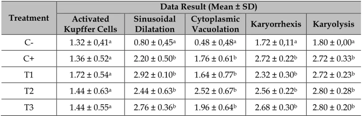
Table 2 Data result of Liver Histopathological Scoring (p>0.05)
Treatment
Data Result (Mean ± SD) Activated
Kupffer Cells Sinusoidal Dilatation Cytoplasmic Vacuolation Karyorrhexis Karyolysis C- 1.32 ± 0,41a 0.80 ± 0,45a 0.48 ± 0,48a 1.72 ± 0,11a 1.80 ± 0,00a C+ 1.36 ± 0.52a 2.20 ± 0.50b 1.76 ± 0.61b 2.72 ± 0.22b 2.72 ± 0.33b T1 1.72 ± 0.54a 2.92 ± 0.10b 1.64 ± 0.77b 2.32 ± 0.30b 2.72 ± 0.23b T2 1.44 ± 0.63a 2.44 ± 0.63b 2.52 ± 0.67b 2.56 ± 0.22b 2.80 ± 0.28b T3 1.44 ± 0.55a 2.76 ± 0.36b 1.96 ± 0.64b 2.68 ± 0.30b 2.80 ± 0.20b
Statistical analysis result from Table 2, activated kupffer cells variable showed there is no significant difference between each group C-, C+, T1, T2 and T3.
It also can be seen from sinusoidal dilatation, cytoplasmic vacuolation, karyorrhexis and karyolysis variable, the C- group is significantly difference with C+, T1, T2 and T3 group. Positive control C+ group did not show significantly difference compared to treatment group T1, T2 and T3. Treatment group T1, T2 and T3 also did not show significantly difference.
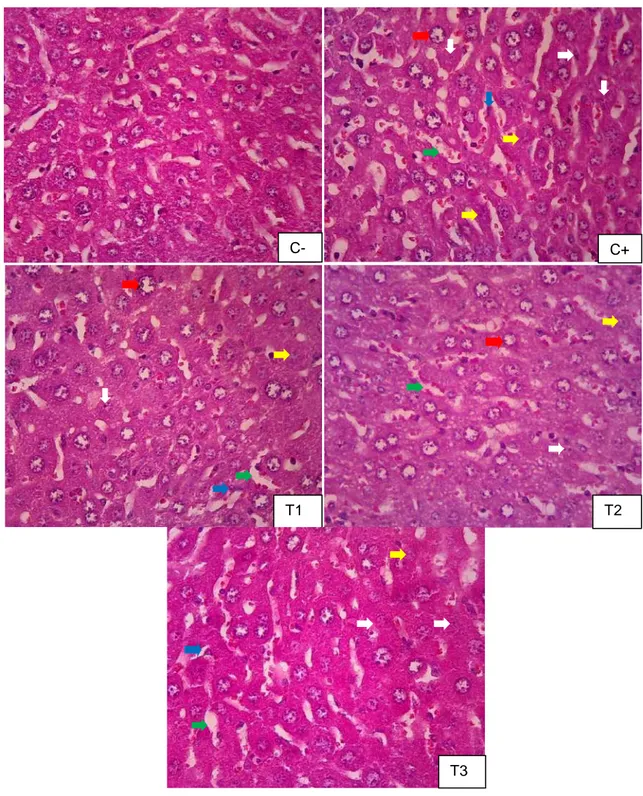
Wahyu Agung Nurdewantoro. dkk. Efektivitas Ekstrak Etanol Daun Kelor ...………. 18 C+ T1 T2 T3 C-
Figure 1 Histopathological features of intralobules liver. Pictures taken using Olympus®
CX-21 Microscope 400x magnification. Features showed tissues abnormalities in the liver: 1) activated kupffer cells (blue arrow), sinusoidal dilatation (green arrow), cytoplasmic vacuolation (red arrow), karyorrhexis (white arrow) and karyolysis (yellow arrow).
19 Negative control group that only
receive water had the significant difference with positive control group that received 0.4 mg/kg bw methylmecury. It exhibited the evidence of liver damage caused by methylmecury exposure.
All nutrients and liquids that are absorbed in the intestines enter the liver through the hepatic portal vein, except the complex lipid products, which are transported by the lymph vessels (Eroschenko, 2005), including methylmercury that go directly to liver from intestine (Desphande, 2002).
Moreover, binding of mercuric ions to sulfhydryl groups ROS and thus it increases the antioxidant may cause decreased glutathione levels, leading to by ameliorating the deleterious effects of free radical increases in levels of reactive oxygen species (ROS), such as superoxide anion radicals, hydrogen peroxide and regulates the intracellular contents of the reduced hydroxyl radicals, which provoke lipid, protein and DNA oxidation (Oda and Ashmawy, 2012).
According to Saleem (1995) in Mbikay (2012), among the major classes of phytochemicals found in the plant, flavonoids appear to carry most of free radical scavenging. Leave extracts were capable of scavenging peroxyl and superoxyl radicals. The ethanolic extract of Moringa oleifera leaves have some degree of hepatoprotective ability by reducing damage in liver. It was also reported that the major bioactive compounds of phenolics such as quercetin and kaempferol are responsible for antioxidant activity (Ezejindu, 2014; Siddhuraju and Becker, 2003).
There is also some disturbances in the negative control result, it showed in the C- group histopathological features. Localized necroses in some area of liver lobules occur. It could happen because of the high stressor from the environment (physical or
environ-mental) or from the procedure also from another variable such as feed for the mice that we used in this research. This research used Broiler feed for the experimental mice.
Stress could be one of the factor that can affect the health condition by any aversive stimulus, it is a state of threatened and the ability to cope with such stressful stimuli is a crucial determinant of health and disease. Stress responses are regulated by interactions between physiological and neurochemical factors. Physical or psychological stress induce changes in hypothalamic–pituitary–adrenal axis which culminates with glucocorticoids and chemical mediators release including adrenocorticotropic hormone (ACTH), norepinephrine (NE), serotonin, dopamine, and acetylcholine (Nadeem et al., 2006; Chakraborti et al., 2008; Goncalves et al., 2008). The metabolism of norepinephrine and dopamine leads to production of free radicals and reactive oxygen species (ROS). Glucocorticoids may increase the basal level of ROS in cells and also increase the toxicity of oxygen radical generators (Uysal et al., 2005).
CONCLUSION
Based on this research, it could be concluded Moringa oleifera leaves extract did not enough to protect hepatocytes from cytoplasmic vacuolation, sinusoidal dilatation, karyorrhexis and karyolysis also activated kupffer cells in mice (Mus musculus).
REFERENCES
Arsad, S.S., N.M. Esa and H. Hamzah. 2014. Histophatologic Changes in Liver and Kidney Tissues from Male Sprague Dawley Rats Treated with Rhaphidophora Decursiva (Roxb.) Schott Extract. J Cytol Histol S4: 001.
Wahyu Agung Nurdewantoro. dkk. Efektivitas Ekstrak Etanol Daun Kelor ...……….
20
Aschner, M., T. Syversen, D.O. Souza, J.B.T. Rocha and M. Farina. 2007. Involvement of Glutamate and Reactive Oxygen Species in Methylmercury Neurotoxicity. Brazilian Journal of Medical and Biological Research. 40: 285-291. Buraimoh, A.A., I.G. Bako and F.B.
Ibrahim. 2011. Hepatoprotective Effect of Ethanolic Leave Extract of Moringa oleifera on The Histology of Paracetamol Induced Liver Damage in Wistar Rats. Int. J. Anim. Veter. Adv., 3(1): 10-13.
Chakraborti, A., K. Gulati and A. Ray. 2008. Age Related Differences In Stress Induced Neurobehavioral Responses In Rats: Modulation By Antioxidants And Nitrergic Agents. Behav Brain Res; 194: 86 – 91.
Dalla Corte, C.L., C. Wagner, J.H. Sudati, B. Comparsi, G.O. Leite, A. Busanello, F.A.A. Soares, M. Aschner and J.B.T. Rocha. 2013. Effects of Diphenyl Diselenide on Methylmercury Toxicity in Rats. Biomed Res Int. 1-13.
Desphande, S.S. 2002. Handbook Of Food Toxicology. Marcel Dekker Inc. New York. United States of America.
Eroschenko, V.P. 2008. diFiore’s Atlas of
Histology with Functional Correlations. 11th Edition. Lippincott Williams and Wilkins. USA. 313-321.
Ezejindu, D.N., O.O. Udemezue and K.C. Chinweife. 2014. Hepatoprtective Effects of Moringa oleifera Extract on Liver of Wistar Rats. International J. of Res. In Med. and Health Sci. 3(5): 23-27.
Sevillano, M.A., T. García-Barrera, F. Navarro, N. Abril, C. Pueyo, J. López-Barea and J.L. Gómez-Ariza. 2015. Combination of Direct Infusion Mass Spectrometry and Gaschromatography Mass Spectrometry for Toxicometabolomic Study of Red Blood Cells and Serum of Mice Mus
Musculus After Mercuryexposure. Journal of Chromatography B, 985: 75–84.
Goncalves, L., A.L. Dafre, C.S. Goncalves, G.O. Cezar. 2008. A Temporal Analysis of The Relationships Between Social Stress, Humoral Immune Response and Glutathione-Related Antioxidant Defenses. Behav Brain Res. 192: 226
– 231.
Hodgson, E. 2010. A Textbook Of Modern Toxicology. John Wiley & Sons, Inc., Hoboken, New Jersey, USA. Chapter 4: 52; Chapter 7: 161-162.
Kesharwani, S., P. Prasad, A. Roy and R.K. Sahu. 2014. An Overview on Phytochemistry and Pharmacological Explorations of Moringa oleifera. UK Journal of Pharmaceutical and Biosciences Vol. 2(1): 34 – 41.
Mbikay, M. 2012. Therapeutic Potential of Moringa oleifera Leaves in Chronic Hyperglycemia and Dyslipidemia: a Review. Frontiers in Pharmacology. 3: 24.
Mucillo-Baisch, A.L., N. Mirlean, D. Carrazzoni, M.C.F. Soares, G.P. Goulart and P. Baisch. 2012. Health Effects of Ingestion of Mercury-Polluted Urban Soil: An Animal Experiment. Environ Geochem Health 34: 43 – 53.
Nadeem, A., A. Masood, N. Masood, R. Afzal Gilani and Z. Ahmad Shah. 2006. Immobilization Stress Causes Extracellular Oxidant-Antioxidant Imbalance in Rats: Restoration by L-NAME and Vitamin E. Eur Neuropsychopharmacol. 16: 260 - 267.
Newman, M.C. and W.H. Clements. 2007. Ecotoxicology : A Comprehensive Treatment. Chapter 13: 224-254.
Oda, S.S. and I.M. El-Ashmawy. 2012. Protective Effect of Silymarin on Mercury-Induced Acute
Nephro-21 Hepatotoxicity in Rats. Global
Veterinaria, 9 (4): 376-383
Rice, K.M., E.M. Walker Jr, M. Wu, C. Gillette and E.R. Blough. 2014. Environmental Mercury and Its Toxic Effects J Prev Med Public Health. 47(2): 74–83.
Saalu, L.C., B. Ogunlade, G.O. Ajayi, A.O. Oyewopo, G.G. Akunna and O.S. Ogunmodede. 2012. The Hepato-Protective Potentials of Moringa oleifera Leaf Extract on Alcohol-Induced Hepato-Toxicity in Wistar Rat. Am. J. Biotechnol. Mol. Sci. 2(1): 6-14.
Saleem, R. 1995. Study of Chemical Constituents of MoringaOleifera Lam. Ph.D.thesis, University of Karachi. Karachi.
Siddhuraju, P. and K. Becker. 2003. Antioxidant Properties of Various Solvent Extracts of Total Phenolic Constituents From Three Different Agroclimatic Origins of Drumstick Tree (Moringa Oleifera Lam.) Leaves. J Agric Food Chem. 51(8): 2144-55.
Sreelatha, S. and P.R. Padma. 2009. Antioxidant Activity and Total henolic Content of Moringa oleifera Leaves in Two Stages of Maturity. Plant Foods Hum. Nutr. 64: 303–
311.
Uysal, N., O. Acikgoz, S. Gonenc, B.M. Kayatekin, M. Kiray and A. Sonmez. 2005. Effects of Acute Footshock Stress on Antioxidant Enzyme Activities in The Adolescent Rat Brain. Effects Physiol Res. 54: 437-442.
WHO. 2007. Exposure to Mercury: A Major Public Health Concern. World Health Organization. Geneva. 1-4.